간질환자 두피뇌파의 발작사이간질모양방전과 배경파 델타영역: 전류원분포의 해부학적 연관성
경상대학교 의학전문대학원 신경과학교실1, 경상대학교 건강과학연구원2
정승욱1·권오영1,2·강종수1·하은옥1·정석원1 강규식1,2·강희영1,2·박기종1,2·최낙천1,2·임병훈1,2
Interictal Epileptiform Discharges and Background Delta Frequency Bands in Scalp EEGs of Epileptic Patients: Anatomical Correlation
between the Current Source Distributions
Seunguk Jung, M.D.1, Oh-Young Kwon, M.D.1,2, Jongsoo Kang, M.D.1,
Eun-ok Ha, M.D.1, Seokwon Jung, MPA1, Kyusik Kang, M.D.1,2, Heeyoung Kang, M.D.1,2, Ki-Jong Park, M.D.1,2, Nack-Cheon Choi, M.D.1,2, Byeong Hoon Lim, M.D.1,2
1
Department of Neurology, Gyeongsang National University School of Medicine;
2
Gyeongsang Institute of Health Science, Gyeongsang National University, Junju, Korea
Received 31 December 2008; received in revised form 31 March 2009; accepted 8 April 2009.
Background: The intermittent delta activity in electroencephalographies (EEGs) of patients with focal brain lesions has been reported to be a marker of an epileptogenic focus. This study investigated the concordance between the current source dis- tribution (CSD) of the interictal epileptiform discharges (IEDs) and that of the background delta frequency bands (DFBs) of the scalp EEG. Methods: We collected scalp EEGs of 13 patients with focal epilepsy that contained uniregional IEDs and unilateral delta to theta slow waves. We applied a distributed source model using LORETA
®to determine the CSD of the peak points of the IEDs and the DFBs of the background activity. Results: The CSDs of the DFBs were ipsilateral to the CSDs of the peak point of the averaged IEDs in ten patients, and bilateral with ipsilateral predominance in three patients.
In the cases with an ipsilateral CSD of the DFB, 8 of 10 patients had concordance of the CSD localization between the averaged IED and the DFB. In the cases with bilateral CSD of the DFB, 2 of 3 patients had concordance of the CSD local- ization between the averaged IED and the DFB. Conclusions: The CSD localization and lateralization appear to be con- cordant between the IEDs and the DFB of background activity in epileptic patients. Therefore, the CSD of the DFB in EEGs with visually observable slow activities may predict those of IEDs.
Key Words: Current source localization, Interictal epileptiform discharges, Delta frequency band, LORETA
Address for correspondence;
Oh-Young Kwon, M.D., PhD.
Department of Neurology,
Gyeongsang National University School of Medicine, 90 Chiram-dong, Jinju 660-702, Korea
Tel: +82-55-750-8077 Fax: +82-55-755-1709 E-mail: [email protected]
서 론
두피 뇌파(scalp electroencephalography, scalp EEG)
에서 지속적이거나 혹은 간헐적으로 관찰되는 국소 서파
는 뇌의 해부학적 병터와 관련이 있는 것으로 알려졌다.
1국소 서파는 간질환자의 배경파에서도 흔히 관찰되는데,
간질환자 두피뇌파의 발작사이간질모양방전과 배경파 델타영역: 전류원분포의 해부학적 연관성
이는 뇌에 존재하는 병리 소견에 의해 뇌 기능이 비특이적 으로 저하된 것을 의미한다.
2-5그러나 부분간질환자에게 서 한 쪽의 대뇌반구에서 관찰되는 서파가 단순히 뇌기능 저하에 대한 비특이적 증거만을 제공하는지 아니면 간질병 소의 존재를 반영할 수 있는지에 대해서는 명확하지 않다.
6부분간질환자의 뇌파에서 관찰되는 비대칭 델타파는 발 작간극파의 위치와 일치하는 경향이 있다.
5측두엽 간질환 자의 뇌파에서 관찰되는 측두엽의 간헐적인 델타활성은 복합부분간질의 진단에 높은 특이도와 예측가치를 가진 다.
7발작간 측두엽 델타활성은 측두엽간질 환자의 30~
90%에서 관찰되며 간질병소와 같은 쪽에서 관찰되었다.
8뇌파의 시각적 분석은 전류원의 위치를 어느 정도 추정할 수 있지만 한계가 있다. 다양한 위치에 존재하고 다양한 전위강도를 가지고 있는 전류원이 두피 전극에는 같은 뇌 파 신호를 발생시킬 수 있다. 따라서 단순한 시각적 판독 으로 뇌파의 발작사이간질모양방전과 델타파의 전류원을 찾는 것은 역부족이다. 디지털 뇌파 데이터를 이용한 전류 원분석은 수학적 계산을 통해 가장 적절한 전류원의 위치 를 찾아내는 방법이다.
9전류원분석을 이용한 이전의 연구들에서 국소 델타파의 전류원이 간질병소를 반영한다고 보고하였다. 뇌병터가 없는 잠복부분간질 환자의 뇌파에서 관찰되는 국소 델타 파전류원은 발작간극파전류원과 근접하게 위치하였다.
10뇌병터가 있는 부분간질 환자의 뇌파에서 관찰되는 국소 델타파전류원과 발작간극파전류원은 대부분 뇌병터 근처 피질에 위치하였다.
10,11이 연구들은 전류원 분석방법으로 모두 쌍극자전류원모델을 이용하였다. 부분간질환자 뇌파 에서 관찰되는 국소 델타파전류원과 발작간극파전류원의 위치관계를 분산전류원모델을 통해 관찰한 연구는 찾아보 기 힘들다. 이전의 연구에서는 적용되지 않았던 분산전류 원모델을 통한 분석에서도 일치하는 결과가 나온다면 그 결과의 신뢰도는 더욱 높아질 것이다.
발작간 두피 뇌파는 비침습적이고 비용이 낮아 간질의 평가를 위해 쉽게 접근할 수 있는 검사이다. 부분간질환자 의 발작간 두피 뇌파에서 발작사이간질모양방전의 낮은 민감도에 비해 배경서파는 상대적으로 민감도가 높다.
7,12따라서 배경서파의 전류원분포가 발작사이간질모양방전 의 전류원분포의 위치가 어느 정도 일치한다면 배경서파 가 간질병소를 파악하는데 사용될 잠재적 가능성이 더욱 높아질 것이다. 본 연구에서는 발작사이간질모양방전과 서파의 전기생리학적인 공통 부분을 분산전류원분석을 통해 확인해 보고자 하였다. LORETA (low-resolution brain electromagnetic tomography)를 이용해 부분간질
환자의 뇌파에서 관찰되는 발작사이간질모양방전과 일측 성 서파를 분석하여 각각의 전류원을 3차원으로 영상화하 여 해부학적 위치를 관찰하였다. 이러한 분석을 통해 두 전류원의 위치를 비교하여 배경파에 존재하는 델타활성 전류원이 발작사이간질모양방전 전류원의 위치를 추정하 는데 도움이 되는지 알아보았다.
방법과 대상
1. 연구대상
뇌파에서 일측성 발작사이간질모양방전과 같은 쪽 배경 활동에서 일측성 서파가 관찰되는 13명의 부분간질환자의 뇌파를 모았다(Fig. 1). 남자가 9명, 여자가 4명이었으며, 평균 연령은 45.5±18.0세(mean±SD)였다. 13명의 환자 가 2차 전신발작을 동반하거나(환자 1, 2, 3, 4, 5, 6, 7, 8, 10, 11, 12, 13) 혹은 동반하지 않는(환자 9) 복합부분발 작을 보였으며, 그 중 4명(환자 6, 7, 9, 11)은 단순부분발 작의 형태를 보이기도 하였다.
한 명(환자 1)에서는 뇌자기공명영상(MRI, magnetic resonance imaging)의 이상 소견이 발견되지 않았으나, 나머지 12명의 환자에서 다양한 뇌병터가 뇌컴퓨터단층촬 영(CT, computer tomography)이나 뇌자기공명영상에서 관찰되었다. 5명(환자 7, 9, 11, 12, 13)에서 사고에 의한 뇌타박상과 출혈에 의한 조직결손이 관찰되었으며, 2명 (환자 2, 3)에서 뇌출혈, 3명(환자 4, 8, 10)에서 뇌경색, 1명에서(환자 5) 대뇌피질이형성증에 의한 병터가 관찰되 었다. 나머지 1명은(환자 6) 일측성의 조직 결손이 있으나 병력상 그 원인을 알 수 없었다. 12명의 환자 중 10명에서 는 일측성 병터가 관찰되었다.
2. 뇌파 기록
각각의 환자에게 국제 10-20 체계에 따른 19개의 기본 전극(Fp1/2, F7/8, T7/8, P7/8, F3/4, C3/4, P3/4, O1/2, Fz, Cz, Pz)에 전측두엽전극(T1/T2) 또는 측두밑 전극(F9/10, T9/10, P9/10)을 포함한 21 혹은 25개의 전 극을 환자의 두피에 부착하였다. 뇌파는 32채널 Grass Telefactor
®Version 3.5 디지털 뇌파기를 이용하여 최소 30분 이상 기록하였다. 저주파여과는 1 Hz, 고주파여과는 70 Hz, 노치여과는 60 Hz, 표본율은 400 Hz였다.
3. 뇌파의 시각적 분석과 뇌파구간(EEG epoch)의 선택
기록된 뇌파는 1명의 신경과 전문의와 1명의 신경과 전
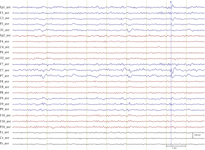
Figure 1. A segment of EEG obtained from an enrolled patient. A sharp wave (1) is seen on the temporal area of the left cerebral hemisphere and the continuous background slow activities are also seen on the same hemisphere during the EEG segment.
공의에 의해 시각적으로 분석되었다. 발작사이간질모양방 전의 분석을 위해 각 환자의 뇌파에서 뚜렷한 극파 또는 예파의 최대정점으로부터 전후 500 msec를 포함하는 구 간을 지정하였다. 각 환자의 뇌파에서 이러한 절편 20개를 모아 BESA
®(brain electrical source analysis) 소프트 웨어의 자료처리모듈을 이용해 평균한 발작사이간질모양 방전을 얻었다. 배경파 델타영역의 전류원분석을 위해서 는 각 환자의 뇌파에서 시각적으로 발작사이간질모양방전 과 잡파가 없이 일측성의 서파가 관찰되는 5초 길이의 뇌 파구간을 5군데 선택하였다.
134. LORETA를 이용한 전류원 국지화
발작사이간질모양방전의 전류원분포(current source distribution at the peak point of an averaged inter- ictal epileptiform discharge, CSD-IED)와 배경파의 델
타영역 전류원분포(current source distribution of the delta frequency band of the background activity, CSD-D)의 LORETA 영상을 얻기 위해서는 LORETA- KEY
®(KEY Institute for Brain-Mind Research, Switzerland) 소프트웨어를 사용하였다. 이를 통해서 각 환자에서 얻어진 뇌파절편을 컴퓨터로 계산하여 Talair- ach human brain atlas (digitized magnetic resonance images from the Brain Imaging Center, Montreal Neurologic Institute)에 등록된 세껍질둥근머리모형 (three shell spherical head model)을 이용해 전류밀도 의 분포를 3차원으로 영상화하였다. LORETA
®영상은 7 mm의 해상도를 가진 2,394 화소 안에 전류밀도를 그려 낸다.
발작사이간질모양방전의 전류원밀도의 분포와 해부학
적 위치를 확인하기 위해서는 각 환자의 평균한 발작사이
간질환자 두피뇌파의 발작사이간질모양방전과 배경파 델타영역: 전류원분포의 해부학적 연관성
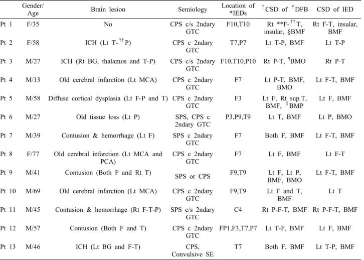
Table 1. Dermographic data and current source distribution of the enrolled patients Gender/
Age Brain lesion Semiology Location of
*IEDs
†CSD of
‡DFB CSD of IED
Pt 1 F/35 No CPS c/s 2ndary
GTC F10,T10 Rt **F-
††T,
insular, §BMF Rt F-T, insular, BMF
Pt 2 F/58 ICH (Lt T-
‡‡P) CPS c 2ndary
GTC T7,P7 Lt T-P, BMF Lt T-P
Pt 3 M/27 ICH (Rt BG, thalamus and T-P) CPS c/s 2ndary
GTC F10,T10,P10 Rt P-T,
¶BMO Rt P-T Pt 4 M/13 Old cerebral infarction (Lt MCA) CPS c 2ndary
GTC F7 Lt P-T, BMF,
BMO Lt F-T, BMF
Pt 5 M/58 Diffuse cortical dysplasia (Lt F-P and T) CPS c 2ndary
GTC F3 Lt F, Rt sup.T,
BMF,
∥BMP Lt F, BMF
Pt 6 M/27 Old tissue loss (Lt P) SPS, CPS c
2ndary GTC P3,P9,T9 Lt T, BMF Lt P, BMO Pt 7 M/39 Contusion & hemorrhage (Lt F) SPS c 2ndary
GTC F7 Both F, BMF Lt F-T, BMF
Pt 8 F/77 Old cerebral infarction (Lt MCA and
PCA) CPS c 2ndary
GTC F7 Lt F, BMF Lt F-T
Pt 9 M/41 Contusion (Both F and Rt T) SPS or CPS F9,T9 Lt F, Lt P,
BMF, BMO Lt F-T, BMF Pt 10 M/69 Old cerebral infarction (Lt MCA) CPS c 2ndary
GTC F9,T9 Lt F and T,
BMF Lt T
Pt 11 M/45 Contusion & hemorrhage (Rt F-T-P) SPS c/s 2ndary
GTC C4 Rt P-F-T, BMF Rt P-F-T, BMF
Pt 12 M/57 Contusion (Both F and T) CPS c 2ndary
GTC FP1,F3,T7,P7 Lt T-F, BMF Lt F, BMF
Pt 13 M/46 ICH (Lt BG and F-T) CPS,
Convulsive SE T7 Both F, BMF Lt T-P, BMF
*IED; interictal epileptiform discharge,
†CSD; current source distribution,
‡DFB; delta frequency band, SPS; simple partial seizure, CPS; complex partial seizure, GTC; generalized tonic-clonic seizure, SE; status epilepticus, Rt; right, Lt; left,
§BMF;
both medial frontal area,
∥BMP; both medial parietal area,
¶BMO; both medial occipital area, **F; frontal lobe,
††T; temporal lobe,
‡‡P; parietal lobe, BG; basal ganglia, MCA; middle cerebral artery, PCA; posterior cerebral artery, ICH; intracerebral hemorrhage, c; with, c/s; with or without.
Table 2. Concordance of localization between the current source distribution of delta frequency band and that of the averaged interictal epileptiform discharge
Lateralization of *CSD-D to
†CSD-IED Relationship between CSD-D and CSD-IED
Concordant Not concordant
Ipsilateral 8 2
Bilateral with ipsilateral predominance 2 1
*CSD-D; current source distribution of the delta frequency band of background activity,
†CSD-IED; current source distribution at the peak point of an averaged interictal epileptiform discharge.
간질모양방전의 음전위 최고정점에서의 전류원밀도를 영 상화하였다. 평균발작사이간질모양방전 정점의 전류원밀 도를 의미 있게 객관화하기 위해서 전류원밀도가 95‰ 이 상인 영역을 유의전류밀도구간(significant current den- sity areas)으로 정하여 LORETA 영상에 표시하였다. 배
경파 델타영역의 전류원밀도의 분석을 위해서는 일측 서
파가 관찰되는 뇌파 구간에서 얻어진 5개의 5초간의 뇌파
절편을 교차스펙트럼분석(cross spectrum analysis)으로
분석하여 1~3 Hz 범위의 델타영역을 영상화하였다. 3차
원 영상으로 만들어진 평균발작사이간질모양방전의 음전
Figure 2. Current source distribution of the background delta frequency band (DFB) and the averaged interictal epileptiform discharge of the eight patients with the CSD-D ipsilateral to CSD-IED. The locations of CSD-Ds are con- cordant with the CSD-IEDs in all eight patients.
In three (patient 9, 10, 12) of the eight patients, CSD-Ds are also located in the different areas from the locations of CSD-IEDs. The numbers mean the study numbers of the enrolled patients.
*CSD-D; current source distribution of the delta frequency band of background activity,
†CSD- IED; current source distribution at the peak point of an averaged interictal epileptiform discharge.
위 정점의 전류원밀도와 배경파 델타영역의 전류원밀도 분포를 비교하였다.
결 과
뇌파상의 극파와 예파의 위치는 다양하였으며, 일측의 전두엽과 측두엽, 두정엽과 중심부에 위치하였다. 한쪽 대
뇌반구에만 병터가 있는 10명의 환자에서는 모두 같은 대
뇌반구 안에서 발작사이간질모양방전과 배경활동 서파가
관찰되었다. 양쪽 대뇌반구에 병변이 있었던 2명의 환자
중 1명은(환자 9) 상대적으로 더 병터가 심한 대뇌반구 쪽
에 발작사이간질모양방전과 배경활동 서파가 관찰되었으
나, 다른 1명에서는(환자 12) 상대적으로 뇌병터가 심하지
않는 좌측 대뇌반구에 발작사이간질모양방전과 배경활동
간질환자 두피뇌파의 발작사이간질모양방전과 배경파 델타영역: 전류원분포의 해부학적 연관성
서파가 관찰되었다.
평균발작사이간질모양방전 정점의 전류원밀도분포(CSD- IED)는 모두에게서 일측성이었으며, 뇌파상 간질모양방전 이 나타난 전극과 같은 편측의 대뇌반구 안에 위치하였다.
CSD-D는 13명 중 10명에서 한쪽 대뇌반구에서만 관찰되 었고, CSD-IED와 같은 대뇌반구에 위치하였다. 이를 동 측델타전류원군(ipsilateral delta source group, I-DG) 이라 하였다. 나머지 3명은 모두 양측에서 CSD-D가 관찰 되었으나, 비교적 CSD-IED와 동일한 편측 대뇌반구에서 더 두드러지게 나타났다. 이를 동측우세양측델타전류원군 (bilateral delta source group with ipsilateral pre- dominancy, B-DG)이라고 하였다(Table 1, 2 and Fig.
2). 편측성과 위치에 대한 결과 판정에 양측 중앙부위에 대칭적으로 나타난 전류원밀도는 고려하지 않았다.
대상 환자 13명 중 10명(환자 1, 2, 3, 5, 7, 8, 9, 10, 11, 12)에서 CSD-IED가 CSD-D와 일치하거나 서로 겹쳐 있었다. 이 중 2명(환자 5, 7)은 B-DG에 속하였으며 나머 지 8명은 I-DG에 속하였다. I-DG에 속한 8명의 환자 중 3명(환자 9, 10, 12)에서는 CSD-D는 CSD-IED가 포함하 는 위치와 동떨어진 위치에서도 CSD가 관찰되었다. 9번 환자에서 CSD-IED는 왼측 전두엽과 측두엽에서 나타났 으며, CSD-D는 왼측 전두엽과 두정엽에서 관찰되었다.
10번 환자에서는 CSD-IED는 왼측 측두엽에서 나타난 반 면, CSD-D는 왼측 측두엽과 전두엽에서 관찰되었다. 12 번 환자에서는 CSD-IED가 왼측 전두엽에서 관찰되었으 며, CSD-D는 왼측 전두엽과 측두엽에서 관찰되었다 (Table 1, 2 and Fig. 2).
대상 환자 13명 중 3명(환자 4, 6, 13)에서는 CSD-IED 와 CSD-D가 일치하지 않았다. 4번 환자는 CSD-IED는 왼측 전두엽-측두엽에 나타났으나, CSD-D는 왼측 두정 엽에 위치하였다. 6번 환자는 왼측 두정엽에 CSD-IED가 위치하였으나, CSD-D는 왼측 측두엽에 위치하였다. 13번 환자는 CSD-IED는 왼측 측두엽과 두정엽에서 관찰되었 으나, CSD-D는 양측 전두엽에서 관찰되었다(Table 1, 2).
모든 환자에서 측향화된 CSD외에도 내전두엽 또는 내 후두엽과 내두정엽에서 추가적인 CSD가 대칭적으로 관찰 되었다. 13명 중 11명(환자 1, 2, 5, 6, 7, 8, 9, 10, 11, 12, 13)에서는 내전두엽에서 대칭적 CSD-D가 관찰되었다. 그 중 1명(환자 9)에서는 내후두엽에서도 관찰되었다. 나머지 2명은(환자 3, 4) 내후두엽에서 대칭적 CSD-D가 관찰되 었다. 8명(환자 1, 4, 5, 7, 9, 11, 12, 13)에서는 CSD-IED 가 양측 내측두엽에서 대칭적으로 관찰되었고, 1명(환 자 6)에서는 CSD-IED가 내후두엽에 대칭적으로 관찰
되었다.
고 찰
본 연구에서는 부분간질환자에서 관찰되는 배경파에 존 재하는 델타활성의 전류원이 발작사이간질모양방전의 전 류원 위치를 추정하는데 유용한지 알아보고자 하였다. 대 상이 된 환자 13명 중 10명의 환자에게서 발작사이간질모 양방전 정점의 전류원 분포와 델타 활동파의 전류원 분포 가 일치하거나 겹쳤다(Table 1). 이는 뇌파의 배경활동에 서 보이는 비대칭성의 일측성 델타파를 통해 극파의 발생 위치를 추정할 수 있다는 점을 시사한다.
수술 전 자기뇌파에서 관찰된 발작간극파를 분석하여 간질유발병소가 밝혀진 12명의 환자를 대상으로 서파와 발작간극파의 쌍극자전류원밀도(density of current di- pole source)의 해부학적 위치를 비교한 연구에서도 서파 의 쌍극자전류원밀도가 극파의 발생 영역과 근접한 위치 에서 증가하는 것이 관찰되었다.
10이 연구에서 서파의 활 성은 극파가 발생하는 영역의 가장자리 위치하였다. 이는 서파가 간질병소화(epileptogenic precoss)에 의해 손상 된 주변 조직에서 발생하는 것을 의미한다. 따라서 부분간 질환자의 뇌파에서 관찰되는 비대칭 서파는 간질 병소화 에 따른 조직손상과 국소 뇌기능 저하에 의해 발생한다는 것을 알 수 있다. 간질환자의 뇌파에서 관찰되는 비특이적 인 서파가 간질병소화의 영향에 따른 기능변화 때문에 발 생한다는 사실은 이전의 여러 연구에서도 제시되었다.
14-17부분간질환자의 뇌파에서 관찰되는 발작간극파와 비대 칭 델타파의 위치관계를 시각적으로 비교한 연구가 있다.
이 연구에서는 비대칭 델타파를 대조군과 비교하여 관찰
하였다. 대상이 된 부분간질 환자 22명 중 20명에서 델타
파의 비대칭이 관찰되었다. 결과에서 17명에서 델타파의
비대칭이 간질병소와 연관성이 있었다.
5내측두엽위축증
이 있는 측두엽간질 환자에서 관찰되는 국소 델타파와 간
질모양방전의 위치를 시각적으로 분석한 연구에서도 델타
파는 측두엽위축증이 상대적으로 더 심한 위치보다 간질
모양방전의 위치와 일치하는 경향이 관찰되었다. 이 연구
에서 병터가 양쪽 대뇌반구에 모두 존재하는 경우에도 간
질모양방전이 한쪽에서만 관찰되는 경우는 같은 쪽 대뇌
반구 안에서 델타파가 나타났다. 또한 간질모양방전이 양
측성인 경우 델타파도 양측에서 나타났다. 양쪽 대뇌반구
에 내측두엽위축증이 있으며 상대적으로 덜 심한 쪽에서
간질모양방전이 나타난 환자의 경우에도 델타파는 병터의
위축 정도가 심한 쪽보다는 간질모양방전이 나타난 쪽에
서 관찰되었다.
12뇌종양 환자에서 자기뇌파로 기록된 자 발적 뇌활성의 분석에서도 델타활성의 쌍극자들은 병변 과 같은 쪽의 대뇌반구에서 관찰되었다.
1864채널의 뇌파 에서 관찰된 국소 델타파와 발작간극파의 등가쌍극자모 델(equivalent dipole medeling)을 통한 분석에서 델타파 의 전류원은 병터 자체 보다는 근접한 피질에서 관찰되었 으며, 발작간극파의 전류원과 위치가 일치하는 경향을 보 였다.
11이는 국소 델타파가 해부학적 병터보다는 간질병 소에 의한 뇌 기능저하를 더 민감하게 반영하는 것을 의미 한다.
뇌파에서 얻어진 데이터를 통해서 전류원을 찾는 데는 서로 차이가 있는 접근법이 있다. 뚜렷하게 구별이 되는 쌍극자전류원의 위치와 방향을 찾아내는 방법이 쌍극자모 델(discrete dipole model)이고, 전류원의 대뇌 회백질에 서의 분포를 찾아내는 방법이 분산전류원국지법(dis- tributed source localization)이다. LORETA
®는 분산전 류원국지법을 이용해 전류밀도의 분포를 3차원으로 영상 화하여 찾아내는 분석방법이다.
9,19,20본 연구에서는 부 분간질환자의 뇌파에서 관찰되는 발작사이간질모양방전 과 배경파 델타영역의 전류원밀도분포를 찾아내기 위해 이 전의 연구에서는 관심이 적었던 분산전류원국지법인 LORETA
®를 이용하였다.
본 연구의 결과에서 3명의 환자에서는 CSD-D와 CSD- IED가 일치하지 않고 동떨어져 있었다. 간질유발병소가 밝혀진 12명의 환자를 대상으로 수술 전에 시행한 자기뇌 파(MEG)에서 관찰된 서파와 발작간극파의 쌍극자전류원 밀도의 해부학적 위치를 비교한 연구에서도 이러한 증례 가 포함되어 있었다. 이 연구의 대상 환자 12명 중에 11예 에서는 두 가지 전류원의 위치의 거리가 3.3 cm 이내에서 존재하였지만 한 예에서는 거리가 5.7 cm 떨어져 분석에 서 제외되었다.
10이 경우에서는 간질수술 후에도 간질발 작이 조절되지 않았다. 두 전류원분포가 일치하지 않은 예 의 이유를 언급하기에는 본 연구 자료나 이전의 자료로서 는 어렵지만, 전기생리학적으로 뇌기능을 저하시키는 병 리현상이 발작사이간질모양방전의 전류원과 동떨어져서 형성된 경우로 해석할 수는 있을 것으로 판단한다.
발작간극파와 배경파 델타영역의 전류원분석에 있어 중 앙부 대칭 활성은 피하기 어렵다. 본 연구의 배경파 델타 영역과 발작사이간질모양방전의 분석 결과에서도 뇌의 중 앙 부위에 대칭적으로 관찰되는 전류원밀도분포가 관찰되 는 경우가 많았다. 전류원국지화를 통한 분석에서 나타나 는 중앙부위 전류원의 우세(interhemispheric prepon- derance)는 다양한 원인에 의해 형성될 수 있어 판단에 주
의를 필요로 한다. 눈깜빡임을 포함한 생리적인 신호 및 무작위노이즈(random noise), 10 Hz 사인파(sine wave) 와 같은 아티팩트는 중앙 부위에 잘못된 전류원밀도분포 를 형성할 수 있다.
21,22부분간질환자에서 발작사이간질모양방전의 낮은 민감 도를 고려할 때 상대적으로 높은 민감도를 가지는 일측성 서파가 간질의 진단과 간질병소의 추정에 도움이 될 수 있 으나 아직 증거가 충분하지 않다. 본 연구에서는 발작사이 간질모양방전과 서파의 전기생리학적인 공통 부분을 분산 전류원분석을 통해 확인해 보았다. 본 연구의 결과에서 배 경서파의 전류원분포가 발작사이간질모양방전의 전류원 분포의 위치와 어느 정도 일치한 점은 배경서파가 간질병 소를 파악하는데 사용될 잠재적 가능성을 더욱 높였다고 판단한다.
REFERENCES
1. Gloor P. Brain lesions that produce delta waves in the EEG.
Neurology 1977;27:326-333.
2. Reinikainen KJ, Keranen T, Lehtinen JM, Kalviainen R, Saari T, Riekkinen PJ. CT brain scan and EEG in the diagnosis of adult onset seizures. Epilepsy Res 1987;1:178-184.
3. Nuwer MR. Frequency analysis and topographic mapping of EEG and evoked potentials in epilepsy. Electroencephalogr Clin Neurophysiol 1987;69:118-126.
4. Gibbs J, Appleton RE, Carty H, Beirne M, Acomb BA. Focal electroencephalographic abnormalities and computerised to- mography findings in children with seizures. J Neurol Neuro- surg Psychiatry 1993;56:369-371.
5. Panet-Raymond D, Gotman J. Asymmetry in delta activity in patients with focal epilepsy. Electroencephalogr Clin Neur- ophysiol 1990;75:474-481.
6. Engel J JR. Recent advances in surgical treatment of temporal lobe epilepsy. Acta Neurol Scand Suppl 1992;86:71-80.
7. Reiher J, Beaudry M, Leduc CP. Temporal intermittent rhyth- mic delta activity (TIRDA) in the diagnosis of complex partial epilepsy: sensitivity, specificity and predictive value. Can J Neurol Sci 1989;16:398-401.
8. Di Gennaro G, Quarato PP, Onorati P, et al. Localizing sig- nificance of temporal intermittent rhythmic delta activity (TIRDA) in drug-resistant focal epilepsy. Clinical Neurophysi- ology 2003;114:70-78.
9. Koles ZJ. Trends in EEG source localization. Elec- troencephalography and Clin Neurophysiol 1998;106:127-137.
10. Kaltenhauser M, Scheler G, Rampp S, Paulini A, Stefan H.
Spatial intralobar correlation of spike and slow wave activity localisations in focal epilepsies: A MEG analysis. NeuroImage 2007;34:1466-1472.
11. Huppertz H-J, Hof E, Klisch J, Wagner M, Lucking CH,
Kristeva-Feige R. Localization of interictal delta and epilepti-
간질환자 두피뇌파의 발작사이간질모양방전과 배경파 델타영역: 전류원분포의 해부학적 연관성