하악골 절제술후 간단한 과두재위치 소형금속판을 이용한 재건 금속판의 적용: 증례보고
Abstract (J. Kor. Oral Maxillofac. Surg. 2002;28:231-233)
231
Ⅰ. 서 론
법랑아세포종, 점액종, 악성종양과 같은 하악골 종양의 수술은 하악골 절제술을 필요로하게 된다. 절제술 후 하악골의 연속성 소실을 일시적으로 재건하기 위해 재건판(reconstruction plate)을 사용함으로써 정상적인 안모 외형을 유지시키고 정기적 관찰을 통해 질환의 재발 여부를 평가하게된다. 재건판은 하악골을 절 제하기 전에 골 표면에 적합시키고 몇 개의 나사로 임시 고정하 게 된다
1,2). 하악골 표면에 정확히 외형을 적합시키는 것은 골편 의 전위와 상부 피부의 압박성 궤양 형성을 방지하기 위해 대단 히 중요하다. 어떤 학자는 상방 피부의 긴장성 압박과 궤양 발생 을 최소화하기 위해 재건판의 설측 적용을 소개한 바 있다
3). 그 러나 하악골에 거대한 팽창성 협측 종괴가 존재한다면 종물과 하악골을 절제하기 전에 재건판을 미리 외형에 맞게 구부려 적 합시키는 것이 어렵거나 불가능할 수 있다.
저자는 단순한 과두재위치 소형금속판을 사용하여 하악골 절
제술 시행 후 재건판을 적절히 적용하는 술식을 소개하고자 한 다.
Ⅱ. 증례보고
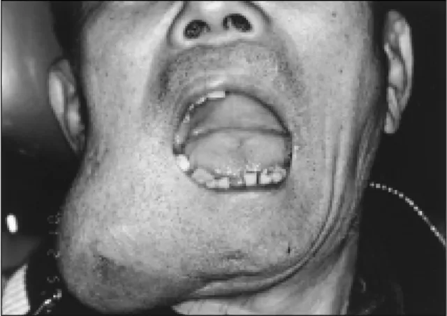
거대한 법랑아세포종을 보유한 64세 남자환자가 전신마취 하 에 협측 거대종물을 포함한 하악골 절제술을 시행 받았다(Fig. 1).
하순 분할 절개와 악하부 절개를 통해 종물과 하악골을 노출시 켰으며 강선으로 악간고정을 시행하였다. 하악골을 절제하기 전 에 티타늄 금속판(1.5-mm)을 구부려서 상악골 후방부의 협측면 과 하악골 상행지 협측면에 적합시킨 후 보관하였다. 종물을 분 리하고 하악골을 절제하기 직전에 미리 구부린 소형 금속판을 다시 적합시켜 양측에 3개의 단방피질성 나사(drill-free screws)들 로 고정하여 과두 골편의 위치를 유지시킨 후 종물의 경계부로 부터 1cm 부위까지 하악골을 절제하였다. 원심부를 먼저 절단한 후 종물과 함께 근심부를 신속히 절단하였고 전기소작기와 골왁 스로 즉시 지혈처치를 하였다. 반대측 하악골의 외형을 참고하 면서 유연한 알루미늄 template를 적합시킨 후 재건 금속판을 template에 맞추어 구부렸고 하악골 결손부에 적합시키면서 최 종 외형을 수정하였다(Fig. 2, 3, 4, 5). 양측에 2.4-mm 직경의 4개 의 양측 피질성 나사로 금속판을 고정한 후 악간고정과 과두재 위치 금속판을 제거하였다. 자연스런 하악골의 운동을 확인한 후 구내 연조직 결손 재건을 위해 전방기저 측방 설피판을 형성
하악골 절제술후 간단한 과두재위치 소형금속판을 이용한 재건 금속판의 적용: 증례보고
김 영 균
대진의료재단 분당제생병원 구강악안면외과
김 영 균
463-050, 경기도 성남시 분당구 서현동 255-2 대진의료재단 분당제생병원 치과 구강악안면외과 Young-Kyun Kim
Dept. of OMFS, Pundang Jesaeng Hospital, Dejin Medical Center, Korea 255-2,
Tel : 82-31-779-0731 Fax : 82-31-779-0744 E-mail : [email protected]
APPLICATION OF RECONSTRUCTION PLATE USING SIMPLE CONDYLAR REPOSITIONING MINIPLATE AFTER SEGMENTAL RESECTION OF MANDIBLE
Young-Kyun Kim, D.D.S., M.S.D., Ph.D.
Department of Oral and Maxillofacial Surgery, Pundang Jesaeng Hospital, Dejin Medical Center, Korea
Temporary reconstruction of the mandibular continuity defect resulting from the ablative tumor surgery with a reconstruction plate can be used for the preservation of normal facial contour and oral function and for periodic follow up of recurrence. Reconstruction plates are adapted to the bone before the resection and provisionally fixated with some screws. Accurate contouring and adaptation are very important for the prevention of displacement of bony stumps and decubituous skin ulcer. However, if there is large expanding buccal tumor mass in mandible, it is very difficult or even impossible to contour the plate before resection. I, therefore, introduce the reconstruction plate application technique using a simple condylar repositioning miniplate after segmental mandibular resection.
Key words : Reconstruction plate, Condylar repositioning
대구외지 2002;28:231-233
232
Fig. 1. Preoperative clinical photograph of amelob- lastoma. Buccal fluctuant swelling and expansile mass are observed.
Fig. 2. Buccal expansile mass was isolated and proximal resection line was marked with reciprocating saw.
Fig. 3. 1.5-mm miniplate was adapted between the condylar segment and maxillary buccal surface.
Miniplate was fixated temporarily with 3 monocortical drill-free screws on each side.
Fig. 4. The reconstruction plate was bent to conform to the shape of the template. The final adjustments were made directly on the mandible and fixated with screws.
Fig. 5. Drawing of application of reconstruction plate using simple condylar repositioning miniplate after segmental resection of mandible.
crp: condylar repositioning miniplate, m: tumor mass, rp: reconstruction plate
Fig. 6. Postoperative orthopantomogram. Mandibular
condyle position was maintained.
하악골 절제술후 간단한 과두재위치 소형금속판을 이용한 재건 금속판의 적용: 증례보고
233 하여 층별 봉합을 시행하였다. 술후 정상적인 악골의 기능과 안
면 외형을 회복할 수 있었다(Fig. 6).
Ⅲ. 총괄 및 고찰
술후 근심 과두골편의 안정성을 유지하고 인위적인 악관절 손 상을 피하기 위해 다양한 과두재위치 장치들이 악교정 수술에서 사용되어 왔다
4-7). 악교정 수술중 과두의 위치변화는 술후 재발과 악관절 장애를 유발할 수 있기 때문에 술전의 과두 위치를 유지 하는데 특별한 주의를 기울여야 한다. 이 등
7)은 고정적인 두개 골의 어떤 부위에 기반을 둔 새로운 과두재위치 장치를 개발하 여 임상에 사용하였다.
하악골 절제술 후 과두의 위치를 안정시키면서 재건판을 적용 시키는 일반적인 방법은 하악골 분절골절제술 전에 template로 하악골 외형을 채득하여 재건금속판을 하악골 외형에 맞게 정확 히 적합시킨다. 금속판의 양 끝에 최소한 4개의 양측피질성 나사 로 고정한 후 제거하여 보관하고 하악골 절제술을 시행한다. 이 런 과정을 거치는 목적은 술중 과두-관절와의 위치를 그대로 유 지시키고자 하는 것이다. 그러나 협측의 거대한 팽창성 병소가 존재하는 경우엔 골절제술 시행 전에 금속판을 하악골 표면에 적합시키는 것이 불가능하다. 따라서 종물을 절제한 후에 재건 금속판을 적용해야 하며 골편의 전위 및 함몰을 최소화하기 위 해선 아치바를 이용한 술중 악간고정법에 의존할 수 밖에 없다.
본 증례에서는 하악골 협측에 거대한 팽창성 종괴가 존재하는 법랑아세포종 절제술을 시행하기 전에 재건 금속판을 미리 하악 골 외형에 맞추어 적합시키는 것이 불가능하였다. 따라서 항상 일정한 위치를 유지하는 상악골 협측 후방부와 하악골 근심 과 두골편을 1.5-mm 소형 금속판으로 고정 연결함으로써 과두골편
의 위치를 유지하였으며 하악골 절제술 시행 후 재건판을 정확 히 적합시킴으로써 양호한 수술 결과를 얻을 수 있었다.
IV. 결 론
하악골 절제술 시행 후 재건금속판을 적용할 때 단순 소형금속 판을 상악골 후방부와 근심 과두골편을 연결함으로써 과두골편 의 위치 변화를 최소화하고 원심골편의 함몰을 방지하면서 재건 술을 시행할 수 있었으며 임상에서 적절히 적용할 수 있는 증례 라 사료되어 기술적 술기를 보고하는 바이다.
참고문헌