RESEARCH ARTICLE
Importance of tear volume for positivity of tear matrix metalloproteinase-9
immunoassay
Jong Hwa JunID*, You Hyun Lee, Myeong Jin Son, Harim Kim
Department of Ophthalmology, Dongsan Medical Center, Keimyung University School of Medicine, Daegu, Korea
Abstract
The tear matrix metalloproteinase-9 (MMP-9) immunoassay (Inflammadry) exhibits variable results in dry eye (DE) patients. We investigated if the tear volume in DE patients affects the results of MMP-9 immunoassay in clinical and in vitro settings. This cross-sectional study enrolled 188 eyes of 188 DE patients. The clinical symptoms and signs of DE were assessed using the Ocular Surface Disease Index and visual analog scale, strip menisco- metry, tear break-up time, and tear meniscus height (TMH), area (TMA), and depth (TMD) using swept-source optical coherence tomography and corneal and conjunctival staining scores. For quantitative evaluation, the bands produced by the InflammaDry test were ana- lyzed with ImageJ. DE subjects were grouped according to MMP-9 positivity and TMH. The InflammaDry-positive group showed greater TMH, TMA, and TMD than the MMP-9-nega- tive group (p<0.05). InflammaDry test band density in the high TMH group was significantly greater than that in the low and normal TMH groups (p<0.05). InflammaDry test band den- sity correlated positively with TMH, TMA, and TMD (all p<0.05). InflammaDry test results were influenced by tear volume. Low tear volume in aqueous tear-deficient DE may induce false-negative results, and reflex tearing during the test may induce false-positive results.
Introduction
Dry eye (DE) is a multifactorial disease caused by changes in the quality and/or quantity of the precorneal tear film [1]. Current diagnosis of DE is based on a combination of clinical signs, such as those determined by the Schirmer test, tear break-up time (tBUT), and staining scores, as well as symptoms, which are determined by formalized questionnaires. Although the Schir- mer test results, tBUT, and corneal staining scores have long been used as the main indicators of DE, these tests lack objectivity and show low reproducibility due to user-dependent errors [2,3]. Therefore, efforts are being made to develop new objective methods for DE diagnosis, such as tear osmolarity tests and the tear matrix metalloproteinase (MMP)-9 immunoassay [4,5].
a1111111111 a1111111111 a1111111111 a1111111111 a1111111111
OPEN ACCESS
Citation: Jun JH, Lee YH, Son MJ, Kim H (2020) Importance of tear volume for positivity of tear matrix metalloproteinase-9 immunoassay. PLoS ONE 15(7): e0235408.https://doi.org/10.1371/
journal.pone.0235408
Editor: Carlos Zaragoza, Universidad Francisco de Vitoria, SPAIN
Received: December 2, 2019 Accepted: June 15, 2020 Published: July 10, 2020
Copyright:© 2020 Jun et al. This is an open access article distributed under the terms of theCreative Commons Attribution License, which permits unrestricted use, distribution, and reproduction in any medium, provided the original author and source are credited.
Data Availability Statement: All relevant data are within the manuscript and its Supporting Information files.
Funding: This research was supported by the Keimyung University Research Grant of 2017 from Keimyung University (http://www.kmu.ac.kr/uni/
main/main.jsp). JHJ received this fund. The funders had no role in study design, data collection and analysis, decision to publish, or preparation of the manuscript.
Competing interests: The authors have declared that no competing interests exist.
MMP-9 plays two key roles in ocular pathophysiology. In tears, MMP-9 expression is nor- mally below 40 ng/mL and is secreted from the ocular surface epithelium [6]. MMP-9 main- tains the epithelial barrier function by cleaving epithelial basement membrane components and tight-junction proteins, such as ZO-1 and occludin [7,8]. On the other hand, MMP-9 is also related to the pathogenesis of various diseases, such as sterile ulceration, ocular allergy, keratoconus, conjunctivochalasis, and DE [3,9–11]. In the pathology of DE, from the initiation of tear hyperosmolarity in the early phase of DE, a vicious cycle of DE likely induces inflamma- tory processes and triggers the release of MMP-9 in a relatively late phase of DE [12]. As detec- tion of elevated tear MMP-9 could be an ideal tool for diagnosis and management of DE, a tear MMP-9 immunoassay could be valuable to clinicians [13].
Most studies have reported superior sensitivity and specificity of MMP-9 immunoassay compared to conventional diagnostics, and the MMP-9 immunoassay reflected the clinical signs and symptoms well in those reports [5,6,14]. However, Lanza et al. failed to find differ- ences in clinical symptoms, underlying diseases, and clinical signs of DE between MMP-9-pos- itive and -negative groups [15,16]. In addition, although not statistically significant, Schargus et al. showed that the Schirmer score was higher in the MMP-9-positive group than in the -negative group [3]. Further, a recentin vitro study that investigated the pre-form and active- form of human MMP-9 in a commercially available MMP-9 immunoassay revealed depen- dency of the result on the loading volume [17]. Thus, not only severely tear-deficient DE con- ditions, such as Sjo¨gren syndrome and ocular graft-versus-disease, but also mild to moderate aqueous tear-deficient DE may produce false-negative results.
Herein, we aimed to elucidate the impact of tear volume on positivity in a commercially available MMP-9 immunoassay (Inflammadry, Quidel Corporation, San Diego, CA, USA) in DE patients and investigated changes in MMP-9 immunoassay-positivity according to the tear volume, as measured by swept-source optical coherence tomography (OCT).
Materials and methods Study population and setting
This study was conducted in accordance with the ethical principles of the Declaration of Hel- sinki. The study protocol and written informed consent were approved by the Keimyung Uni- versity Dongsan Medical Center Institutional Review Board (IRB no. DSMC 2017-06-008- 005). This cross-sectional study was performed in DE patients who visited our ophthalmic department from April 1, 2017, through June 30, 2019. Written informed consent was
obtained from all adult patients and two patients under 19 received consent from their parents after a detailed explanation of the study was provided. The study investigator collected clinical data and MMP-9 immunoassay (InflammDry) results from the right eye of each enrolled patient to avoid a coupling effect.
The clinical diagnosis of DE was made according to the following three criteria in the right eye of each patient: Ocular Surface Disease Index (OSDI) score of more than 12, tBUT of less than 10 s, and corneal fluorescein staining results of 1 or more by the Oxford scheme. The patients who met all the three criteria were classified as having DE. Exclusion criteria were as follows: (1) active or recent keratitis or conjunctivitis within the previous 4 weeks, (2) moder- ate to severe conjunctivochalasis, (3) any stage of keratoconus, (4) lacrimal drainage disorders, such as lacrimal punctal stenosis, deformed lacrimal punctum, canalicular anomalies, and nasolacrimal duct obstruction, or temporary or permanent punctal occlusion, (5) topical or systemic corticosteroid treatment or immunomodulatory therapy within the previous 4 weeks, (6) fluorescein, cornstarch, or Dacron allergy, (7) undergone ocular surgery within the
previous 6 months, or had ocular trauma in the previous 3 months, and (8) use of contact lenses within the previous 72 h.
Clinical assessment
Clinical examinations were performed in the following order: tear meniscus measurements by anterior swept-source OCT, OSDI questionnaire/visual analog scale (VAS), tear MMP-9 immunoassay (InflammaDry test), strip meniscometry (SM tube1, Echo Electricity Co., Ltd., Fukushima, Japan), tBUT, and both corneal and conjunctival staining scores by fluorescein staining.
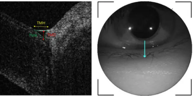
The anterior segment OCT image was acquired with a corneal module mounted on a swept-source OCT device (DRI OCT Triton; Topcon, Tokyo, Japan). All tear parameters were measured by a single experienced technician. Participants were instructed to blink three times, followed by gazing straight forward, and the reference was aligned to the inferior cornea and the center of the lower eyelid. A single vertical raster image of the right eye was obtained 3 s after voluntary blinking in a dark room. Tear meniscus height (TMH) was measured as the distance between the cornea–meniscus junction and the lower eyelid–meniscus junction. Tear meniscus depth (TMD) was taken as the vertical distance from the interface of the cornea with the lower eyelid to the TMH line. Tear meniscus area (TMA) was calculated proportionally using ImageJ software (ImageJ 1.44p; National Institutes of Health, Bethesda, MD, USA) [18,19]. First, the anterior segment OCT image with the measured TMH value was captured.
Then, the captured image was mounted to the ImageJ program and the TMH and TMA were measured. After the ratio of the ImageJ-based TMH to the anterior segment OCT-based TMH was obtained, the anterior segment OCT-based TMA was calculated by the square of the ratio multiplied by the ImageJ-based TMA (Fig 1).
Strip meniscometry was performed using a commercially available kit composed of a poly- ethylene terephthalate panel and centrally embedded 8 um nitrocellulose membrane filter
Fig 1. Measurements of TMH, TMD, and TMA. TMH (yellow line) was measured as the distance from the cornea–meniscus junction to the lower eyelid–meniscus junction. TMD (red line) was measured as the vertical distance from the interface of the cornea with the lower eyelid to the yellow line. TMA (area bounded by green lines) was calculated proportionally using ImageJ software from the OCT measured TMH value, TMA was calculated by the square of the ratio multiplied by the ImageJ-based TMA.
https://doi.org/10.1371/journal.pone.0235408.g001
paper strip. The strip was positioned adjacent to the right lower palpebral conjunctiva and the examiner tried to absorb the tear lake of each participant during 5 seconds. Tear volume was estimated based on the length of wetted strip.
After tear meniscus measurements, participants’ symptoms were evaluated using the OSDI questionnaire [20]. This consisted of 12 questions on eye-related symptoms, vision-related function, and environment-related symptoms. The scores range from 0 to 100, where 0 indi- cates no discomfort, and 100 indicates the most severe symptom and disability. In addition, the severity of ocular pain was analyzed using a VAS, where 0 indicates no pain, and 10 indi- cates the worst possible pain [21].
The tear MMP-9 immunoassay (InflammaDry test) was performed according to the manu- facturer’s instruction by a single examiner (JHJ). The sampling fleece was contacted three times at each location of the inferior palpebral conjunctival (temporal, middle, nasal; from nasal to temporal direction) and was rested against the temporal inferior palpebral conjunctiva for an additional 5 s. After assembling the sampling fleece to a sample collector, the test strip was dipped into a buffer solution for 20 s. To evaluate the result-band’s density, the result win- dow was photographed with a slit-lamp biomicroscope-mounted single-lens reflex camera (Canon EOS 700D, setting: ISO 400, shutter speed 1/200 s) at 10 min after test initiation. For quantitative evaluation of the result band of the InflammaDry test, band densitometry was per- formed with ImageJ. Qualitative evaluations (positive/negative) were also performed by a sin- gle examiner (JHJ), and faint to strong bands were judged as positive, and the absence of a band was considered as negative, according to the manufacturer’s instruction.
tBUT was evaluated after touching a fluorescein strip wetted with normal saline (Haag- Streit AG, Koenig, Switzerland) to the lower inferotemporal palpebral conjunctiva. After blinking three times, the interval from the last blink to the first appearance of dark spots on the corneal surface was measured using a stopwatch.
Corneal and conjunctival staining was performed using fluorescein stain, and the score was measured under the Oxford grading system. The staining score ranges from 0 to 5 for each panel and from 0 to 15 for the total exposed interpalpebral conjunctiva and cornea [22]. The conjunctival stain score was also checked at both the nasal and temporal sides of the right eye.
Grouping of participants according to TMH and InflammaDry-positivity
To evaluate the influence of tear volume on InflammaDry test positivity, we grouped the par- ticipants according to InflammaDry positivity and the TMH value measured by anterior OCT.According to the results of the Inflammadry test, we divided the DE patients into Inflamma- Dry-positive and -negative groups. DE patients were also allocated to three groups (low, nor- mal, and high) according to their TMH results. A TMH value of 77 to 210μm was indicated as a low TMH, 211 to 310μm as a normal TMH, and over 311 μm as high TMH.
Statistical analysis
Data are expressed as means± standard deviation (SD) unless otherwise specified. Statistical analyses were performed using SPSS version 12.0 (IBM, Chicago, IL, USA). The between- group differences in age, OSDI/VAS score, tBUT, corneal or conjunctival staining, TMH, TMD, and TMA were compared using independent t-tests and one-way analysis of variance.
Post-hoc tests of between-group analyses were performed using the Tukey HSD test. Pearson’s correlation test was performed between the MMP-9 assay band densitometry results and the OSDI/VAS score, tBUT, corneal or conjunctival staining score, TMH, TMD, and TMA.P val- ues of 0.05 or less were considered statistically significant.
Results
Demographics of study populations
One-hundred-and-eighty-eight DE patients (188 eyes) were enrolled in the study. The patients’ mean age was 58.8± 13.0 years (range: 17–86 years), and 47 (33.3%) were men. Posi- tive MMP-9 tests were confirmed in 120 patients, and negative results were noted in 68 patients. There were 64 patients in the low TMH group, 76 in the normal TMH group, and 48 in the high TMH group. Demographic data of patients according to InflammaDry-positivity and TMH measurements are shown inTable 1.
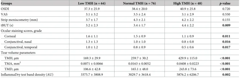
Comparisons of clinical parameters between the three TMH groups
There was no difference in OSDI/VAS and strip meniscometry results between the three TMH groups. tBUT differed significantly among the three TMH groups in the post-hoc test (low vs high TMH group: p = 0.012; normal vs high TMH group: p = 0.024). In the high TMH group, tBUT was longer than that in the low TMH group. Corneal and conjunctival staining scores showed significant differences among the three TMH groups (Table 2). The corneal staining score showed significant differences between the low and high TMH groups (p = 0.014) and between the normal and high TMH groups (p = 0.029). The staining score of both the nasal and temporal conjunctiva in the low TMH group was higher than those in the high TMH groups (p = 0.014 and 0.013, respectively). There was no difference between the low and nor- mal TMH, or normal and high TMH, in corneal and conjunctival staining.
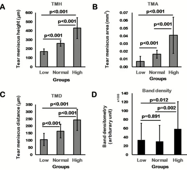
Tear volume measurements by assessing TMH, TMD, and TMA differed significantly in post-hoc tests among the three TMH groups (p < 0.001;Fig 2A–2C). In the comparisons of InflammaDry band density, there were significant differences between the low and high TMH groups (p = 0.012) and between the normal and high TMH groups (p = 0.002). The mean band density was markedly higher in the high TMH group than that in both the low and nor- mal TMH groups (Table 2,Fig 2D).
Comparisons between InflammaDry-positive and -negative groups
There were no significant differences in ODSI/VAS, strip meniscometry, tBUT, and conjuncti- val/corneal staining scores between the InflammaDry-positive and -negative groups
Table 1. Patient demographics of TMH groups and InflammaDry positivity groups.
Low TMH (n = 64) Normal TMH (n = 76) High TMH (n = 48) p-value
Age (mean± SD), years 58.3± 11.2 56.9± 15.2 62.4± 10.9 0.177
Sex (M/F) 9/55 18/58 20/28 0.004
Diabetes (Y/N) 4/60 7/69 1/47 0.286
Hypertension (Y/N) 10/54 20/56 19/29 0.017
Sjo¨gren syndrome (Y/N) 7/57 12/64 3/45 0.266
InflammaDry-positive (n = 120) InflammaDry-negative (n = 68) p-value
Age (mean± SD), years 59.2± 13.2 58.0± 12.8 0.548
Sex (M/F) 35/85 12/56 0.080
Diabetes (Y/N) 10/110 2/66 0.146
Hypertension (Y/N) 36/84 13/55 0.102
Sjo¨gren syndrome (Y/N) 17/103 5/63 0.163
TMH = tear meniscus height, SD = standard deviation, Y/N = Yes/No.
https://doi.org/10.1371/journal.pone.0235408.t001
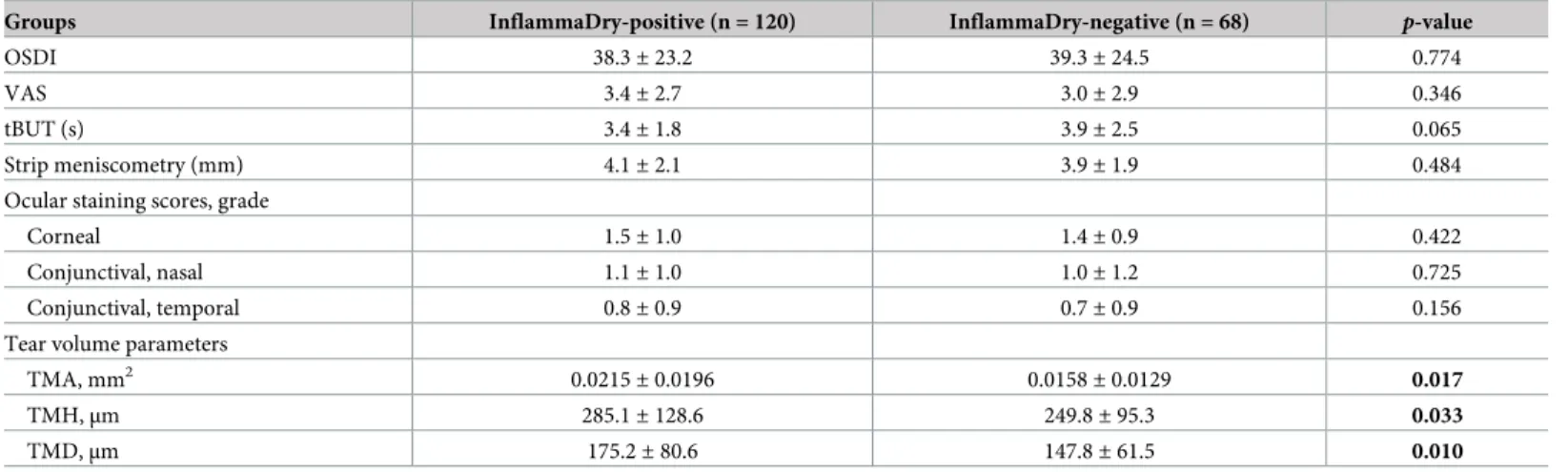
(p > 0.05). In comparisons between the InflammaDry-positive and -negative groups, there were significant differences in terms of TMH, TMA, and TMD (p = 0.033, 0.017, and 0.010, respectively). The positive group had greater TMH, TMA, and TMD values than the negative group (Table 3).
Correlation between InflammaDry band density and tear meniscus parameters and clinical signs/symptoms
Pearson’s correlation analysis revealed that InflammaDry band density and tear meniscus parameters, such as TMH, TMA, and TMD, were positively correlated (Table 4). The OSDI/
VAS score, tBUT, and corneal or conjunctival staining score showed no correlation with band density (p > 0.05, respectively).
Discussion
MMP-9 is considered a nonspecific and late phase inflammatory marker in DE patients [23–
25]. On this basis, an immunoassay was developed to detect and confirm the elevation of tear MMP-9 in DE patients, and recent validation studies reported good diagnostic performance and correlation with clinical severity of DE [5,6,14]. Among these reports, Sambursky et al.
reported that this test had 85% sensitivity and 94% specificity for diagnosing DE [14]. How- ever, two separate reports from Schargus et al. and Lanza et al. showed moderate to low posi- tivity in the MMP-9 immunoassay in DE study subjects [3,15,16]. In addition, Lanza et al.
reported no difference in subjective symptoms and clinical signs of DE between MMP-9-posi- tive and -negative groups [15,16].
Inflammadry, a point-of-care MMP-9 immunoassay, is based on the principle of lateral- flow immunoassay (LFIA); this type of assay has advantages of low cost, rapid analysis, and ease of device preparation. However, in LFIAs, if the sample volume is small, the reliability of the test result may be significantly impaired. Therefore, despite these advantages, performance of LFIA could be seriously affected by the volume of the loaded analytes [26]. On this basis, in
Table 2. Comparisons of clinical parameters within three TMH groups.
Groups Low TMH (n = 64) Normal TMH (n = 76) High TMH (n = 48) p-value
OSDI 37.3± 25.9 38.4± 20.0 40.9± 25.8 0.720
VAS 3.1± 3.2 3.5± 2.4 3.1± 2.9 0.550
Strip meniscometry (mm) 3.7± 1.7 4.3± 2.1 4.2± 2.2 0.155
tBUT (s) 3.2± 2.3 3.4± 1.7 4.4± 2.2 0.009
Ocular staining scores, grade
Corneal 1.6± 1.1 1.5± 0.9 1.1± 0.9 0.011
Conjunctival, nasal 1.3± 1.3 1.0± 1.0 0.8± 0.8 0.016
Conjunctival, temporal 1.0± 1.2 0.8± 0.9 0.5± 0.6 0.017
Tear volume parameters
TMH,μm 169.3± 29.9 259.7± 30.2 429.9± 115.0 <0.001
TMA, mm2 0.0071± 0.0066 0.0163± 0.0052 0.0408± 0.0223 <0.001
TMD,μm 106.6± 42.8 165.1± 48.0 243.8± 73.6 <0.001
InflammaDry test band density (AU) 3375.7± 3808.9 3029.7± 3618.4 5876.2± 6206.7 0.002
OSDI: Ocular Surface Disease Index, VAS: Visual analogue scale, tBUT: tear break-up time, TMH: tear meniscus height, TMA: tear meniscus area, TMD: tear meniscus depth, AU: arbitrary unit, data are expressed as the mean± standard deviation.
https://doi.org/10.1371/journal.pone.0235408.t002
the Inflammadry kit instructions, the manufacturer has also pointed out that less than 6μl of sample volume could produce false-negative results.
In the present study, there was a tendency for participants having higher tear volumes to show higher band densities, but the subjects who had lower tear volumes, indicating aqueous tear-deficient DE, showed lower band densities on MMP-9 immunoassay. Therefore, a highly inflamed ocular surface with decreased tear volume, such as that found in Sjo¨gren syndrome, could show negative results because of the markedly decreased tear secretion, despite the highly elevated tear MMP-9 concentration. However, clinically, the opposite is possible.
Among the participants of the present study, a strong positive band was identified even in patients with mild or nearly no fluorescein staining of the cornea and conjunctiva, who are expected to have very mild inflammatory eye surface inflammation.
Nevertheless, there was no difference in the strip meniscometry results between the MMP- 9-positive and -negative groups. In particular, the MMP-9 test is usually performed first, after noninvasive tests, such as TMH, and thus, the volume of tears stored on the ocular surface and the amount of tear volume after stimulation of the strip meniscometry may be fundamentally
Fig 2. Post-hoc analysis of the three TMH groups. TMH (A), TMA (B), and TMD (C) showed statistically significant differences within the three TMH groups. D. Matrix metalloproteinase (MMP)-9 band density analysis of the three TMH groups showed significantly higher band density in the high TMH group than that in the low and normal TMH groups.
https://doi.org/10.1371/journal.pone.0235408.g002
different. In addition, the strip meniscometry was performed after the MMP-9 immunoassay, and recovery of tears depleted by the sampling apparatus of the MMP-9 immunoassay may take time. Since the low TMH group represents aqueous tear-deficient DE, subjects belonging to this group would require more time for recovery than those in the other groups. In terms of concentration, since MMP-9 is secreted not by the lacrimal gland, but by the ocular surface epithelium, reflex tearing may dilute the solutes dissolved in tears. Therefore, tear parameters that were measured by anterior OCT would better reflect the actual loading volume than the strip meniscometry in the MMP-9 immunoassay.
The present study has some limitations. As we only included participants who showed dry eye symptoms and signs at initial examination, there was no control group for comparisons of InflammaDry results with dry eye groups. Thus, further analysis between dry and not-dry eye patients would be helpful for understanding the clinical features of the InflammaDry test. In addition, because classification according to the tear meniscus value is very artificial, there may be significant limitations based on whether the three groups are appropriate. However, variability in TMH values may be inevitable because these can be very different depending on race, measurement equipment, and subtype of the dry eye.
In conclusion, we found the volume dependency of the MMP-9 immunoassay, which could induce false-negative results clinically. In particular, moderate to severe forms of aqueous tear- deficient DE conditions, such as Sjo¨gren syndrome, could be underdiagnosed by the commer- cially available tear MMP-9 immunoassay. In addition, our results suggest that appropriate tear collection by the sampling fleece of the MMP-9 immunoassay is crucial to the appropriate detection of ocular surface inflammation.
Table 3. Comparisons of clinical characteristics between InflammaDry-positive and -negative groups of dry eye patients.
Groups InflammaDry-positive (n = 120) InflammaDry-negative (n = 68) p-value
OSDI 38.3± 23.2 39.3± 24.5 0.774
VAS 3.4± 2.7 3.0± 2.9 0.346
tBUT (s) 3.4± 1.8 3.9± 2.5 0.065
Strip meniscometry (mm) 4.1± 2.1 3.9± 1.9 0.484
Ocular staining scores, grade
Corneal 1.5± 1.0 1.4± 0.9 0.422
Conjunctival, nasal 1.1± 1.0 1.0± 1.2 0.725
Conjunctival, temporal 0.8± 0.9 0.7± 0.9 0.156
Tear volume parameters
TMA, mm2 0.0215± 0.0196 0.0158± 0.0129 0.017
TMH,μm 285.1± 128.6 249.8± 95.3 0.033
TMD,μm 175.2± 80.6 147.8± 61.5 0.010
OSDI: Ocular Surface Disease Index, VAS: visual analogue scale, tBUT: tear break-up time, TMH: tear meniscus height, TMA: tear meniscus area, TMD: tear meniscus depth, data are expressed as the mean± standard deviation.
https://doi.org/10.1371/journal.pone.0235408.t003
Table 4. Correlation analysis between tear meniscus parameters (Height, Depth, and Area) and InflammaDry test band density.
Parameters TMH TMD TMA
r-value 0.218 0.243 0.216
p-value 0.003 0.001 0.003
TMA: tear meniscus area, TMD: tear meniscus depth, TMH: tear meniscus height.
https://doi.org/10.1371/journal.pone.0235408.t004
Supporting information
S1 File. The raw excel file of dry eye parameters.
(XLSX)
Author Contributions
Conceptualization: Jong Hwa Jun.Data curation: Jong Hwa Jun, You Hyun Lee, Myeong Jin Son, Harim Kim.
Formal analysis: Jong Hwa Jun.
Funding acquisition: Jong Hwa Jun.
Investigation: Jong Hwa Jun.
Methodology: Jong Hwa Jun.
Project administration: Jong Hwa Jun.
Resources: Jong Hwa Jun.
Software: Jong Hwa Jun.
Visualization: Jong Hwa Jun.
Writing – original draft: Jong Hwa Jun, You Hyun Lee.
Writing – review & editing: Jong Hwa Jun.
References
1. Yazdani M, Elgstøen KBP, Rootwelt H, Shahdadfar A, UtheimØA, Utheim TP. Tear metabolomics in dry eye disease: a review. Int J Mol Sci. 2019; 20: 3755.
2. Korb DR. Survey of preferred tests for diagnosis of the tear film and dry eye. Cornea. 2000; 19: 483–
486.https://doi.org/10.1097/00003226-200007000-00016PMID:10928763
3. Schargus M, Geerling G, Joachim SC. [Significance of new methods of examining the tear film in dry eye disease: tear film osmolarity and matrix metalloproteinases (MMP-9)]. Klin Monbl Augenheilkd.
2018; 235: 597–602. German.https://doi.org/10.1055/s-0042-121424PMID:28147407
4. Lemp MA, Bron AJ, Baudouin C, Benı´tez Del Castillo JM, Geffen D, Tauber J, et al. Tear osmolarity in the diagnosis and management of dry eye disease. Am J Ophthalmol. 2011; 151: 792–798.e1.https://
doi.org/10.1016/j.ajo.2010.10.032PMID:21310379
5. Messmer EM, von Lindenfels V, Garbe A, Kampik A. Matrix metalloproteinase 9 testing in dry eye dis- ease using a commercially available point-of-care immunoassay. Ophthalmology. 2016; 123: 2300–
2308.https://doi.org/10.1016/j.ophtha.2016.07.028PMID:27665213
6. Sambursky R, Davitt WF 3rd, Friedberg M, Tauber S. Prospective, multicenter, clinical evaluation of point-of-care matrix metalloproteinase-9 test for confirming dry eye disease. Cornea. 2014; 33: 812–
818.https://doi.org/10.1097/ICO.0000000000000175PMID:24977985
7. Asahi M, Wang X, Mori T, Sumii T, Jung JC, Moskowitz MA, et al. Effects of matrix metalloproteinase-9 gene knock-out on the proteolysis of blood-brain barrier and white matter components after cerebral ischemia. J Neurosci. 2001; 21: 7724–7732.https://doi.org/10.1523/JNEUROSCI.21-19-07724.2001 PMID:11567062
8. Sternlicht MD, Werb Z. How matrix metalloproteinases regulate cell behavior. Annu Rev Cell Dev Biol.
2001; 17: 463–516.https://doi.org/10.1146/annurev.cellbio.17.1.463PMID:11687497
9. Acera A, Vecino E, Duran JA. Tear MMP-9 levels as a marker of ocular surface inflammation in conjunc- tivochalasis. Invest Ophthalmol Vis Sci. 2013; 54: 8285–8291.https://doi.org/10.1167/iovs.13-12235 PMID:24255042
10. Shetty R, Ghosh A, Lim RR, Subramani M, Mihir K, Reshma AR, et al. Elevated expression of matrix metalloproteinase-9 and inflammatory cytokines in keratoconus patients is inhibited by cyclosporine A.
Invest Ophthalmol Vis Sci. 2015; 56: 738–750.https://doi.org/10.1167/iovs.14-14831PMID:25648341
11. Kaufman HE. The practical detection of mmp-9 diagnoses ocular surface disease and may help prevent its complications. Cornea. 2013; 32: 211–216.https://doi.org/10.1097/ICO.0b013e3182541e9aPMID:
22673852
12. Baudouin C, Messmer EM, Aragona P, Geerling G, Akova YA, Benı´tez-del-Castillo J, et al. Revisiting the vicious circle of dry eye disease: a focus on the pathophysiology of meibomian gland dysfunction. Br J Ophthalmol. 2016; 100: 300–306.https://doi.org/10.1136/bjophthalmol-2015-307415PMID:
26781133
13. Park JY, Kim BG, Kim JS, Hwang JH. Matrix metalloproteinase 9 point-of-care immunoassay result pre- dicts response to topical cyclosporine treatment in dry eye disease. Transl Vis Sci Technol. 2018; 7: 31.
14. Sambursky R, Davitt WF 3rd, Latkany R, Tauber S, Starr C, Friedberg M, et al. Sensitivity and specificity of a point-of-care matrix metalloproteinase 9 immunoassay for diagnosing inflammation related to dry eye. JAMA Ophthalmol. 2013; 131: 24–28.https://doi.org/10.1001/jamaophthalmol.2013.561PMID:
23307206
15. Lanza NL, McClellan AL, Batawi H, Felix ER, Sarantopoulos KD, Levitt RC, et al. Dry eye profiles in patients with a positive elevated surface matrix metalloproteinase 9 point-of-care test versus negative patients. Ocul Surf. 2016; 14: 216–223.https://doi.org/10.1016/j.jtos.2015.12.007PMID:26807724 16. Lanza NL, Valenzuela F, Perez VL, Galor A. The matrix metalloproteinase 9 point-of-care test in dry
eye. Ocul Surf. 2016; 14: 189–195.https://doi.org/10.1016/j.jtos.2015.10.004PMID:26850527 17. Huh J, Choi SY, Eom Y, Kim HM, Song JS. Changes in the matrix metalloproteinase 9 point-of-care test
positivity according to MMP-9 concentration and loading volume. Cornea. 2019 Jul 31.https://doi.org/
10.1097/ICO.0000000000002096PMID:31369458
18. Czajkowski G, Kaluzny BJ, Laudencka A, Malukiewicz G, Kaluzny JJ. Tear meniscus measurement by spectral optical coherence tomography. Optom Vis Sci. 2012; 89: 336–342. PMID:22282222 19. Hartig SM. Basic image analysis and manipulation in ImageJ. Curr Protoc Mol Biol. 2013;Chapter 14:
Unit14.15.
20. Schiffman RM, Christianson MD, Jacobsen G, Hirsch JD, Reis BL. Reliability and validity of the Ocular Surface Disease Index. Arch Ophthalmol. 2000; 118: 615–621.https://doi.org/10.1001/archopht.118.5.
615PMID:10815152
21. McCormack HM, Horne DJ, Sheather S. Clinical applications of visual analogue scales: a critical review.
Psychol Med. 1988; 18: 1007–1019.https://doi.org/10.1017/s0033291700009934PMID:3078045 22. Bron AJ, Evans VE, Smith JA. Grading of corneal and conjunctival staining in the context of other dry
eye tests. Cornea. 2003; 22: 640–650.https://doi.org/10.1097/00003226-200310000-00008PMID:
14508260
23. Aragona P, Aguennouz M, Rania L, Postorino E, Sommario MS, Roszkowska AM, et al. Matrix metallo- proteinase 9 and transglutaminase 2 expression at the ocular surface in patients with different forms of dry eye disease. Ophthalmology. 2015; 122: 62–71.https://doi.org/10.1016/j.ophtha.2014.07.048 PMID:25240629
24. Chotikavanich S, de Paiva CS, Li de Q, Chen JJ, Bian F, Farley WJ, et al. Production and activity of matrix metalloproteinase-9 on the ocular surface increase in dysfunctional tear syndrome. Invest Ophthalmol Vis Sci. 2009; 50: 3203–3209.https://doi.org/10.1167/iovs.08-2476PMID:19255163 25. Pinto-Fraga J, Enrı´quez-de-Salamanca A, Calonge M, Gonza´ lez-Garcı´a MJ, Lo´pez-Miguel A, Lo´pez-
de la Rosa A, et al. Severity, therapeutic, and activity tear biomarkers in dry eye disease: an analysis from a phase III clinical trial. Ocul Surf. 2018; 16: 368–376.https://doi.org/10.1016/j.jtos.2018.05.001 PMID:29772277
26. Koczula KM, Gallotta A. Lateral flow assays. Essays Biochem. 2016; 60: 111–120.https://doi.org/10.
1042/EBC20150012PMID:27365041