Journal of Trauma and Injury Vol. 26, No. 4, December, 2013
[ J Trauma Inj 2013;26:273-278 ]
� Address for Correspondence : Ho-Seong Han, M.D.
Department of Surgery, Seoul National University Bundang Hospital,
82, Gumi-ro 173 Beon-gil, Bundang-gu, Seongnam-si, Gyeonggi-do, 463-707, Korea Tel : 82-31-787-7099, Fax : 82-31-787-4078, E-mail : [email protected]
Submitted : October 29, 2013 Revised : October 30, 2013 Accepted : October 30, 2013
�These authors equally contributed to this paper and should be considered co-first authors
CT를 이용한 혈복강의 등급: 비장 손상의 정도 및 회복의 객관화
서울대학교 의과대학 분당서울대학교병원 외과
신홍경�, 송라영�, 한호성, 윤유석, 조재영, 황대욱, 정규환, 김영기, 이우형
- Abstract -
CT Based Hemoperitoneum Scoring for Clinicians:
Objectifying the Severity of Splenic Injury and Recovery
Hong Kyung Shin, M.D.
�, Ra-Yeong Song, M.D.
�, Ho-Seong Han, M.D., Yoo-Seok Yoon, M.D., Jai Young Cho, M.D., Dae Wook Hwang, M.D., Kyuwhan Jung, M.D., Young Ki Kim, M.D.,
Woo Hyung Lee, M.D.
Department of Surgery, Seoul National University Bundang Hospital, Seoul National University College of Medicine
Purpose: In patients with splenic trauma, Computed Tomography (CT) scan is helpful in selecting treatment options and evaluating resolution after NOM (Non-Operative Management). The purpose of this study was to suggest a CT based hemoperitoneum (HP) scoring system that can easily be used by clinicians to evaluate the severity of injury and recovery.
Methods: A retrospective review of patients with splenic trauma admitted to our hospital between May 2003 and January 2013 was conducted. Patients diagnosed with isolated spleen injury who had a CT scan on admis- sion were included. 1 or 2 points were given according to location and amount of hematoma in the CT image.
Using the existing ultrasonography (US) based HP scoring system, the same method was applied to obtain our CT based HP scoring (CBHS) system, which ranges from 0 to 8 points. The CBHS system can be easily used by clinicians for a quick assessment of splenic injury.
Results: Of the 39 patients meeting the inclusion criteria, 6 patients were managed operatively and 33 non- operatively. There was a significant difference in CBHS between the OM (operative management) group and the NOM group.(p=0.03) CBHS showed correlation with Hb (hemoglobin), Hct (hematocrit), spleen injury grade(AAST), and Hounsfield unit of ROI (Region of interest). (p=0.17, p=0.18, p<0.000, p=0.02, respectively)
After successful NOM with stabilized Hb level, the amount of hemoperitoneum was scored in the follow-up CT. CBHS demonstrated correlation with decreased spleen injury grade, decreased Hounsfield unit of ROI (Region of interest) (p=0.039, p=0.049, respectively), and also objectively reflected patient recovery.
Conclusion: CBHS can be used as an objective and intuitive tool for clinicians in grading the severity of splenic injury by scoring the amount of hemoperitoneum, and in assessing recovery.
Key Words: Hemoperitoneum, Trauma, Spleen injury
I. 서 론
비장손상은 복부 둔상 환자에게 가장 많이 발생하는 손상 중의 하나이며 신속히 진단하여 치료하면 특이 합병증 없이 회복 할 수 있는 반면 진단과 치료가 늦어지는 경우 순식간 에 목숨을 잃을 수 있는 외상 질환이다. 비장은 간과 더불어 혈관분포가 가장 많은 장기이며 매우 약한 조직으로 이루어 져 있어 쉽게 손상받으며 손상시 대량출혈을 일으키게 된 다.(1-3)
비장손상이 의심되는 환자 또는 혈복강이 의심되는 환자는 진단 및 치료방법을 정하기 위하여 대부분의 환자에게서 주 로 복부컴퓨터단층촬영술(CT)을 시행하게 된다. CT는 과거 부터 혈역학적 안정상태의 환자에게만 시행하는 것을 원칙으 로 생각하여 왔으나 외상 환자에게 있어 CT 검사의 유용성 이 매우 높으며 진단영상 기술의 발전으로 검사시간을 단축 시킬 수 있어, 수액 및 수혈, 승압제의 사용 등으로 혈역학적 안정을 유지 할 수 있는 상태의 환자에게서도 선택적으로 사 용되고 있다. 특히 검사 시간의 단축으로 때에 따라서는 단 순한 혈액검사인 CBC 검사 보다도 빨리 검사 결과를 얻을
수 있어 외상환자 초기 평가 검사로 그 범위와 빈도가 증가 하고 있다.(4-7)
대부분의 응급실에서는 이렇게 얻어진 CT를 사용하여 혈 복강 및 비장손상을 진단하고 손상등급을 분류하여 왔으며 주로 American Association for the Surgery of Trauma (AAST)에서 제안한 비장손상등급을 주로 사용하 여 손상등급이 낮은 경우 비수술적 치료를 주로 시행하고 손 상등급이 높은 경우에는 수술적 치료를 주로 시행하여왔다.
이렇게 치료를 달리해온 근거는 손상등급에 따라 다른 예후 를 보여 등급이 높은 경우에 더욱 신속하고 적극적인 치료가 요구되었기 때문이다. 수술적 치료로는 주로 시험적 개복술 을 통한 평가와 더불어 비장절제술이 시행되어 왔으며 splenorrhapy 등의 수술 방법은 이러한 고식적 수술방법으 로 지혈 될 만한 환자들에게 있어 수술 빈도가 줄어들고 있 는 추세로 거의 시행되고 있지 않다.(1,8-15)
그러나 비장손상 환자에게 있어 가장 중요한 병태 생리적 현상은 저혈량성 상태(hypovolemic state) 또는 저혈량성 쇼크인데, 비장손상등급은 비장의 실질손상 정도에 따라 주 로 중증도를 분류하고 있어 저혈량성 상태를 평가하기에 한
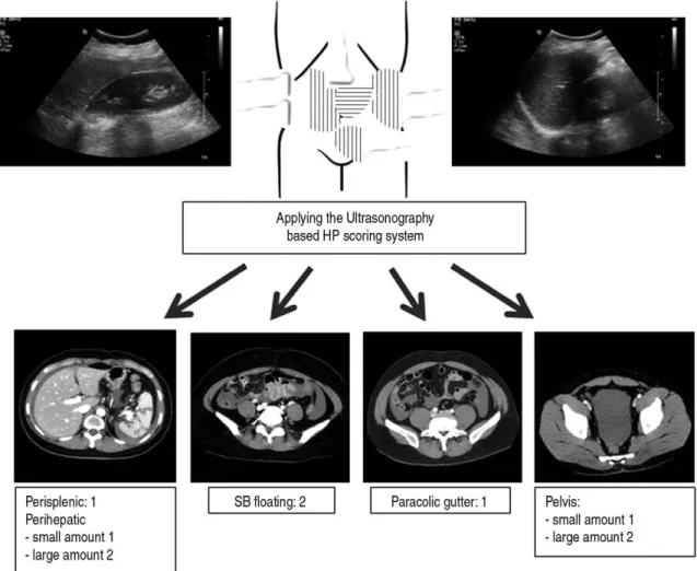
Fig. 1. Applying the Ultrasonography-based hemoperitoneum scoring system.
계가 있을 수 있다. 실질 손상의 정도는 비수술적 치료시 회 복될 가능성을 보일 수는 있으나 현재 실혈 상태를 잘 반영 한다고 할 수 없기 때문이다. 따라서 비장손상후 실혈에 의 한 저혈량성 상태를 객관적으로 알 수 있는 지표인 혈색소, 헤마토크리트와 함께 흔히 빠르게 시행할 수 있는 CT와 같 은 영상검사를 통해 이를 객관화 할 수 있는 방법이 필요하 게 되었다.
Huang 등에 의하여 초음파를 이용한 혈복강 등급이 제안 되어 왔으며 이는 등급에 따라 수술적 치료의 빈도를 잘 반영 하였다.(6,11,16,17) 그러나 외상 환자에게 있어 CT의 비중이 증가함에 따라 응급 초음파의 빈도는 줄어들고 있으며 이와
같은 초음파를 이용한 혈복강 등급은 근래 들어 많이 사용되 고 있지 않는 추세이다. 이에 저자는 Huang 등의 초음파를 이용한 혈복강 등급을 CT에 적용하여 비장손상에서 실혈의 정도를 객관화 할 수 있는 지표를 제안하고자 하였다.
II. 대상 및 방법
2003년 5월부터 2013년 2월까지 분당서울대병원 응급실 로 내원하여 복부 CT를 시행 비장손상으로 진단받은 39명의 환자를 대상으로 후향적 의무기록 분석을 시행하였다. 영상 의학과 의사에 의하여 비장손상 및 혈복강을 진단받은 환자
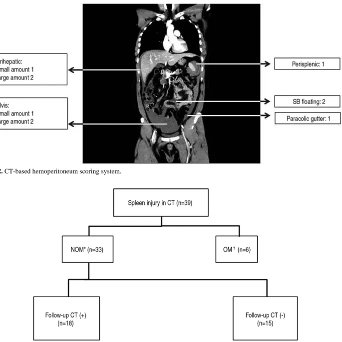
Fig. 2. CT-based hemoperitoneum scoring system.
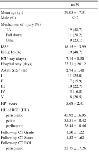
Fig. 3. Management and follow-up of patients with spleen injury.
Fig. 3. * NOM: Non-operative management Fig. 3. �OM: Operative management
를 대상으로 하였으며 간손상, 장천공등 복부 동반 손상이 있는 환자는 배제하였다.
CT를 이용한 혈복강 등급은 Huang 등에 의하여 제안된 초음파를 활용한 혈복강 등급과 동일한 방법으로 CT에 적용 하여 점수화 하였으며 임상의가 응급실에서 직관적이고 신속 하게 시행할 수 있는 혈복강 등급을 고안하고자 하는 본 연 구의 목적상 외상외과 임상의에 의하여 이루어 졌다. CT를 활용한 혈복강 등급은 perisplenic area, perihepatic area, small bowel (SB) floating, paracolic gutter, pelvic cavity 등으로 구역을 나누고 각각 구역의 혈복강 여 부에 따라서 1점 또는 2점을 부여하여 단순합으로 등급화 하 였다(Fig. 1, 2). Perihepatic area의 혈종은 1 cm을 기준 으로, 그 이하일 때 1점(small amount), 1 cm 보다 클 때 2 점(large amount)을 부여하였다. Pelvic cavity의 경우에 는 골반 내에만 국한된 상태를 1점, 골반 밖으로 넘친 상태를 2점으로 정하였다.
혈복강등급이 실혈의 정도를 잘 반영하는지 알아보기 위하 여 혈복강 등급과 혈색소, 헤마토크리트, 비장손상등급, Hounsfield Unit of Region of interest (HU of ROI)와 연관성을 분석하였고, 비장손상등급과 연관성을 분석하였다.
또 경과관찰 후 CT를 시행한 18명의 환자에 대하여 혈복강 등급을 측정하여 회복정도를 점수화하였고 회복후 비장손상 등급, 회복후 HU of ROI와 상관관계를 분석하였다(Fig. 3).
여기서 HU of ROI는 혈복강 부위의 Hounsfield Unit를 측정한 것을 의미하고, 급성혈종이 만성혈종보다 HU 값이 높다.(18)
통계분석을 위하여 SPSS 18 (Chicago, USA)을 사용하였 으며 실혈을 나타내는 알려진 지표와 CBHS와의 상관관계 분석을 위해 단순회기분석 및 다중회귀분석을 시행하였다.
통계적 유의성은 p값이 0.05미만일 경우로 한정하였다.
III. 결 과 1. Demographics
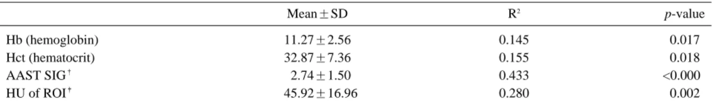
환자군 평균 나이는 29.0세로 성비는 남성이 69.2%로 남 성비율이 높았다. 손상기전은 운수사고가 가장 많았으며 낙 상이 그 다음 순이었다. 평균 Injury Severity Score(ISS) 는 18.15였으며 비교적 중상으로 생각되는 16점이상은 19명 으로 48.7%였다. 중환자실 입원기간 평균은 7.14일이었으며 총 입원기간평균은 23.31일이었다. 평균 AAST 비장손상등 급은 2.74였으며, 평균 CBHS 3.68이었다. 평균 HU of ROI는 perisplenic, pelvis, perihepatic 부위별로 45.92.
35.51, 28.44로 비장에서 멀어질수록 낮아지는 경향을 보여 비장 근처일수록 급성출혈에 의한 혈종임을 반증하였다. 평 균 11.4일 후 경과관찰 CT를 시행하였고 평균 손상등급은
1.50로, 평균 CBHS는 1.52로, 평균 perisplenic HU of ROI는 22.75로 각각 감소하였다(Table 1).
2. Difference in CBHS between NOM group and OM group
비수술적 치료를 시행한 군과 수술적 치료를 시행한 군에 서 CT-based Hemoperitoneum Scoring (CBHS)를 비교 한 결과 두 군간에 통계적으로 유의한 차이를 보여 (p=0.009) CBHS는 손상의 중증도를 잘 반영하여 비장손상 등급과 더불어 치료 계획을 수립하는데 좋은 근거로 사용될 수 있다(Table 2).
3. Correlation of CBHS with variables
실혈을 반영하는 대표적인 지표인 혈색소, 헤마토크리트와
Table 1. Demographics
n=39
Mean age (yr) 29.03±17.31
Male (%) 69.2
Mechanism of injury (%)
TA 19 (48.7)
Fall down 11 (28.2)
Other 09 (23.1)
ISS* 18.15±13.99
ISS≥16 (%) 19 (48.7)
ICU stay (days) 7.14±8.50
Hospital stay (days) 23.31±26.12
AAST SIG�(%) 2.74±1.48
I 11 (25.0)
II 07 (15.9)
III 10 (22.7)
IV 03 (06.8)
V 08 (20.5)
HP�score 3.68±2.41
HU of ROI§(HU)
perisplenic 45.92±16.95
pelvis 35.51±10.42
perihepatic 28.44±10.46
Follow-up CT Grade 1.50±1.22
Follow-up CT Score 1.52±1.62
Follow-up CT ROI
perisplenic 22.75±17.26
* ISS: Injury Scale Score
�AAST SIG: American Association for the Surgery of Trauma Spleen Injury Grade
�HP: hemoperitoneum
§HU of ROI: Hounsfield Unit of Region of interest
비장손상의 정도를 반영하는 비장손상등급, 출혈 및 혈종의 급성도를 판단할 수 있는 지표를 잘 알려져 있는 HU of ROI 등과 선형회귀분석을 통해 상관관계를 분석해본 결과 각 지 표들과 통계적으로 유의한 결과를 보였다(Table 3). CBHS 는 실혈의 정도, 비장손상의 정도, 출혈의 급성도 등을 잘 반 영하여 상기한 변수들과 더불어 비장손상의 중증도를 평가할 수 있는 좋은 지표로 사용될 수 있다.
4. Correlation of follow-up CBHS with variables
일정한 간격을 두고 회복여부를 판단하기 위해 시행한 경 과관찰 CT에서 CBHS와 비장손상등급, HU of ROI와 통계 적으로 유의한 상관관계를 보여 회복의 정도를 평가하는데도 좋은 지표로 사용될 수 있다(Table 4).
IV. 고 찰
기계공학적 기술의 발달로 비장손상, 간손상 환자 등 외상 환자에게서 CT검사의 비중은 점점 높아지고 있으며 CT검사 가 이와 같은 질병에서 갖는 진단적 가치는 절대적이다. 혈 복강의 양에 따른 분류는 Federle에 의한 HP scoring 이 주
로 사용되어 왔으나 소량, 중등도, 다량 등으로 모호하고 세 분화되지 못하여 임상적으로나 학문적으로 활용도에 한계를 지녀왔으며 영상의학적 계산방법은 중증외상환자에게 응급 실에서 신속하게 적용하여 치료방침을 결정하는데 제한점이 있었다.(9)
한편 최근처럼 multidetector CT가 발달되지 않아 시간 이 오래 걸려 생체징후가 불안정한 환자에게 주로 FAST(Focused Assessment Sonography for Trauma) 가 시행되던 시기에 사용되었던 Huang 등의 scoring sys- tem은 외상 환자의 초음파 사용 빈도가 줄고 있으며 CT에 비해 객관성이 떨어져 사용빈도가 감소하는 추세이다. 그러 나 그 연구에서는 식염수를 직접 주입하여 객관적인 액체 양 을 평가하였었고 등급화에 따라 중증도를 잘 반영하였던 등 급화 방법으로 이를 CT에 사용한다면 비슷한 결과를 기대할 수 있을 것이라 생각하여 적용해 보게되었다.(6)
최근 들어 비장손상환자에게 수술적 치료보다 비수술적 치 료의 유용성이 더욱 커지고 있으며 비교적 심한 손상에서도 비수술적 치료가 주된 치료방법으로 부각되고 있다.(10,13- 15,19) 이러한 추세가운데 과거필요성을 인정받지 못해왔던 경과관찰 CT검사의 유용성 또한 증가하고 있어 혈액검사, 손상등급과 더불어 회복의 정도를 평가하기 위한 또 다른 지
Table 3. Correlation of CBHS* with variables
Mean±SD R2 p-value
Hb (hemoglobin) 11.27±2.56 0.145 0.017
Hct (hematocrit) 32.87±7.36 0.155 0.018
AAST SIG� 02.74±1.50 0.433 <0.0000
HU of ROI� 045.92±16.96 0.280 0.002
* CBHS: CT based hemoperitoneum scoring
�AAST SIG: American Association for the Surgery of Trauma Spleen Injury Grade
�HU of ROI: Hounsfield Unit of Region of interest
Table 4. Correlation of follow-up CBHS* with variables
Mean±SD R2 p-value
Recovered AAST SIG� 1.50±1.22 0.196 0.039
Recovered HU of ROI� 22.76±17.26 0.286 0.049
* CBHS: CT based hemoperitoneum scoring
�AAST SIG: American Association for the Surgery of Trauma Spleen Injury Grade
�HU of ROI: Hounsfield Unit of Region of interest
Table 2. Difference in CBHS between NOM group and OM group
NOM* group (n=33) OM�group (n=6) p-value
CBHS 3.26±2.48 5.57±1.51 0.009
* NOM: Non-operative management
�OM: Operative management
표의 필요성도 증가하게 되었다.(2,4,7,12,20) 이와 같은 추 세에 맞춰 CBHS은 비수술적 치료 후 회복의 정도를 평가하 는데 있어서 좋은 지표로 활용될 수 있을 것이다.
CBHS은 중증도의 판단, 회복 정도의 객관화 지표라는 유 용성과 더불어 응급실에서 쉽게 임상의에 의하여 신속히 등 급화할 수 있다는 장점을 지니고 있다. 물론 각종 수학적, 공 학적 방법을 동원하면 보다 정확한 혈복강의 양을 측정해 낼 수 있을 것이고 이 결과가 더욱 환자의 실혈량을 평가하는데 정확한 척도로 활용될 수 있을 것이다. 그러나 본 연구의 서 두에서 언급한 바와 같이 CBHS은 응급상황에서 신속히 외 상 외과의 및 응급의학과의사에 의해서 쉽게 직관적으로 계 산해 낼 수 있는 scoring system이라는 것에 가장 중요한 가치를 두고 있는 것이므로 영상의학과 의사에 의해 계산되 고 또한 이 때문에 시간이 지체된다면 혈복강등급의 가치는 오히려 떨어지게 될 것이다. 본 CBHS의 가장 중요한 장점은 임상의가 신속히 직관적으로 쉽게 등급화한 지표가 비교적 환자의 손상정도를 잘 반영하여 환자의 평가 및 치료계획 수 립을 위한 근거로 사용될 수 있다는 것이다.
본 연구의 제한점은 비교적 적은 환자수를 대상으로 하여 필자들이 제시한 지표들이 모집단으로서의 가치가 떨어진다 는 점이며, 최근까지 여느 대학병원들과 같이 외상환자를 전 담해서 진료하는 의사가 없이 경험이 적은 의사들에 의해서 주로 치료가 이루어져 치료방침의 일관성이 없어 이러한 데 이터를 통해 결론을 도출하는 것에 오류를 가질 수 있다고 생각된다. 그리고 회복정도를 평가함에 있어 CT의 유용성을 평가하기에도 시행한 환자가 적고 회복정도에 대한 기준이 없어 향후 대규모 연구를 통해 CBHS의 임상적의의 및 활용 의 가치를 도출해 낼 수 있어야 하겠다.
V. 결 론
비장손상 환자에서 비수술적 치료를 시행한 군과 수술적 치료를 시행한 군에서 CBHS를 비교했을 때 CBHS는 손상 의 중증도를 잘 반영하였다. 또 실혈을 반영하는 혈색소, 헤 마토크리트 등과의 상관관계에서도 CBHS는 실혈의 정도, 비장손상의 정도와 출혈의 급성도를 잘 반영하였다. CBHS 는 비장손상을 동반한 혈복강 환자에게 있어 신속히 중증도 및 회복을 평가하는 객관적이고 직관적인 지표로 사용될 수 있다.
REFERENCES
01) Charbit J, Mahul M, Roustan JP, Latry P, Millet I, Taourel P, et al. Hemoperitoneum semiquantitative analysis on admission of blunt trauma patients improves the prediction of massive transfusion. Am J Emerg Med. 2013 Jan; 31(1): 130-6.
02) Minarik L, Slim M, Rachlin S, Brudnicki A. Diagnostic imag-
ing in the follow-up of nonoperative management of splenic trauma in children. Pediatr Surg Int. 2002 Sep; 18(5-6): 429-31.
03) Olthof DC, Joosse P, van der Vlies CH, de Haan RJ, Goslings JC. Prognostic factors for failure of nonoperative management in adults with blunt splenic injury: a systematic review. J Trauma Acute Care Surg. 2013 Feb; 74(2): 546-57.
04) Sharma OP, Oswanski MF, Singer D. Role of repeat comput- erized tomography in nonoperative management of solid organ trauma. Am Surg. 2005 Mar; 71(3): 244-9.
05) Emery KH, Babcock DS, Borgman AS, Garcia VF. Splenic injury diagnosed with CT: US follow-up and healing rate in children and adolescents. Radiology. 1999 Aug; 212(2): 515-8.
06) Huang MS, Liu M, Wu JK, Shih HC, Ko TJ, Lee CH.
Ultrasonography for the evaluation of hemoperitoneum during resuscitation: a simple scoring system. J Trauma. 1994 Feb;
36(2): 173-7.
07) Mizzi A, Shabani A, Watt A. The role of follow-up imaging in paediatric blunt abdominal trauma. Clin Radiol. 2002 Oct;
57(10): 908-12.
08) Bhullar IS, Frykberg ER, Siragusa D, Chesire D, Paul J, Tepas JJ, 3rd, et al. Selective angiographic embolization of blunt splenic traumatic injuries in adults decreases failure rate of nonoperative management. J Trauma Acute Care Surg. 2012 May; 72(5): 1127-34.
09) Charbit J, Millet I, Martinez O, Roustan JP, Merigeaud S, Taourel P, et al. Does the size of the hemoperitoneum help to discriminate the bleeding source and guide therapeutic deci- sions in blunt trauma patients with pelvic ring fracture? J Trauma Acute Care Surg. 2012 Jul; 73(1): 117-25.
10) Haan JM, Bochicchio GV, Kramer N, Scalea TM.
Nonoperative management of blunt splenic injury: a 5-year experience. J Trauma. 2005 Mar; 58(3): 492-8.
11) McKenney KL, McKenney MG, Cohn SM, Compton R, Nunez DB, Dolich M, et al. Hemoperitoneum score helps determine need for therapeutic laparotomy. J Trauma. 2001 Apr; 50(4): 650-4; discussion 4-6.
12) Bhangu A, Nepogodiev D, Lal N, Bowley DM. Meta-analysis of predictive factors and outcomes for failure of non-operative management of blunt splenic trauma. Injury. 2012 Sep; 43(9):
1337-46.
13) Sinha S, Raja SV, Lewis MH. Recent changes in the manage- ment of blunt splenic injury: effect on splenic trauma patients and hospital implications. Ann R Coll Surg Engl. 2008 Mar;
90(2): 109-12. PubMed PMID: 18325207.
14) Stassen NA, Bhullar I, Cheng JD, Crandall ML, Friese RS, Guillamondegui OD, et al. Selective nonoperative manage- ment of blunt splenic injury: an Eastern Association for the Surgery of Trauma practice management guideline. J Trauma Acute Care Surg. 2012 Nov; 73(5 Suppl 4): S294-300.
15) Stein DM, Scalea TM. Nonoperative management of spleen and liver injuries. J Intensive Care Med. 2006 Sep-Oct; 21(5):
296-304.
16) McKenney K, McKenney M, Nu?ez D, McDowell L, Martin L. Interpreting the trauma ultrasound: Observations in 62 posi- tive cases. Emergency Radiology. 1996 May/June; 3(3): 113-7.
17) Sirlin CB, Casola G, Brown MA, Patel N, Bendavid EJ, Hoyt DB. Quantification of fluid on screening ultrasonography for blunt abdominal trauma: a simple scoring system to predict severity of injury. J Ultrasound Med. 2001 Apr; 20(4): 359- 64.
18) Pablo R. Ros KJM. CT and MRI of the abdomen and pelvis: a teaching file. 2 ed. Boston, MA: Lippincott Williams &
Wilkins; 2007.
19) Velmahos GC, Toutouzas KG, Radin R, Chan L, Demetriades D. Nonoperative treatment of blunt injury to solid abdominal organs: a prospective study. Arch Surg. 2003 Aug; 138(8):
844-51.
20) Weinberg JA, Magnotti LJ, Croce MA, Edwards NM, Fabian TC. The utility of serial computed tomography imaging of blunt splenic injury: still worth a second look? J Trauma. 2007 May; 62(5): 1143-7; discussion 7-8.