굴 패각 구연산칼슘이 Papain으로 유도된 골관절염 C57BL/6J Mice에 미치는 영향
최은영1․김학주2․한지숙1
1부산대학교 식품영양학과
2㈜서진바이오텍
Effects of Calcium Citrate Isolated from Oyster Shell on Papain-induced Osteoarthritis in C57BL/6J Mice
Eun-Young Choi1, Hak-Ju Kim2, and Ji-Sook Han1
1Department of Food Science and Nutrition, Pusan National University
2Seojin Biotech Co., Ltd.
ABSTRACT This study investigated the effects of calcium citrate on papain-induced osteoarthritis in C57BL/6J mice.
Osteoarthritis was induced by injecting 6 μL of papain into the knee joints of mice. Calcium citrate was made by crushing the centrifuged precipitate after reacting 0.5 M citric acid with 1 kg of oyster shell extract. The mice were divided into five groups (n=8). The normal group was untreated, whereas the papain group was induced to have osteoarthritis and treated with 200 μL of water per day. The papain+DS group was treated with diclofenac sodium.
The papain+calcium citrate groups were treated with calcium citrate at 150 and 300 mg/kg/bw for 28 days. Proteoglycan contents in articular cartilages were measured by safranin O/fast green staining and hematoxylin & eosin staining.
Histopathological changes in cartilages were analyzed by the Rudolphi score approach. Contents of pro-inflammatory cytokines including TNF-α, IL-1β and IL-6 in plasma, were measured by the ELISA method. Body weights among the treated groups were not significantly different compared with that of the normal group. Cartilage loss and joint instability in the calcium citrate group improved significantly (P<0.05) in a dose-dependent manner compared with the papain group. Further, proteoglycan content of the calcium citrate group was considerably (P<0.05) higher than that of the papain group. Osteoarthritis scores in the calcium citrate group were considerably (P<0.05) reduced compared with the papain group. In the group treated with calcium citrate, contents of TNF-α, IL-1β and IL-6 in plasma were significantly (P<0.05) reduced in a dose-dependent manner in comparison with the normal group. Based on these results, we suggest that calcium citrate is effective for treatment of osteoarthritis.
Key words: osteoarthritis, calcium citrate, C57BL/6J mice, oyster shell
Received 16 June 2014; Accepted 17 July 2014
Corresponding author: Ji-Sook Han, Department of Food Science and Nutrition, Pusan National University, Busan 609-735, Korea E-mail: [email protected], Phone: +82-51-510-2836
서 론
골관절염은 관절연골이 닳아 없어지면서 비롯되는 질환 으로 일차적으로 국소적인 퇴행성 변화가 일어나고 이로 인 해 관절 통증, 골극 형성, 근육 위축 및 근력 약화 등의 이차 적인 변화를 초래한다. 특히 체중 부하를 많이 받는 슬관절 및 고관절, 수지관절, 척추 등에서도 발생하며 여성에게는 더욱 빈번하게 나타나는 질환이다. 골관절염의 원인은 정확 히 규명되지 않았지만 대사성, 염증성 인자 등이 나이, 성별, 유전적 인자들과 관련되어 관절연골을 파괴함으로써 골관 절염을 유발하는 것으로 알려져 있다(1,2). 특히 50세 이상 에서 갑자기 발생이 증가하여 60세 이상 노인에서 약 80%,
75세 이상의 노인에게 약 95% 이상의 높은 발병률을 나타 내는 가장 흔한 골관절계 질환 중 하나이기도 하다(3). 하지 만 노화에 따른 퇴행 현상으로 보는 시각으로 인해 치료나 질병 관리 면에서 간과되고 있는 실정이다. 이러한 골관절염 의 치료방법에는 체중조절, 운동치료, 약물치료, 수술치료 등이 있다. 흔히 사용되는 약물치료 방법으로는 스테로이드 관절 내 주사, 비 스테로이드성 소염진통제 등이 있다(4).
스테로이드 관절 내 주사는 강력한 항염증 작용이 있지만 2차 감염, 연골파괴, 전신 부작용 등의 위험이 있어 반복적 으로 사용하는 것을 제한하며(5), 소염진통제를 장기간 사용 하는 경우에는 프로스타글란딘의 합성이 억제되어 신장애, 위장관 장애 등의 합병증을 초래한다고 알려져 있다(6). 수 술치료의 경우에는 수술 후 관절의 강직, 감염 등의 부작용 이 나타날 수 있어 시술이 제한적으로 이루어진다(7). 이처 럼 골관절염의 적절한 치료법은 아직 미흡한 수준이며 검증 된 과학적 근거가 없는 약물이나 민간요법 등에 의존하는
경우가 많은 실정이다(8). 따라서 보다 안전하고 효과적인 골관절염의 새로운 치료제의 개발이 요구되는 실정이다.
본 실험의 시료인 구연산칼슘은 구연산의 칼슘염으로 보 존제, 연수제 등의 식품첨가제로 흔히 사용되는 물질이다.
최근에는 탄산칼슘과 더불어 칼슘보충제의 구성성분으로도 사용되어 관절염 및 골다공증 예방의 용도로 사용되기도 한 다. 칼슘보충제로 사용되는 칼슘의 종류에는 탄산칼슘 (calcium carbonate), 구연산칼슘(calcium citrate), 젖산 칼슘(calcium lactate), 글루콘산칼슘(calcium gluconate), 코랄칼슘(coral calcium), 염화칼슘(calcium chloride), 수 산화칼슘(calcium hydroxide) 등이 있으며, 가장 많이 사용 되는 것은 탄산칼슘과 구연산칼슘이다(9). 그중에서도 구연 산칼슘은 중량당 칼슘의 보유율은 적으나 칼슘의 흡수율이 탄산칼슘보다 22∼27% 정도 더 높다. 또한 흡수에 위산이 필요하여 식사 중 또는 식후에 복용해야 하는 탄산칼슘과는 달리 구연산칼슘은 위산이 필요하지 않으므로 식사와 상관 없이 복용할 수 있다(10). 이러한 장점들을 근거로 하여 본 실험에서는 구연산칼슘을 사용하였다. 선행연구에서 구연 산칼슘이 폐경기 여성의 골다공증 위험도를 알 수 있는 골흡 수 표지자를 낮췄다는 결과가 밝혀져 골다공증에는 효과가 있음을 증명하였다(11). 또한 구연산의 동물모델에서의 염 증세포 감소 작용(12), 구연산이 변형된 형태인 구연산염의 염증 사이토카인 발현 억제 작용(13) 등 구연산의 항염증에 관한 연구 등은 시행되었으나 구연산칼슘의 항염증 효과에 대한 연구는 현재 부족한 실정이다. 따라서 본 연구에서는 papain으로 유도된 골관절염 C57BL/6J 마우스를 사용하여 구연산칼슘의 골관절염 완화 효과를 밝히고자 하였다.
재료 및 방법
시료의 제조
본 실험에 사용한 구연산칼슘은 굴 패각으로부터 제조하 였다. 굴 패각은 경남 통영시 (주)덕연수산에서 굴 패각시료 를 구입하여 사용하였다. 굴 패각은 세척하여 60°C에서 24 시간 건조기(VS-1202D3, Vision Scientific Co., Ltd., Dae- jeon, Korea)에서 건조하여 핀밀(HK-50, Powder Tech- nology & System Engineering Co., Incheon, Korea)로 100 mesh 크기로 굴 패각 분말을 제조하였다. 구연산칼슘 제조를 위하여 100 mesh로 분쇄된 굴 패각 분말 1 kg에 0.5 M 구연산(Daejung Chemicals & Metals Co., Ltd., Gyeonggi, Korea)을 30°C에서 12시간 반응시킨 후 원심분 리기(VS-24SMT, Vision Scientific Co., Ltd.)를 사용하여 4°C 8,000 rpm에서 20분간 원심분리 하였다. 침전물을 회 수하여 60°C에서 24시간 동안 건조한 후 핀밀로 400 mesh 크기로 분쇄하여 실험에 사용하였다. 구연산칼슘의 순도 검 증을 위해 식품첨가물 공전의 방법(14)에 따라 원자흡광분 광광도계(AA-6300, Shimadzu Co., Kyoto, Japan)를 사용 하여 분석한 결과 3회 평균 0.0906±0.001로 측정되었으며
표준 흡광도와 비교해본 결과 순도 98.48%의 구연산칼슘을 회수하였으며, 이것을 본 실험에 사용하였다.
실험동물
8주령의 골관절염 동물 모델인 C57BL/6J mouse 40마리 를 (주)중앙실험동물(Seoul, Korea)에서 구입하여 1주일간 일반식이로 적응시킨 후 실험에 사용하였다. 사육실의 온도 및 습도는 20±2°C, 50±10%로 유지하고, 명암은 12시간 간격으로 점등 및 소등하며 07:00∼19:00에 불을 켜둔다.
실험기간 동안 사료(Orientbio Seoul, Korea)와 음수는 자 유로이 섭취할 수 있도록 하였다. 본 연구계획은 부산대학교 동물실험윤리위원회의 승인(PNU-2013-0413)을 받았다.
실험군의 분류 및 골관절염 유발
실험동물을 실험환경에 일주일간 적응시킨 후 체중을 측 정하여 각 군당 체중이 고르게 8마리씩 배정하였다. 정상군 은 우측 슬관절강에 생리식염수 6 μL 주사하고 28일간 1일 1회 생리식염수 0.2 mL씩 경구 투여하였다. 대조군, 약물투 여군, 샘플 투여군은 우측 슬관절강에 papain 6 μL를 주사 하여 골관절염을 유발 후 대조군은 생리식염수를, 약물투여 군은 diclofenac sodium(Sigma-Aldrich Co., St. Louis, MO, USA) 2 mg/kg/bw를, 구연산칼슘 투여군의 경우 150, 300 mg/kg/bw를 28일 동안 매일 1회 정해진 시간에 경구 투여하였다.
골관절염은 papain(type Ⅳ, double crystallized, 15 units/mg, Sigma-Aldrich Co.)은 농도가 1.0%(w/v)가 되 도록 조정하여 papain이 활성화되도록 0.03 M L-cystein HCl(Sigma-Aldrich Co.)을 보충한 다음 슬개골 인대를 통 과하여 우측 슬관절강에 6 μL를 투여하여 골관절염을 유발 시켰다. Papain 용액은 투여 전 0.2 μm syringe filter에 통과시켜 박테리아를 제거하였다.
체중의 측정
실험개시일, 7, 14, 21, 28일에 모든 개체의 체중을 전자 저울(KB-500, Kyung-in Ind., Gyeonggi, Korea)을 이용 하여 g 단위로 측정하였다.
표본의 처리
실험 종료일에 모든 개체를 ether로 가볍게 마취시켜 심 장 채혈하고, 혈액을 원심분리기(Union 32R Plus, Hanil Science Ind Co., Ltd, Incheon, Korea)를 사용하여 2,600 rpm으로 15분간 원심분리 하여 혈청을 분리하였다. 심장 채혈 직후에 우측 고관절 및 족근관절을 분리하여 슬관절 부위가 손상되지 않도록 적출하였다. 우측 슬관절의 경골은 10% 중성 포르말린액에 24시간 고정한 후 관절의 육안 관 찰 및 병리학적 관찰에 사용하였다. 경골 근위단의 관절 표 면이 노출되도록 주위 조직을 세밀하게 제거한 후 safranin O 염색에 사용하였다.
Table 1. Criteria for histopathological score of osteoarthritic lesions
Matrix structure Cellularity
0 1 3 6 8
Normal
Surface irregularities Superficial fibrillation Clefts in deep zone Massive loss of cartilage
0 2 5 8
Normal Reduced Strongly reduced
Total loss of cartilage and cells
Subchondral bone Synovial membrane
0 3 8
Normal
Remodeling processes Thickening, strong sclerosis
0 3 5 8
Normal
Mild hyperplasia Severe hyperplasia
Hyperplasia with infiltration of inflammatory cells
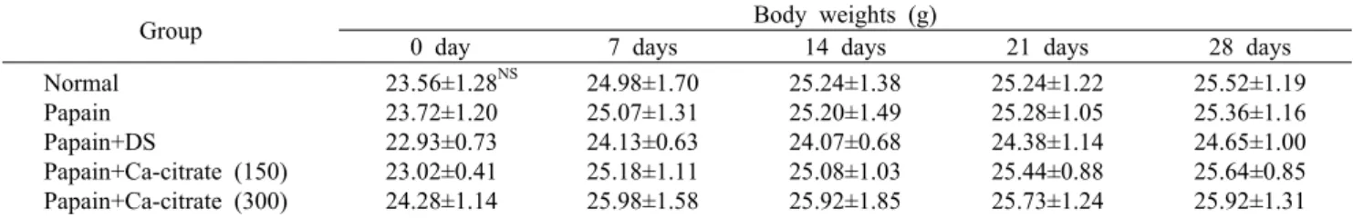
Table 2. Effects of calcium citrate treatment on the body weights of papain-induced osteoarthritic mice
Group Body weights (g)
0 day 7 days 14 days 21 days 28 days
Normal Papain Papain+DS
Papain+Ca-citrate (150) Papain+Ca-citrate (300)
23.56±1.28NS 23.72±1.20 22.93±0.73 23.02±0.41 24.28±1.14
24.98±1.70 25.07±1.31 24.13±0.63 25.18±1.11 25.98±1.58
25.24±1.38 25.20±1.49 24.07±0.68 25.08±1.03 25.92±1.85
25.24±1.22 25.28±1.05 24.38±1.14 25.44±0.88 25.73±1.24
25.52±1.19 25.36±1.16 24.65±1.00 25.64±0.85 25.92±1.31 Each value is expressed as mean±SD (n=8).
Normal: saline injected and treated with water group. Papain: papain injected and treated with water group. Papain+DS: papain injected and treated with diclofenac sodium (2 mg/kg/bw) group. Papain+Ca-citrate (150): papain injected and treated with Papain+
Ca-citrate (150 mg/kg/bw) group. Papain+Ca-citrate (300): papain injected and treated with Papain+Ca-citrate (300 mg/kg/bw) group.
NS: not significant.
혈액 내 사이토카인 함량 측정
혈액 내 tumor necrosis factor-α(TNF-α), interleukin- 1β(IL-1β), interleukin-6(IL-6)의 함량을 각각 enzyme- linked immunosorbent assay kit(Komabiotech, Seoul, Korea)를 사용하여 측정하였다.
관절연골의 proteoglycan 함유율 평가
채취한 슬관절은 10% formalin에 24시간 고정하고 그 후 파라핀 포매 전 10% formic acid로 72시간 동안 탈회하 였다. 탈회된 경골 근위단의 정중 관상단면을 만들고 일반적 인 조직 처리 과정을 거쳐 파라핀 포매하고 5 μm 두께로 박절하여 슬라이드에 부착시켰다. 경골 근위단 관절연골 내 의 proteoglycan 변성을 확인하기 위하여 박절편된 표면을 safranin O 및 hematoxylin 대조염색을 실시하였다. 칼라 영상분석기(Media Cybernetics, Image-ProPlusm, Rock- ville, MD, USA)를 이용하여 모든 관절연골에서의 평균 투 과도를 측정하였다. 정상군에서 측정된 평균 투과도의 평균 을 100%로 설정하고 대조군, 약물 투여군, 샘플 투여군의 투과도를 환산하여 proteoglycan 함유율을 평가하였다.
병리조직학적 관찰 및 골관절염 지수의 평가
대퇴골 및 경골의 내측과(medial condyle) 관절면과 슬 관절낭이 포함되도록 슬관절 시상단면을 만들고 일반적인 조직 처리 과정을 거쳐 파라핀 포매하여 5 μm 두께로 박절
하여 슬라이드에 부착시켰다. 슬관절 박절편을 hematox- ylin과 eosin(H&E) 및 safranin O-fast green으로 염색하 고 광학 현미경으로 관절연골 및 활막의 병리조직학적 변화 를 관찰하였다. 골관절염 지수는 관절연골 및 활막에서 관찰 된 병리조직학적 병변을 Rudolphi 등(15)의 방법에 준하여 계수화 하였다(Table 1).
통계분석
대조군과 각 시료들로부터 얻은 실험 결과들은 mean±
SD로 표시하였고, 각 시료 간의 유의성은 one-way ANOVA (analysis of variance)로 사전 검증한 후 Duncan's multi- ple range test에 의해 사후 검정하였다.
결 과
체중 변화
실험기간 동안 마우스의 체중 변화를 Table 2에 나타내 었다. Papain 투여군, DS 및 구연산칼슘 처치군은 실험기간 동안 정상군과 비교하여 유의적 차이를 나타내지 않았으며, papain 투여로 인한 마우스 간의 체중 변화는 없는 것을 확 인할 수 있었다.
혈액 내 사이토카인 변화
Papain 주입에 의해 관절염이 유발된 동물에서 다량 생성
ab ab b
a
b
0 20 40 60 80 100
Normal Papain Papain
+DS
Papain+
calcium citrate(150)
Papain+
calcium citrate(300)
TNF-α (pg/mL) .
Fig. 1. Effects of calcium citrate on the TNF-α contents in plas- ma of papain-induced osteoarthritic mice. Each value is expre- ssed as mean±SD (n=8). Values with different letters above the bars are significantly different at P<0.05 as analyzed by Dun- can's multiple range test. Groups are the same as in Table 2.
b b c
a
c
0 20 40 60 80 100 120
Normal Papain Papain
+DS
Papain+
calcium citrate(150)
Papain+
calcium citrate(300)
IL-1β (pg/mL) .
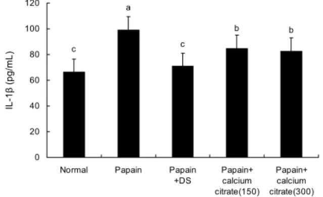
Fig. 2. Effects of calcium citrate on the interleukin-1β contents in plasma of papain-induced osteoarthritic mice. Each value is expressed as mean±SD (n=8). Values with different letters above the bars are significantly different at P<0.05 as analyzed by Dun- can's multiple range test. Groups are the same as in Table 2.
b b c
a
c
0 20 40 60 80 100 120
Normal Papain Papain
+DS
Papain+
calcium citrate(150)
Papain+
calcium citrate(300)
IL-6 (pg/mL) .
Fig. 3. Effects of calcium citrate on the interleukin-6 contents in plasma of papain-induced osteoarthritic mice. Each value is expressed as mean±SD (n=8). Values with different letters above the bars are significantly different at P<0.05 as analyzed by Dun- can's multiple range test. Groups are the same as in Table 2.
Table 3. Effects of calcium citrate treatment on the proteoglycan contents in articular cartilages of papain-induced osteoarthritic mice
Group1) Proteoglycan contents (%) Normal
Papain Papain+DS
Papain+Calcium citrate (150) Papain+Calcium citrate (300)
100.00±4.51a2) 46.51±3.02e 87.24±5.31b 68.46±3.52d 77.41±6.03c
1)Groups are the same as in Table 2.
2)Each value is expressed as mean±SD (n=8). Values with differ- ent letters are significantly different at P<0.05 as analyzed by Duncan's multiple range test.
되는 염증 사이토카인인 TNF-α, IL-1β, IL-6에 대한 구연 산칼슘의 효과를 측정하기 위하여 ELISA 방법으로 측정하 였다.
혈액 내 TNF-α의 함량은 papain 투여군이 정상군에 비 해서 유의성 있게 높았으며, DS 및 구연산칼슘 투여군은 papain 투여 대조군에 비해서 유의성 있게 낮았다. 구연산칼 슘-300 투여군이 구연산칼슘-150 투여군에 비해 낮게 나 타났지만 유의성은 없었다(Fig. 1).
혈액 내 IL-1β 함량은 papain 투여군이 정상군에 비해서 유의성 있게 높았으며, DS 및 구연산칼슘 처치군은 papain 투여군에 비해서 유의성(P<0.05) 있게 낮았다. 구연산칼슘 처치군 간의 유의성은 없었으나 구연산칼슘-300 처치군이 구연산칼슘-150 처치군에 비해 낮았다(Fig. 2).
혈액 내 IL-6의 함량은 papain 투여군이 정상군에 비해서 유의성 있게 높았다. DS 및 구연산칼슘 처치군이 papain 투여군에 비해서 유의성 있게 낮았다. 구연산칼슘-300 처 치군이 구연산칼슘-150 처치군에 비해 IL-6 함량이 낮게 나타났지만 유의성은 없었다(Fig. 3).
관절연골의 proteoglycan 함유율
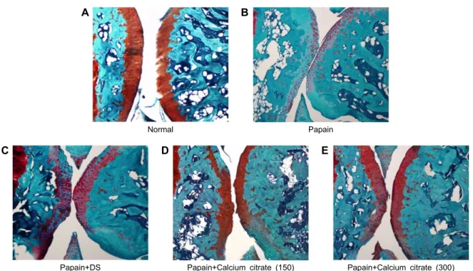
실험 종료 후 safranin O 염색을 실시하여 관찰한 결과는 Fig. 4와 같다. 정상군의 관절연골은 전 층이 짙은 적색으로 염색되어 proteoglycan의 함량이 풍부한 것을 확인할 수 있다. 그에 비해 papain 투여 대조군은 정상군과 비교하였을 때 훨씬 엷게 염색되거나 거의 염색이 되지 않아 proteo- glycan의 손실이 많음을 알 수 있다. DS 처치군은 정상군보 다는 엷지만 papain 투여군보다는 진하게 염색되어 있어 proteoglycan의 함량이 회복되었음을 알 수 있다. 구연산칼 슘 처치군의 경우 정상군과 비교 시에는 관절연골의 염색 범위가 감소하였으나, papain 투여 대조군과 비교 시에는 보다 짙은 적색으로 염색되어 DS 처치군처럼 proteoglycan 의 함량이 회복됨을 관찰할 수 있었다. 특히 구연산칼슘 -300 처치군이 구연산칼슘-150 처치군보다 좀 더 진하게 염색된 것을 확인할 수 있었다.
각 군에서 칼라영상분석기를 통해 측정한 safranin O 염 색 정도를 관절연골 내 proteoglycan 함유율로 평가하였으 며 그 결과는 Table 3에 나타내었다. Proteoglycan 함유율 은 papain 투여 대조군이 정상군에 비하여 유의성(P<0.05) 있게 낮았으며, DS 처치군은 papain 투여 대조군에 비해
A B
Normal
Papain
C D E
Papain+DS
Papain+Calcium citrate (150)
Papain+Calcium citrate (300)
Fig. 4. Safranin O stain intensity of articular cartilage of tibial platues. (A) Stain intensity of red color increased in proportion to proteoglycan content. (B) The red color disappeared in most of articular cartilage. (C) The red color remained in periphery of necrotic area with papain+DS group. (D) Proteoglycan contents in articular cartilages of the Ca-citrate 150 treated group were increased compared with papain group. (E) Proteoglycan contents in articular cartilages of the Ca-citrate 300 treated group were increased compared with Ca-citrate 150 group or papain group.
유의성(P<0.05) 있게 높았다. 구연산칼슘 처치군 역시 pa- pain 투여 대조군에 비해 proteoglycan 함유율이 유의하게 (P<0.05) 높았으며, 구연산칼슘-300 처치군이 구연산칼슘 -150 처치군보다 높았다.
병리조직학적 소견
본 연구에서는 papain으로 유도된 골관절염에 구연산칼 슘이 미치는 영향을 확인하기 위하여 동물을 희생시킨 후 동물의 관절조직을 H&E 염색을 하여 연골세포와 활액 주변 변화를 관찰하였고, 그 결과는 Fig. 5와 같다.
정상군은 모든 개체의 슬관절을 구성하는 대퇴골 및 경골 의 관절연골이 정상적인 구조를 유지하고 있었고 관절낭의 활막도 활막세포로 피복된 정상적인 상태를 유지하고 있었 으며, 퇴행성 변화를 관찰할 수 없었다. Papain 투여군에서 는 모든 개체에서 관절연골의 퇴행성 변화가 관찰되었다.
슬관절을 구성하는 대퇴골 및 경골의 관절연골이 변성 및 괴사되었으며, 연골 내 존재하는 연골세포의 손실도 관찰되 었다. 또한 대부분의 연골층이 탈락되어 연골하 골조직이 노출되어 있거나 연골세포의 윤곽이 사라졌다. 연골층 탈락 으로 관절강 내로 노출되어 있는 연골하 골조직은 파골세포 의 활성으로 인한 골조직 침습 및 섬유조직의 증식이 관찰되 었다. DS 처치군은 연골의 표층에서 연골 및 연골세포의 탈락이 관찰되었으나 변성 괴사된 관절연골의 범위가 감소 되어 있었다. Papain 투여군과 비교하였을 때 염증세포의 침윤과 활막의 비후가 억제되었으며 관절면의 연골손실이
적은 것을 관찰할 수 있었다.
구연산칼슘 처치군에서 슬관절을 구성하는 관절연골의 병리조직학적 병변은 관절연골의 변성 및 괴사된 범위가 papain 투여군에 비해 감소되어 있었다. 또한 활막세포의 증식으로 인한 활막의 비후 정도도 억제된 것을 관찰할 수 있었다. 구연산칼슘-300 처치군은 구연산칼슘-150 처치군 보다 관절연골의 표층에서 연골 및 연골세포의 탈락이 적게 관찰되었으며, 연골 내 존재하는 연골세포의 손실도 적게 관찰되었다.
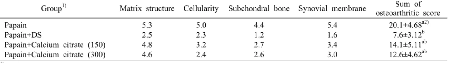
Rudolphi 등(15)의 방법에 준하여 계수화한 골관절염 지 수의 결과는 Table 4와 같다. Papain 투여 대조군의 골관절 염 지수가 유의성 있게 가장 높았으며, DS 및 구연산칼슘 투여군은 papain 투여 대조군에 비해 골관절염 지수가 유의 성 있게 낮았다. 특히 구연산칼슘-300 투여군이 구연산칼 슘-150 투여군보다 골관절염 지수가 낮았지만 유의적 차이 는 없었다.
고 찰
골관절염은 물리적인 힘, 생화학적 요인, 유전 등 여러 요인에 의한 지속적인 관절연골 손상으로 관절 간격의 감소, 연골하 골의 퇴행성 변화를 유발하는 질환으로 통증, 관절 변형, 기능 약화를 일으키는 만성질환이며, 퇴행성 관절염 또는 퇴행성 관절질환이라고도 한다(16). 골관절염의 병태 생리에는 여러 주장과 가설들이 있지만 크게 두 가지로 설명
A B
Normal
Papain
C D E
Papain+DS
Papain+Calcium citrate (150)
Papain+Calcium citrate (300)
Fig. 5. Microphotograph of articular structure of knee joint. (A) In normal group, articular cartilages (*) are well developed. (B) In papain group, most of articular cartilage was degenerated and desquamated. (C) In papain+DS group, most of articular cartilages (*) are degenerated and appeared homogeneous, but chondrocytes in periphery of degenerated area are remained. (D) In papain+Ca-cit- rate 150 group, most of articular cartilages (*) are degenerated, and appeared homogeneous. But chondrocytes are remained. (E) In papain+Ca-citrate 300 group, most of articular cartilages (*) are degenerated, and appeared homogeneous. But many of chondrocytes are remained. And proliferation of synovial cell layer is more mild than papain group.
Table 4. Effects of calcium citrate on the histopathological osteoarthritic scores of the knee joints of papain-induced osteoarthritic mice
Group1) Matrix structure Cellularity Subchondral bone Synovial membrane Sum of osteoarthritic score Papain
Papain+DS
Papain+Calcium citrate (150) Papain+Calcium citrate (300)
5.3 2.5 4.8 4.6
5.0 2.3 3.2 2.4
4.4 1.2 2.7 2.6
5.4 1.6 3.4 3.0
20.1±4.68a2) 7.6±3.12b 14.1±5.11ab 12.6±4.62ab
1)Groups are the same as in Table 2.
2)Each value is expressed as mean±SD (n=8). Values with different letters are significantly different at P<0.05 as analyzed by Duncan's multiple range test.
할 수 있다. 첫 번째는 연골기질의 손상과 변화이고, 두 번째 는 조직의 손상에 의한 연골세포의 감소이다. 여러 기계적인 자극이나 염증으로 인한 효소반응, 대사의 변화 등으로 손상 이 시작되어 기질 고분자의 격자가 파괴되고 수분함량의 변 화가 일어난다. 연골세포가 조직손상이나 변화를 감지하게 되면 동화 및 이화작용이 증가되어 주로 IL-1β 및 TNF-α 와 같은 cytokine에 의해 이뤄지는 파괴와 재형성 과정이 일어나게 된다. 이러한 점진적인 관절연골의 소실과 세포사 멸에 의해 골관절염의 진행이 촉진된다(17). 최근 감초부자 탕, 부자탕, 가감소속명탕 등의 한약 재료와 봉독 등 다양한 재료를 이용한 골관절염에 대한 실험적 연구들이 많이 시행 되었으나(18-20), 기능성 소재를 이용한 연구는 미비한 실 정이다.
칼슘보충제의 종류에는 여러 가지가 있으며 그중에서도
탄산칼슘과 구연산칼슘이 가장 많이 쓰인다. 특히 구연산칼 슘은 흡수율이 탄산칼슘에 비해 22∼27%가 높으며, 흡수 시에 위산을 필요로 하지 않아 식사와 관계없이 복용 가능하 다는 장점을 가지고 있다(10).
이에 본 연구에서는 기능성 소재인 구연산칼슘을 사용하 여 골관절염에 대한 효과를 알아보고자 동물 모델 C57BL/
6J 생쥐의 슬관절강 내에 papain을 투여하여 골관절염을 유 발한 후, 구연산칼슘을 경구 투여하여 연골의 변성, 염증 형 성 정도 등의 변화를 관찰하였다. 본 연구에 사용된 단백분 해효소인 papain은 관절연골 기질의 당단백질 복합체로부 터 콘드로이틴황산을 분비함으로써 골관절염을 야기하는 물질로(21), 동물을 대상으로 한 골관절염 유도 실험에서 조직병리학적 양상이 사람의 골관절염과 유사하다고 보고 되어 흔히 사용되는 방법이다(22,23).
골관절염은 여러 요인에 의한 기계적인 자극이나 염증으 로 인해 연골기질이 손상되거나 변화로 시작되어 이에 대한 연골세포의 반응으로 사이토카인이 작용하여 동화 및 이화 작용이 나타난다(24). 특히 이화작용이 증가하여 관절 기질 의 주요 요소인 proteoglycan의 합성과 분해의 균형이 깨져 발생하게 된다(25,26). TNF-α, IL-1β, IL-6 등은 관절염 진행과정에 관여하는 대표적인 사이토카인이며 특히 IL- 1 β와 TNF-α는 앞서 말한 proteoglycan의 이화작용에 관여 하는 사이토카인이다(27).
TNF-α는 IL-1β, IL-6 등과 같은 하위염증매개물질의 생성을 유발하여 염증을 증폭시키고 활막세포의 증식을 증 가시켜(28) proteoglycan의 소실을 증가시키거나 재합성 을 억제함으로써 관절연골을 손상시키고 골관절염의 진행 을 일으킨다(29). IL-1β는 연골세포, 활액, 활막세포 등으 로부터 기질금속 단백분해효소를 생성하고 분비시켜 연골 기질의 고분자를 파괴시키고 증상 발현에 큰 역할을 하는 prostaglandin E2의 생성을 촉진시켜 proteoglycan의 소실 을 증가시키고 재합성을 억제하게 한다(25,30). IL-6는 IL-1β와 TNF-α에 의해 분비가 촉진되어 골관절의 염증반 응에 주된 매개 작용을 하고, 손상된 관절의 관절액에서 활 성도가 증가하여 골관절염의 활성도와 관련이 있다(31). 본 연구에서 혈액 내 TNF-α, IL-1β, IL-6의 함량은 papain 투여 대조군이 정상군에 비해 유의적으로 증가하였으며 약 물 및 구연산칼슘 투여군은 대조군보다 유의적으로 낮게 나 타났다. 이는 구연산칼슘 투여로 인해 TNF-α, IL-1β, IL-6 의 활성이 억제되어 골관절염의 진행이 억제된 것으로 생각 된다.
관절연골은 collagen과 proteoglycan으로 구성된다.
Collagen은 무정형의 세포 사이 물질인 chondroitin sul- fate, keratin sulfate, hyaluronic acid 등과 같은 glyco- saminoglycan(GAG)에 의해 둘러싸여 결합되어 있다. 이들 GAG가 중심단백질과 결합하여 proteoglycan을 형성한다.
Proteoglycan은 충격 흡수 물질로써 조직의 섬유 성분 및 세포 성분을 보호하며, 조직 표면에 가해지는 압박을 견딜 수 있는 탄성을 가지게 하여 생체역학적인 용수철로도 작용 한다. 또한 연골세포에 영양공급을 하게 되며, proteogly- can membrane을 형성하여 관절면을 보호하는 등 관절을 정상적으로 유지하게 된다(32,33). 그러나 연골에 기계적 과부하가 가해지면 proteoglycan의 합성율이 떨어지고 기 질 내 대사성 산물들은 증가하여 연골의 전 층이 손실되는 골관절염이 초래된다(34). 본 연구에서 측정한 관절연골의 proteoglycan의 함유율은 구연산칼슘 투여군이 대조군에 비해 유의성 있게 높았으며, safranin O 염색을 통해 관찰한 육안적인 관절연골의 변성 정도에서도 대조군에 비해 구연 산칼슘 투여군이 더 진하게 염색되어 proteoglycan의 함량 에 차이가 있음을 알 수 있었다. 따라서 구연산칼슘의 투여 가 골관절염의 진행에서 관절연골 내 proteoglycan의 파괴 를 억제하는 것으로 사료된다.
전통적인 병리 조직 소견 판독법은 hematoxylin과 eosin 색소(H&E)를 이용하여 염색한 표본에 광학 현미경을 이용 하여 관찰하는 방식이다. 이 방법에서 hematoxylin 색소는 형태학적 특징을 파악하게 해주고, eosin 색소는 조직 특성 에 따라 미세한 염색 정도의 차이를 구분할 수 있게 해줌으 로써 일상적인 조직 소견 판독에 충분한 형태학적 변별력을 제공한다(35,36). 본 연구에서 H&E 염색법을 이용해 관찰 한 결과, 구연산칼슘 투여군에서 관절연골의 파괴와 골 침식 등 연골의 변성 괴사된 범위가 감소되거나 활막의 비후 정도 가 억제되는 등 약물 투여군과 유사한 것으로 보아 구연산칼 슘이 골관절염 치료에 효과가 있는 것을 확인할 수 있었다.
이전 연구에서 Kang 등(37)에 의하면 calcium gluconate가 수술로 유도된 골관절염 모델에서 조직, 병리학적 변화를 억제한다는 것이 보고되었으며, 이는 calcium gluconate의 항염증 특성 때문이라고 보고하였다.
이상의 실험 결과를 통해서 구연산칼슘 투여가 papain으 로 유도된 골관절염 동물모델에서 연골파괴와 활막 주변의 proteoglycan의 파괴를 억제하고, TNF-α, IL-1β, IL-6 등 의 염증성 사이토카인의 생성을 억제하는 등 유의한 효과가 있음을 알 수 있다. 따라서 추후 구연산칼슘의 항염증 효과 및 작용기전에 대한 연구와 더불어 임상적인 연구가 이루어 진다면 골관절염의 치료에 구연산칼슘이 활용될 수 있을 것 으로 사료된다.
요 약
본 연구에서는 구연산칼슘 투여가 papain으로 유도된 골관 절염 동물모델에 미치는 영향을 살펴보았다. 8주령의 C57 BL/6J 마우스의 우측 슬관절강에 papain 6 μL를 투여하여 골관절염을 유발하였다. 실험군은 각 군당 8마리씩 체중이 고르게 배정하여 총 5군으로 나누었다. 정상군은 우측 슬관 절강에 생리식염수 6 μL를 주사하였고, 대조군, 약물투여 군, 구연산칼슘 투여군은 papain 6 μL로 골관절염을 유발하 였다. 정상군과 대조군은 0.2 mL의 생리식염수를, 약물투여 군은 diclofenac sodium 2 mg/kg/bw를, 구연산칼슘 투여 군은 150, 300 mg/kg/bw를 28일간 1일 매일 정해진 시간 에 1회 경구 투여하였다. 실험기간 동안 체중을 측정하였으 며, 실험 종료일에는 부검하여 슬관절의 병리조직학적 관찰 을 하였고 관절연골 내 proteoglycan 함유율, 골관절염 지 수의 변화, 혈액 내 TNF-α, IL-1β, IL-6 함량을 측정하였 다. 체중은 실험기간 동안 모든 개체 간의 유의적인 차이는 없었다. 혈액 내 TNF-α, IL-1β, IL-6 함량은 papain 투여 대조군이 정상군에 비해 유의적으로 증가하였으며 약물 및 구연산칼슘 투여군은 대조군보다 유의성 있게 낮았다. 관절 연골 내 proteoglycan의 함유율은 약물 및 구연산칼슘 투여 군이 papain 투여 대조군보다 유의성 있게 높았다. Hemato- xylin과 eosin 염색 관찰 결과, 골관절염이 유발된 마우스에 구연산칼슘을 경구 투여하였더니 관절연골의 파괴와 골 침
식 등 연골의 변성이 약물 투여군과 유사하게 나타났다. 이 와 같은 결과는 구연산칼슘이 papain으로 유도된 골관절염 에서 연골의 변성을 줄이고 염증을 억제함으로써 골관절염 치료에 효과가 있은 것으로 사료되며, 추후 더 자세한 연구 가 필요하다.
감사의 글
본 연구는 국토해양부의 국토해양기술연구개발 사업으로 수 행된 연구 결과임.
REFERENCES
1. Wearing SC, Hennig EM, Byrne NM, Steele JR, Hills AP.
2006. Musculoskeletal disorders associated with obesity: a biomechanical perspective. Obes Rev 7: 239-250.
2. Altman R, Asch E, Bloch D. 1986. Development of criteria for the classification and reporting of osteoarthritis. Arthritis Rheum 29: 1039-1049.
3. Yoo MC. 1995. The latest trend in treating arthritis. J Muscle Joint Health 2: 227-229.
4. Hochberg MC, Altman RD, Brandt KD, Clark BM, Dieppe PA, Griffin MR, Moskowitz RW, Schnitzer TJ. 1995. Gui- delines for the medical management of osteoarthritis. Part
Ⅱ. Osteoarthritis of the knee. American College of Rheu- matology. Arthritis Rheum 38: 1541-1546.
5. Behrens F, Shepard N, Mitchell N. 1975. Alterations of rab- bit articular cartilage by intra-articular injections of gluco- corticoids. J Bone Joint Surg Am 57: 70-76.
6. Leslie M. 2000. Knee osteoarthritis management therapies.
Pain Management Nursing 1: 51-57.
7. Yoh SB, Sul JU, Shin MS. 2011. Research trends on the treatment of knee osteoarthritis in Korean medicine. Korean J Acupuncture 28: 139-155.
8. Won CH, Choi ES, Hong SS. 1999. Efficacy of bee venom infection for osteoarthritis patients. J Korean Rheum Assoc 6: 218-226.
9. Straub DA. 2007. Calcium supplementation in clinical prac- tice: a review of forms, doses, and indication. Nutr Clin Pract 22: 286-296.
10. Sakhaee K, Bhuket T, Adams-Huet B, Rao DS. 1999. Meta- analysis of calcium bioavailability: a comparison of calcium citrate with calcium carbonate. Am J Ther 6: 313-321.
11. Kenny AM, Prestwood KM, Biskup B, Robbins B, Zayas E, Kleppinger A, Burleson JA, Raisz LG. 2004. Comparison of the effects of calcium loading with calcium citrate or calcium carbonate on bone turnover in postmenopausal wo- men. Osteoporos Int 15: 290-294.
12. Zaccaro Scelza MF, da Silva Pierro VS, Chagas MA, da Silva LE, Scelza P. 2010. Evaluation of inflammatory response of EDTA, EDTA-T, and citric acid in animal model. J Endod 36: 515-519.
13. Bryland A, Wieslander A, Carlsson O, Hellmark T, Godaly G. 2012. Citrate treatment reduces endothelial death and in- flammation under hyperglycaemic conditions. Diab Vasc Dis Res 9: 42-51.
14. Ministry of Food and Drug Safety. 2013. Korea Food Addi- tives Code. Chapter 2, p 49.
15. Rudolphi K, Gerwin N, Verziji N, van der Kraan P, van der Berg W. 2003. Pralnacasan, an inhibitor of interleukin-1
β converting enzyme, reduces joint damage in two murine models of osteoarthritis. Osteoarthritis Cartilage 11: 738- 746.
16. Felson DT, Zhang Y. 1998. An update on the epidemiology of knee and hip osteoarthritis with a view to prevention.
Arthritis Rheum 41: 1343-1355.
17. Han TR, Bang MS. 2009. Rehabilitation medicine. 3rd ed.
Konnja Inc., Seoul, Korea. p 905-908.
18. Choi JY, Oh MS. 2012. Effects of Kamchobuja-tang (Gan- caofuzi-tang) on papain-induced osteoarthritis in mice. J Oriental Rehab Med 22: 37-57.
19. Park DS, Jeong SH, Kim SJ, Seo IB. 2011. Effects of Gagamsosokmyeong-tang (Jiajianxiaoxuming-tang) treat- ment on the monosodium iodoacetate-induced early stage osteoarthritis in rats. J Oriental Rehab Med 21: 49-65.
20. An HJ, Lee CK, Park JH, Kim KH, Lee WR, Park IY, Han SM, Lee KG, Park KK. 2012. Effects of bee venom on papain-induced osteoarthritis in an animal model. Kor J Pharmacogn 43: 167-172.
21. Huang MH, Ding HJ, Chai CY, Haung YF, Yang RC. 1997.
Effects of sonication on articular cartilage in experimental osteoarthritis. J Rheumatol 24: 1978-1984.
22. Bentley G. 1971. Papain-induced degenerative arthritis of the hip in rabbits. J Bone Joint Surg Br 53: 324-337.
23. Murray DG. 1964. Experimentally induced arthritis using intra-articular papain. Arthritis Rheum 7: 211-219.
24. The Korean Orthopaedic Association. 2006. Orthopaedics.
6th ed. Newmedical Inc., Seoul, Korea. p 253-261.
25. Hulejová H, Baresová V, Klézl Z, Polanská M, Adam M, Senolt L. 2007. Increased level of cytokines and matrix met- alloproteinases in osteoarthritic subchondral bone. Cytokine 38: 151-156.
26. López-Armada MJ, Caramés B, Martín MA, Cillero-Pastor B, Lires-Dean M, Fuentes-Boquete I, Arenas J, Blanco FJ.
2006. Mitochondrial activity is modulated by TNFalpha and IL-1beta in normal human chondrocyte cells. Osteoarthritis Cartilage 14: 1011-1022.
27. Hwang SI. 1998. The semantics of the immune. Haneul Science Library, Seoul, Korea. p 43, 45, 69, 92.
28. Smolen JS, Aletaha D, Koeller M, Weisman MH, Emery P. 2007. New therapies for treatment on rheumatoid arthritis.
Lancet 370: 1861-1874.
29. Lim CK, Yun YG. 2009. Anti-rheumatiod arthritis effects of I-Myo-San water extract. Kor J Oriental Medical Pre- scription 17: 99-111.
30. Martel-Pelletier J, Pelletier JP, Fahmi H. 2003. Cyclooxyge- nase-2 and prostaglandins in articular tissues. Semin arthri- tis Rheu 33: 155-167.
31. Madson KL, Moore TL, Lawrence JM 3rd, Osborn TG.
1994. Cytokine levels in serum and synovial fluid of pa- tients with juvenile rheumatoid arthritis. J Rheumatol 21:
2359-2363.
32. Kim JD, Park SW, Lee HK. 1990. Effects of cationic ion on proteoglycan synthesis in bovine articular cartilage.
Journal of Korea Medicine University 27: 685-694.
33. Park KA. 1992. Histology. Korea Medicine, Seoul, Korea.
p 169-172.
34. Park IS. 1994. A morphological study of collagenase-in- duced degenerative lesion in knee joints of rats. Kor J Knee Surg Res 6: 3-16.
35. Lyle HM. 1947. An improved tissue technique with hema- toxylin-eosin stain. Am J Med Technol 13: 178-181.
36. Bancroft JD, Gamble M. 2002. Theory and practice of histo- logical techniques. Elsevier Science Health Science Div-
sion, Philadelphia, PA, USA. p 121-134.
37. Kang SJ, Kim JW, Kim KY, Ku SK, Lee YJ. 2014. Protective effects of calcium gluconate on osteoarthritis induced by
anterior cruciate ligament transection and partial medial me- nixcectomy in sprague-dawley rats. J Orthop Surg Res 9:
14-21.