좌측 유방암의 방사선치료 자세와 선량인자의 상관관계 분석
인제대학교 해운대백병원, 동의과학대학교 방사선과*
전재완·박철우*·홍종수·진성진·강정훈
목 적 : 방사선치료의 가장 기본적인 조건은 정상조직의 불필요한 피폭을 방지하는 것이다. 유방암의 경우는 폐 와 심장에 조사되는 선량이 중요하게 평가되는 인자이다. 따라서 유방암 방사선치료 자세에 따른 정상조직에 조사되는 선량을 비교하고 그 연관성을 상관관계 분석을 통하여 결과를 확인하여 보다 효과적인 유방암 방사 선 치료방법을 모색하고자 한다.
대상 및 방법 : 본원을 내원한 좌측 유방암 환자 30명을 대상으로 Supine, Prone Position에서 CT image를 획득하였다. Eclipse Treatment Planning System(Version 11, USA)을 이용하여 전산화치료계획을 수립하였 다. DVH(Dose Volume Histogram)을 통해 Position 별로 정상조직에 조사된 선량을 비교하였다. 그 결과를 바 탕으로 SPSS(Version 18)을 이용하여 각 정상조직의 선량인자를 통계분석하고 항목 간 상관관계 분석 및 독립 표본 t-test를 통하여 그 연관성을 알아보았다. 그리고 MIRADA RTx(Version Advanced 1.6, UK)를 이용하여 HI(Homogeneity Index)와 CI(Conformity Index)를 Supine, Prone Position에서 값을 구하고 비교하였다.
결 과 : 유방암의 전산화치료계획의 결과 폐의 경우는 Supine Position에서 V20은 16.5 ± 2.6 %, V30은 13.8
± 2.2 %, Mean dose는 779.1 ± 135.9 cGy(Absolute value)를 보였다. Prone Position은 위 순서대로 3.1 ± 2.2 %, 1.8 ± 1.7 %, 241.4 ± 138.3 cGy를 보였다. Prone Position이 전반적으로 낮은 선량을 나타내었고 평 균선량 537.7 cGy가 더 적게 폐에 조사되었다. 심장의 경우에는 Supine, Prone 순서대로 V30은 8.1 ± 2.6 %, 5.1 ± 2.5 %, Mean dose는 594.9 ± 225.3 cGy, 408 ± 183.6 cGy를 보였다. Prone Position에서 평균선량 182.6 cGy가 더 적게 조사된다는 것을 확인하였다.
통계분석 결과 신뢰도 분석지표인 Cronbach’s Alpha value는 0.563이였고 변수간의 상관관계분석 결과 치 료자세와 폐의 선량평가인자는 대략 0.89 이상으로 그 상관관계가 높았다. 반면 심장의 경우는 V30은 0.488, Mean dose는 0.418로 상관관계가 다소 적었다. 마지막으로 독립표본 t-test 결과 치료자세와 폐, 심장의 선량 평가인자가 신뢰수준 99 %에서 모두 유의하게 나타났다(p-value≤0.05).
결 론 : 현재 방사선치료는 최첨단 선형가속기와 다양화된 전산화치료계획 기술이 개발되고 있다. 이 발전의 기본전제 조건은 PTV(Planning Target Volume) 주위의 정상조직 보호라고 생각한다. 물론 유방암 환자를 Prone Position에서 치료하면 Set-up의 재현성 문제와 다소 많은 시간이 소요되지만 이 실험결과에서 보듯이 Prone Position에서 폐와 심장에 들어가는 선량을 줄일 수 있으며 그 연관관계도 의미가 있다는 것을 확인하였 다. 결론적으로 Prone Position에서 충분한 치료시간을 확보하고 정확한 치료부위 확인이 이루어진다면 환자 에게 보다 좋은 방사선치료를 제공할 수 있다고 생각된다.
▶ 핵심용어 : 좌측 유방암환자, 방사선 치료자세, 상관관계분석, 신뢰도분석지표
서 론
오늘날 우리나라 여성의 유방암 발생률은 점점 증가하 고 있다. 물론 우리나라에서 뿐만 아니라 여러 선진국에서
책임저자 : 박철우, 동의과학대학교 방사선과 부산광역시 부산진구 양지로54 Tel : 051-860-3532 E-mail : [email protected]
도 유방암은 여성에 나타나는 흔한 암으로 알려져 있다. 통 계적으로 유방암은 1970년 이후로 지속적으로 증가하여 1984년에 전체 여성암 발생건수에서 8.7 %를 차지하였고 1987년도에는 9.8 %로 증가하여 2002년에는 16.8 %로 그 비율이 점점 증가하였다. 이는 2000년대까지 여성의 최고 암 발생률을 보이던 위암을 추월하여 2000년 이후 유방암 이 여성암 중 발생빈도 1위로 기록될 만큼 유방암 발생이 급속도로 증가하고 있음을 보여주고 있다.(1,2)
실례로 2001년 한국 중앙 암 연례 등록보고서에 의하면 유방암이 자궁암, 위암을 제치고 1위를 차지하였고 2012년 국가 암 등록 통계에 따르면 유방암 환자는 전체 암 환자의 7.4 %를 차지하고 있다.(3) 본원에서도 개원한 이후로 꾸준 히 유방암 환자는 증가하여 현재에는 전체 방사선치료 환 자의 대략 40 % 정도를 차지하고 있다.
유방암 환자의 경우는 종양을 제거한 후 방사선치료 를 실시하여 국소 재발률을 낮추고 전반적인 생존율을 높 일 수 있기 때문에 대부분의 환자에게 방사선치료를 실시
한다.(4-6) 본원에서는 IDC(Invasive Ductal Carcinoma)
인 경우에는 Photon Beam으로 1.8 Gy/Daily씩 28회 총 50.4 Gy를 조사 후에 Electron Beam으로 수술한 주변에 1.8 Gy/Daily씩 총 5회를 조사하며 DCIS (Ductal Car- cinoma In Situ)인 경우에는 Photon Beam으로 1.8 Gy/
Daily씩 25회 총 45 Gy를 조사 후에 Electron Beam으로 수술한 주변에 2 Gy/Daily씩 총 5회를 더 조사한다. 또한 유방암 환자가 늘어나는 추세에 발맞춰 방사선치료 기술도 발전을 거듭하고 있다. 과거의 단순한 2-Dimension 대 향 2문 접선치료를 시작으로 Physical/Dynamic Wedge 를 사용한 방사선치료, 근래에는 Step-by-Shot 형태의 FIF(Field-in-Field)와 VMAT(Volumetric Modulated Arc Therapy)까지 선형가속기의 발전에 따라 그 전산화 방사선치료계획 방법도 다양해지고 있다.
유방암 환자의 전산화 방사선치료계획을 수립할 때에 가 장 주의해야 할 사항은 유방 아래에 위치하고 있는 Lung, 왼쪽 유방암의 경우는 Heart에 조사되는 선량이다. Stan- dard Fractionation에서 권고하는 Lung의 Tolerance Dose의 범위는 V5‹42 %, V20‹31 %으로 제한하고 있 으며 QUANTEC Summary에서는 Mean Dose를 20 Gy 이하로 권고하고 있다. 특히 V20은 Radiation Pneumo-
nitis를 예측할 수 있는 유용한 인자로 대부분의 병원에서 유방암 치료 시에 중요한 인자로 보고 있다.(7,8)
반면 Heart의 경우는 V30‹46 %, V40‹30 %, Mean Dose 26 Gy 이하로 권고하고 있다.(7) 특히 Heart에 조사 되는 저 선량의 방사선도 심장 혈관의 장애를 일으키는 요 인이 되기 때문에 좌측 유방암에 방사선치료를 실시하는 경우 Heart에 전달되는 작은 선량도 반드시 고려되어야 한
다.(9,10) 최근의 Darby et al.의 유방에 방사선치료를 실시한
이후 Mean Heart Dose와 Ischemic Heart Disease의 관계에 대한 조사결과, Heart의 피폭선량의 감소가 대략 3.7 %의 Major Coronary Artery Risk의 감소효과가 있 다고 발표하였다.(11)
대부분의 유방암 환자의 경우에는 Breast Board 및 Vac-loc을 이용한 Supine Position에서 방사선치료를 실 시하고 있다. Supine Position의 가장 큰 장점은 환자가 편안한 자세를 유지할 수 있다는 점과 환자 Set-up이 간 결하다는 점이다. 그러나 호흡에 의한 유방의 작은 움직임 과 Tolerance Range에는 들어오지만 정상조직인 Lung, Heart Dose를 줄이는데 한계가 있다는 것이다.(12) 따라서 유방암 방사선치료를 실시할 때 Lung과 Heart에 조사되 는 Dose를 줄이기 위한 방법을 다양한 연구를 통해 지속 적으로 모색 중이며 그 중의 하나의 방법으로 Prone Po- sition에서 방사선치료를 실시하는 방법의 유용성이 여러 논문들을 통해 평가되고 있다.(13)
Prone Position에서 매달린 형태의 유방을 접선조사의 형태로 방사선치료를 실시하면 Lung, Heart에 조사되는 선량이 Supine Position과 비교하였을 때 더 적은 양이 조 사되어 Lung Acute Toxicity 및 Cardiac Toxicity의 Risk 를 줄일 수 있다.(14-17) 또한 Large Breast의 경우에는 흉벽 과 유방조직 간의 분리가 더 용이하며 유방 내부의 균등한 선량분포의 효과 및 Supine Position 보다 호흡에 의한 움 직임이 더 작다는 장점이 있다고 한다.(18)
따라서 이 연구는 좌측 유방암 환자의 방사선치료 시에 Supine, Prone Position의 유용성을 확인해 보고자 본 원에 내원한 좌측 유방암 환자를 대상으로 하여 Supine, Prone Position에서 CT Image를 획득하였다. 그 이후 전 산화 치료계획을 수립하여 각 Treatment Position에서 Lung, Heart의 선량을 구하고 그 선량과 Treatment Po-
sition과의 상관관계 및 통계분석을 통해 그 유의성을 평가 하였다. 이를 통해 유방암 환자에게 보다 좋은 방사선치료 방법을 모색하고자 한다.
대상 및 방법
1. 대상
이 연구의 대상은 2015년 1월에서 12월 사이에 본원에 서 방사선치료를 받은 좌측 유방암 환자 30명으로 선정하 였다. 실험결과와 환자체형의 상관관계를 분석하기 위해서 Table 1에 각 환자의 체중을 기록하고 Eclipse Treatment Planning System(Version.11 Varian, USA)의 Measure Volume Function을 사용하여 각 환자마다 Lung, Heart Volume을 측정하여 기록하였다. 단 Small Breast와 Tu- mor Bed가 Inner 방향에 위치한 환자는 Prone Position 으로 치료하기에 제한이 있어서 대상에서 제외하였다.
2. 실험장비
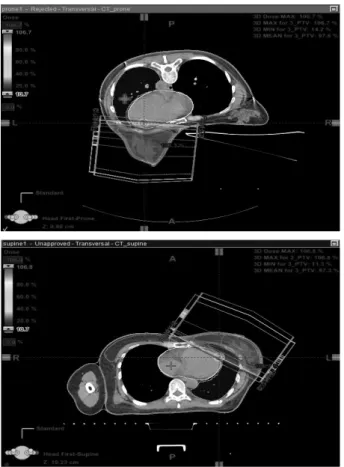
Supine Position은 Breast board(Civoco, USA)에 Vac-loc을 두고 치료하는 쪽의 팔을 거상시킨 상태에서 제작하여 CT를 촬영하였다(Fig. 1).
Table 1. Weight and critical organ volume of subjects
No. Weight(kg) Lung Volume(cm3) Heart Volume(cm3) Supine Prone
1 45 800 1008.3 663.2
2 56 1164.2 1263.9 512.6
3 61 587.8 717.4 587.7
4 48 1069.9 1069.8 520.1
5 53 660.4 828.1 568.8
6 73 1036.2 1302.8 550.8
7 68 1113.7 1356.5 744.9
8 56 972.5 1166.3 521.7
9 46 984 1083.3 507.1
10 44 1157.7 1029.1 437.9
11 62 989.3 1029.7 510.7
12 54 937.7 856.5 348.8
13 68 1162.1 1181.7 553.8
14 61 969.6 1040.9 645.8
15 72 1015 1229.1 408.5
16 53 803.2 900.3 386.5
17 74 1004.5 1039.5 500.2
18 46 956.7 1013.8 489.7
19 43 878.6 900.3 666.2
20 44 843.5 912.5 502.7
21 52 700.2 800.3 555.5
22 56 956.7 1016.2 645.1
23 64 647.8 755.9 435.8
24 70 844.2 1016.8 569.8
25 66 744.8 888.3 645.8
26 47 1022.5 1156.9 422.1
27 40 951.8 1123.5 568.9
28 45 679,7 855.9 455.8
29 50 1059,6 1213.6 410.3
30 51 1122.5 1318.9 455.8
Fig. 1. Civco 社의 Breast board & Vac-loc
Civco 社의 Breast Board는 환자의 체형에 따라 Board 의 높이를 조정할 수가 있다. 이 실험에서는 그 높이를 모두 동일한 단계에 고정한 상태에서 CT Image를 획득하였다.
Prone Position은 상용화된 Prone Breast Board인 Kvue Access 360 RT-4544KV(Q-fix, USA)를 사용하 여 CT Image를 획득하였다(Fig. 2).
실험에 사용한 Prone Breast Board는 Left, Right Breast를 구분하여 사용이 가능하며 다소 불안정한 Prone Position을 보정하기 위한 보조기구로 두 가지 형 태의 Headrests, Hand Grips이 있다. 이 실험에서도 보다 안정적인 자세유지를 위하여 두 가지 보조기구를 사용한 상태에서 CT Image를 얻었다. 이 Prone Breast Board도 역시 환자의 체형에 따라 Accessory의 위치를 단계별로 조정할 수 있으며 모두 동일한 단계에서 CT Image를 획득 하였다.
3. Radiation Treatment Plan and Critical Organ Dosimetric Parameters
본원에 내원한 유방암 환자 중 좌측 유방암 환자 30명 을 선정하여 환자별로 Supine, Prone Position에서 전 산화 방사선치료계획을 수립하였다. 치료계획은 Eclipse Treatment Planning System(Version.11 Varian, USA) 을 사용하였고 선형가속기 Rapid Arc의 (Varian, USA) 6 MV X선 Beam을 사용하여 AAA(Anisotropic Analytic Algorithm) 알고리즘을 이용하여 선량을 계산하였다.
전산화 방사선치료계획은 각 환자에게 적절한 Beam Angle을 선택한 후 모두 Half Beam을 이용한 대향 2문의 FIF 방식으로 계획을 수립하였다. 또한 선량률(Dose rate) 은 400 MU/min으로 설정하였고 PTV(Planing Target Volume)의 처방선량은 1.8 Gy/28 Fx 총 50.4 Gy로 하였다.
유방암 환자의 각 Position에서 Critical Organ의 Do- simetric Effect를 비교하기 위해 선량용적 히스토그램 (Dose Volume Histogram, DVH)을 이용하여 평가하였
Fig. 2. Q-fix 社의 Prone breast board(Kvue Access 360 RT-
4544KV) Fig. 3. Radiation treatment planning image
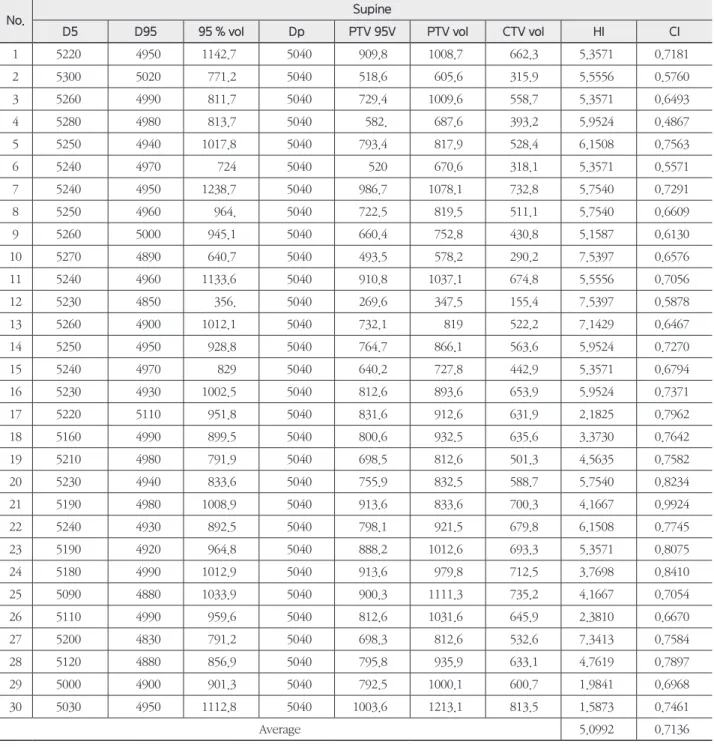
다. 평가인자로는 Lung의 경우는 Ipsilateral Lung의 V20, V30, Mean Dose, Heart의 경우는 V30, Mean Dose, Dmax를 선정하였다. PTV의 처방선량 50.4 Gy을 기준으 로 그 결과를 비교하였다. 그리고 Position에 따른 PTV 내 의 Dose Distribution의 균일성을 비교해보고자 Dose Homogeneity Index 값을 구하였다.
Dose Homogeneity Index는 HI(Homogeneity In-
dex)와 CI(Conformity Index) 값을 계산하여 비교하였 다. HI는 PTV 내의 선량분포 균일성을 나타내는 지표로서 구하는 식은 다음과 같다.(19)
HI = (D5-D95)/ Dp·100
위 식에서 D5는 PTV 전체 부피에서 5 %에 해당되는
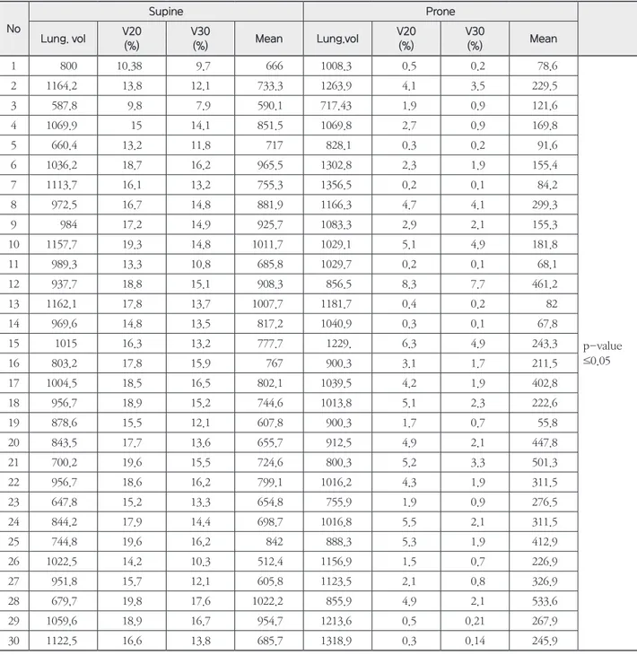
Table 2. Evaluation factors of lung dose according to treatment position
No
Supine Prone
Lung. vol V20 (%)
V30
(%) Mean Lung.vol V20
(%)
V30
(%) Mean
1 800 10.38 9.7 666 1008.3 0.5 0.2 78.6
p-value
≤0.05
2 1164.2 13.8 12.1 733.3 1263.9 4.1 3.5 229.5
3 587.8 9.8 7.9 590.1 717.43 1.9 0.9 121.6
4 1069.9 15 14.1 851.5 1069.8 2.7 0.9 169.8
5 660.4 13.2 11.8 717 828.1 0.3 0.2 91.6
6 1036.2 18.7 16.2 965.5 1302.8 2.3 1.9 155.4
7 1113.7 16.1 13.2 755.3 1356.5 0.2 0.1 84.2
8 972.5 16.7 14.8 881.9 1166.3 4.7 4.1 299.3
9 984 17.2 14.9 925.7 1083.3 2.9 2.1 155.3
10 1157.7 19.3 14.8 1011.7 1029.1 5.1 4.9 181.8
11 989.3 13.3 10.8 685.8 1029.7 0.2 0.1 68.1
12 937.7 18.8 15.1 908.3 856.5 8.3 7.7 461.2
13 1162.1 17.8 13.7 1007.7 1181.7 0.4 0.2 82
14 969.6 14.8 13.5 817.2 1040.9 0.3 0.1 67.8
15 1015 16.3 13.2 777.7 1229. 6.3 4.9 243.3
16 803.2 17.8 15.9 767 900.3 3.1 1.7 211.5
17 1004.5 18.5 16.5 802.1 1039.5 4.2 1.9 402.8
18 956.7 18.9 15.2 744.6 1013.8 5.1 2.3 222.6
19 878.6 15.5 12.1 607.8 900.3 1.7 0.7 55.8
20 843.5 17.7 13.6 655.7 912.5 4.9 2.1 447.8
21 700.2 19.6 15.5 724.6 800.3 5.2 3.3 501.3
22 956.7 18.6 16.2 799.1 1016.2 4.3 1.9 311.5
23 647.8 15.2 13.3 654.8 755.9 1.9 0.9 276.5
24 844.2 17.9 14.4 698.7 1016.8 5.5 2.1 311.5
25 744.8 19.6 16.2 842 888.3 5.3 1.9 412.9
26 1022.5 14.2 10.3 512.4 1156.9 1.5 0.7 226.9
27 951.8 15.7 12.1 605.8 1123.5 2.1 0.8 326.9
28 679,7 19.8 17.6 1022.2 855.9 4.9 2.1 533.6
29 1059,6 18.9 16.7 954.7 1213.6 0.5 0.21 267.9
30 1122.5 16.6 13.8 685.7 1318.9 0.3 0.14 245.9
부피에 전달되는 선량이고 D95은 95 %에 전달되는 선량 을, Dp는 처방선량을 의미한다. Registration Program인 MIRADA RTx(Version, Advanced 1.6 MIRADA Medi- cal, UK)의 Dose and Contour Warping 기능을 이용하
여 Supine, Prone Plan의 DVH를 분석하여 D5, D95 값 을 얻었다. 이 식을 통해 계산된 HI Value가 클수록 선량 분포가 불균등하다는 것을 의미한다.
CI는 환자에게 처방된 선량의 95 %에 해당되는 부피와 PTV 내에서 처방선량의 95 %의 부피와 일치되는 정도를 나타내는 지표로서 다음의 식을 이용한다.(20)
CI = V95PTV2/(VPTV · V95)
위 식에서 VPTV는 PTV의 전체 부피이고 V95는 환자에 서 처방선량의 95 %값이 전달되는 부피를, V95PTV는 PTV 내에서 처방선량의 95 %값이 전달되는 부피를 말한다. CI Value는 0-1의 값을 가지며 그 값이 클수록 환자에서 전 달되는 선량(처방선량)의 부피와 PTV에 전달되는 부피가 일치된다는 것을 의미한다.(21)
결 과
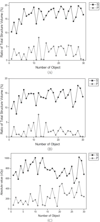
1. Lung Dose of Supine and Prone Position
환자의 Position에 따라 Lung에 입사되는 선량을 Table 2와 Fig. 4에 나타내었다. 그 결과를 살펴보면 Supine Position에서 V20은 16.5 ± 2.6 %, V30은 13.8 ± 2.2 %, Mean Dose는 779.1 ± 135.9 cGy의 값을 보였다. 반면 Prone Position에서는 위의 순서대로 3.1 ± 2.2 %, 1.8± 1.7 %, 241.4 ± 138.3 cGy의 값으로 나타났다. 여기서 cGy로 나타낸 값은 Absolute Value이다. 결과에서 보듯 이 평균적으로 Prone Position에서 V20은 13.4 %, V30은 12 %, Mean Dose는 537.7 cGy 만큼의 선량이 Supine Position에 비해 더 적게 조사된다는 것을 확인할 수 있다.
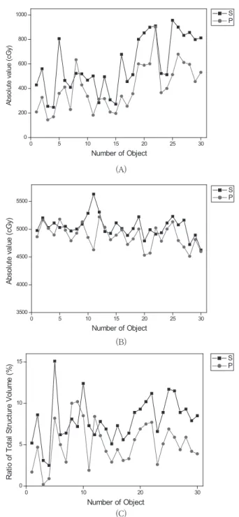
2. Heart Dose of Supine and Prone Position
환자의 Position에 따라 Heart에 입사되는 선량을 Table 3와 Fig. 5에 나타내었다. Heart의 경우는 Supine Position에서 V30은 8.1 ± 2.6 %, Mean Dose는 594.9 ± 225.3 cGy, Dmax는 5042.7 ± 190.7 cGy의 값을 보였다.반면 Prone Position에서는 위의 순서대로 5.1 ± 2.5 %, 408 ± 183.6 cGy, 4879.6 ± 197.3 cGy의 결과를 보였다.
0 10 20 30
0 5 10 15 20
Ratio of Total Structure Volume (%)
Number of Object
S P
0 5 10 15 20 25 30
0 200 400 600 800 1000
Absolute value (cGy)
Number of Object
S P
Fig. 4. Dose distribution of Lung according to Treatment Posi- tion
A. V20 dose of lung B. V30 dose of lung C. Mean dose of lung
0 10 20 30
0 5 10 15 20
Ratio of Total Structure Volume (%)
Number of Object
S P
(A)
(B)
(C)
결과에서 보듯이 평균적으로 Prone Position에서 V30은 3 %, Mean dose는 186.2 cGy, Dmax는 163.1 cGy의 선 량이 더 적게 조사된다는 것을 알 수 있었다.
3. HI, CI Value of Supine and Prone Position
각 Position의 HI, CI value를 Table 4에 나타내었다.그 결과 Supine Position에서 HI Value의 평균과 표준편 차는 5.09 ± 1.5를, CI Value은 0.71 ± 0.1이였다. Prone
Table 3. Evaluation factors of heart dose according to treat- ment position
No
Supine Prone
Mean (cGy)
Dmax (cGy)
V30 (%)
Mean (cGy)
Dmax (cGy)
V30 (%) 1 429 4976.2 5.2 208.3 4864.4 1.7
p-value
≤0.05 2 560.1 5203.2 8.6 327.2 5165 4.7
3 254.6 5029.9 3.1 144.6 5015.5 0.2 4 247.1 5111.8 2.5 169.2 4897.1 0.9 5 805.8 5030.5 15.1 360.3 5180.9 8.2 6 465.2 5051.5 6.2 411.3 4983.3 5 7 409.8 4969.2 6.4 228.6 4792 2.9 8 523.1 5001.9 8.1 634.2 4928.2 10 9 519.4 5091 7.2 429 5132.2 10.2 10 467.9 5287.7 12.4 336.4 4851.7 8.5 11 502.3 5631 7.3 182.4 4629.5 1.9 12 283.8 5307.8 6.2 312.3 5220 8.4 13 495.8 4958 7.8 316.9 5036 6.1 14 307.8 4925.4 6.9 208 4810.9 4.2 15 272.7 5113.2 5.1 196.8 4893 2.9 16 678.2 5012.6 7.3 336.9 4988.2 4.4 17 456.9 4889.2 5.6 255.9 4725.9 3.1 18 512.8 5011.5 6.4 356.9 4825.9 3.3 19 800.6 5222.9 8.9 600.3 5003.6 5.6 20 853.2 4789.2 9.3 589.7 4532.6 6.9 21 899.2 4991.2 10.2 601.3 4569.8 7.5 22 909.6 4912.3 11.2 899.8 5023.6 7.7 23 522.3 4936.5 6.6 365.9 4785.9 2.6 24 512.8 5112.3 8.9 400.3 5003.2 5.1 25 955.9 5231.2 11.7 512.6 5132.6 6.9 26 900.3 5078.6 11.5 679.5 4795.8 5.9 27 833.3 5163.5 8.9 612.5 4678.9 4.4 28 856.9 4725 9.3 596.8 4512.6 5.9 29 799.1 4893.5 7.9 456.9 4812.6 4.2 30 812.3 4623.9 8.5 531.6 4598.2 3.9
(A)
0 5 10 15 20 25 30
3500 4000 4500 5000 5500
Absolute value (cGy)
Number of Object
S P
0 10 20 30
0 5 10 15
Ratio of Total Structure Volume (%)
Number of Object
S P
Fig. 5. Dose distribution of heart according to treatment posi- tion
A. Mean dose of heart B. Maximum dose of heart C. V30 dose of heart
0 5 10 15 20 25 30
0 200 400 600 800 1000
Absolute value (cGy)
Number of Object
S P
(B)
(C)
Position에서는 HI Value는 6.44 ± 1.3 CI Value는 0.58
± 0.1의 결과를 보였다. Supine Position에서 Dose Ho- mogeneity Index의 결과가 작은 차이지만 더 좋음을 확 인할 수 있었다.
4. 통계
수집된 자료를 바탕으로 SPSS (Version 18.0) 통계 프 로그램을 이용하여 신뢰도 분석, 통계 분석 및 독립표 본 t-test 분석을 실시하였다. 신뢰도 분석결과 표준화된 Cronbach’s Alpha 값은 0.563으로 나타났다. 앞으로 더 많은 자료를 수집한다면 더 높은 신뢰도 값을 보일 것으로
Table 4. HI and CI value according to treatment position
No. Supine
D5 D95 95 % vol Dp PTV 95V PTV vol CTV vol HI CI
1 5220 4950 1142.7 5040 909.8 1008.7 662.3 5.3571 0.7181
2 5300 5020 771.2 5040 518.6 605.6 315.9 5.5556 0.5760
3 5260 4990 811.7 5040 729.4 1009.6 558.7 5.3571 0.6493
4 5280 4980 813.7 5040 582. 687.6 393.2 5.9524 0.4867
5 5250 4940 1017.8 5040 793.4 817.9 528.4 6.1508 0.7563
6 5240 4970 724 5040 520 670.6 318.1 5.3571 0.5571
7 5240 4950 1238.7 5040 986.7 1078.1 732.8 5.7540 0.7291
8 5250 4960 964. 5040 722.5 819.5 511.1 5.7540 0.6609
9 5260 5000 945.1 5040 660.4 752.8 430.8 5.1587 0.6130
10 5270 4890 640.7 5040 493.5 578.2 290.2 7.5397 0.6576
11 5240 4960 1133.6 5040 910.8 1037.1 674.8 5.5556 0.7056
12 5230 4850 356. 5040 269.6 347.5 155.4 7.5397 0.5878
13 5260 4900 1012.1 5040 732.1 819 522.2 7.1429 0.6467
14 5250 4950 928.8 5040 764.7 866.1 563.6 5.9524 0.7270
15 5240 4970 829 5040 640.2 727.8 442.9 5.3571 0.6794
16 5230 4930 1002.5 5040 812.6 893.6 653.9 5.9524 0.7371
17 5220 5110 951.8 5040 831.6 912.6 631.9 2.1825 0.7962
18 5160 4990 899.5 5040 800.6 932.5 635.6 3.3730 0.7642
19 5210 4980 791.9 5040 698.5 812.6 501.3 4.5635 0.7582
20 5230 4940 833.6 5040 755.9 832.5 588.7 5.7540 0.8234
21 5190 4980 1008.9 5040 913.6 833.6 700.3 4.1667 0.9924
22 5240 4930 892.5 5040 798.1 921.5 679.8 6.1508 0.7745
23 5190 4920 964.8 5040 888.2 1012.6 693.3 5.3571 0.8075
24 5180 4990 1012.9 5040 913.6 979.8 712.5 3.7698 0.8410
25 5090 4880 1033.9 5040 900.3 1111.3 735.2 4.1667 0.7054
26 5110 4990 959.6 5040 812.6 1031.6 645.9 2.3810 0.6670
27 5200 4830 791.2 5040 698.3 812.6 532.6 7.3413 0.7584
28 5120 4880 856.9 5040 795.8 935.9 633.1 4.7619 0.7897
29 5000 4900 901.3 5040 792.5 1000.1 600.7 1.9841 0.6968
30 5030 4950 1112.8 5040 1003.6 1213.1 813.5 1.5873 0.7461
Average 5.0992 0.7136
생각된다. 여기서 제시된 Cronbach’s Alpha 값은 신뢰도 계수로서 다양한 항목들 간에 일관성이 있는지를 나타내 는 지표입니다.
Table 1에서 보듯이 통계 분석결과 조사 대상의 평균 몸 무게는 55.6 ± 10 kg이였고 Lung, Heart의 Volume은 Supine Position의 경우는 순서대로 1449.6 ± 238.1 cc, 526.4 ± 94.4 cc이였다. Prone Position은 각각 1035.8
± 172.1 cc, 526.5 ± 94.9 cc이였다. Treatment Position 에 따라서 Heart의 Volume은 큰 차이를 보이지 않았지만 Lung의 경우에는 다소 차이를 보였다. 이는 환자자세의 변 화에 따른 호흡의 차이로 인하여 기인된다고 생각된다.
변수 간 상관관계 분석 결과 Treatment Position과
Lung, Heart Volume은 0.144, 0.001으로 그 상관관계 가 낮다. Dosimetric Parameters의 경우 Lung의 V20은 0.943, V30은 0.949, Mean dose는 0.894로 Treatment Position과 그 상관관계가 높다는 것을 알 수 있었다. 반면 Heart dose는 V30은 0.488, Mean dose는 0.418, Dmax 는 0.393으로 그 상관관계가 미미하다고 볼 수 있다.
마지막으로 Treatment Position에 따른 독립표본 t- test 분석결과 Lung의 V20, V30, Mean dose와 Heart의 V30, Mean dose, Dmax가 신뢰수준 99 %에서 모두 유 의하게 나타났다(p-value≤0.05).
고안 및 결론
현재에도 여성의 유방암의 발병률은 점점 증가하고 있 는 추세에 있다. 또한 유방암의 방사선 치료기술도 발전을 거듭하고 있고 보다 효율적인 방법이 지속적으로 제기되고 있다.
이 실험결과에서도 보듯이 유방암의 방사선치료 시에 일 반적으로 취하는 Supine Position 보다는 Prone Posi- tion에서 Lung, Heart에 입사하는 방사선에 더 적은 영향 을 받는 것으로 나타났다. 유방암의 방사선치료에서 가장 크게 고려해야 될 사항은 유방 밑에 위치하고 있는 Lung 에 들어가는 선량과 좌측 유방암의 경우는 Heart에 들어 가는 선량이다. 물론 Supine Position에서 환자가 더 편안 하게 치료를 받을 수 있는 장점도 있지만 최근에 여러 회사 에서 상품화된 Prone Breast Board들도 환자의 편안한 자세유지를 위한 많은 보조기구들이 개발하여 Set-up의 재현성을 보완하고 있다.
다른 부위의 전산화치료계획에 비해 Supine과 Prone Position의 유방암이 전산화치료계획은 비교적 간단하고 많은 시간을 요구하지 않는다. 그러나 Prone Position은 매 달린 형태의 유방을 치료하기 때문에 Supine Position 보 다 Body Contour의 변화가 있을 수 있고, 불안정한 자세로 인한 Set-up Error를 고려해야 한다. 그리하여 본원에서는 OBI(On-Board-Image device)와 EPID(Electric Portal Image Device)를 이용하여 Treatment Isocenter를 확인 하고 CBCT(Cone-Beam CT)를 촬영하여 Body Contour
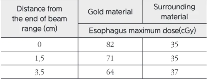
Table 9. Comparison of maximum dose of esophagus accord- ing to position of the gold fiducial marker in the patient study
Distance from the end of beam
range (cm)
Gold material Surrounding material Esophagus maximum dose(cGy)
0 82 35
1.5 71 35
3.5 64 37
Table 10. Comparison of maximum dose of esophagus ac- cording to position of the steel fiducial marker in the patient study
Distance from the end of beam
range (cm)
Steel material Surrounding material Esophagus maximum dose(cGy)
0 79 35
1.5 65 36
3.5 51 37
Table 11. Comparison of maximum dose of esophagus ac- cording to position of the titanium fiducial marker in the patient study
Distance from the end of beam
range (cm)
Titanium material Surrounding material Esophagus maximum dose(cGy)
0 68 35
1.5 55 36
3.5 41 37
의 일치여부를 확인한 후 방사선치료를 실시하였다.
그 결과 치료를 실시하기 전까지 Supine Position에 비 해 많은 시간이 소요되었다. 그러나 Prone Breast Board 에서 환자의 편안한 자세유지를 위한 방법을 연구하고 불 안정한 자세로 인한 Set-up의 재현성을 충분한 치료부위 확인을 통해 보완한다면 이 실험결과에서 보듯이 Lung은 평균선량 537.7 cGy, Heart는 163.1 cGy의 선량을 줄일 수 있고 특히 통계분석 결과 Lung에서 그 상관관계는 매 우 의미가 있다고 보여진다. 이는 유방암 방사선치료 후 초 래할 수 있는 부작용 발생의 감소효과를 가지고 온다고 말 할 수 있다.
결론적으로 안정적인 Prone Position을 유지할 수 있는 방법을 모색하고 환자의 Set-up시에 OBI, EPID을 이용하 여 정확한 치료부위 확인이 이루어진다면 유방암 방사선치 료 시에 Supine Position과 동일하게 PTV에 적정한 선량을 전달하면서 Lung, Heart Dose를 감소시켜 방사선치료로 인한 정상조직의 부작용을 최소화 할 수 있다고 사료된다.
참고문헌
1. Korean Breast Cancer Society. Korean breast can- cer data of 1997. J Korean Cancer Association 1999;31:1202-1209
2. Central Cancer Registry Center in Korea. Cancer facts and figures 2013.Seoul :Ministry of Health and Welfare;2013
3. Ministry of Health and Welfare. Republic of Ko- rea and National Cancer Center. Annual report of cancer registry program in the Republic of Ko- rea.2001.1.1.-2001.12.31.2003
4. NHI Consensus Conference. Treatment of early stage breast cancer. JAMA,1991;265:391-395
5. Poortmans P. Evidence based radiation oncology:
Breast cancer. Radiother Oncol 2007;84:84-101
6. Shafiq J, Delaney G, Barton MB. An evidence- based estimation of local control and survival benefit of radiotherapy for breast cancer. Radiother Oncol 2007;84:11-17
7. Dr Emami B. Tolerance of Normal Tissue to Thera- peutic Radiation. Department of Radiation Oncol- ogy, Loyola University Medical Center. USA
8. Graham MV, Purdy JA, Emami B, et al. Clini- cal dose volume histogram analysis for pneu- monitis after 3D treatment for non-small cell lung cancer(NSCLC). Int J Radiat Oncol Biol Phys.
1999;45:323-329
9. Early Breast Cancer Trialists’ Collaborative Group(EBCTCG): Effects of radiotherapy and of difference in the extent of surgery for early breast cancer on local recurrence and 15-year survival:
an overview of the randomised trials. Lancet 2005;366(9503):2087-2106
10. Sarah CD, Paul MG, Carolyn WT, et al: Long-term mortality from heart disease and lung cancer after radiotherapy for early breast cancer: prospective cohort study of about 300,000 women in USSEER cancer registries. Lancet Oncol 2005;6(8):557-565
11. Darby SC, Ewertz M, McGale P, et al. Risk of isch- emic heart disease in women after radiotherapy for breast cancer. N Engl J Med 2013;368:987-98
12. Remouchamps VM, Vicini FA, Sharpe MB, et al.
Significant reductions in heart and lung doses using deep inspiration breath hold with active breathing control and intensity-modulated radiation therapy for patient treated with locoregional breast irradia- tion. Int J Radiat Oncol Biol Phys. 2003;55:392-406
13. Formeni SC, Gidea-Addeo D, Goldberg JD, et al.
Phase I-II trial of prone accelerated intensity mod- ulated radiation therapy to the breast to optimally spare normal tissue. J Clin Oncol 2007;25:2236-2242
14. Zoltan Varga, Katalin Hideghety,M.D.,Ph D.,외 4 명. INDIVIDUAL POSITIONING: A COMPARATIVE STUDY OF ADJUVANT BREAST RADIOTHERAPY IN THE PRONE VERSUS SUPINE POSITION. Int J.
Radiation Oncology Biol. Phys 2009;75:94-100
15. Liv Veldeman,M.D, Bruno Speleers,Marlies Bak- ker, Filip Jacobs,Ir,Ph.D. PRELIMINARY RESULTS ON SETUP PRECISION OF PRONE-LATERAL PA- TIENT POSITIONING FOR WHOLE BREAST IR- RADIATION. Int J. Radiation Oncology Biol. Phys.
2010;78:111-11
16. Mulliez T, Veldeman L, Van Greveling A, Speleers B, Berwouts D, et al. Hypofractionated whole breast irradiation for patients with large breast : a ran- domized trial comparing prone and supine position . Radiother Oncol 2013;108:203-8
17. Kirby AM, Evans PM, Donovan EM, Convery HM, Haviland JS, Yarnold JR. Prone versus supine posi- tioning for whole and partial- breast radiotherapy : a comparison of non-target tissue dosimetry. Ra- diother Oncol 2010;96:178-84
18. Merchant TE, McCormick B. Prone position breast irradiation. Int J Radiat Oncol Biol Phys 1994;30:197-203
19. Suzy Kim, Yunseok Choi. Dosimetric advantage of the Field-in-field plan compared with the Tan- gential Wedged Beams plan for Whole-Breast Irradiation. Korean Journal of Medical Physics 2014;25(4):199-204
20. Wu Q, Mohan R, Morris M, Lavue A, Schmidt- Ullrich R. Simultaneous integrated boost intensity- modulated radiotherapy for locally advanced head- and-neck squamous cell carcinomas : Dosimetric results. Int J Radiat Oncol Biol Phys. 2003;26:573- 85
21. Sun Young Moon, Myonggeun Yoon, Weon Kuu Chung, Mijoo Chung, Dong Oh Shin, Dong Wook Kim. Comparison of Dosimetric Parameters of Patient with Large and Pendulous Breast Receiv- ing Breast Radiotherapy in the prone versus supine position. Progress in Medical Physics 2015;26(4)
Correlation analysis of radiation therapy position and dose factors for
left breast cancer
Department of Radiation Oncology, Inje University of Haeundae paik Hospital, Busan, Korea Department of radiology, Dongui University of science and technology, Busan, Korea
Jaewan Jeon, Cheolwoo Park, Jongsu Hong, Seongjin Jin, Junghun Kang
Purpose: The most basic conditions of radiation therapy is to prevent unnecessary exposure of normal tissue.
The risk factors that are important o evaluate the dose emitted to the lung and heart from radiation therapy for breast cancer. Therefore, comparing the dose factors of a normal tissue according to the radion treatment posi- tion and Seeking an effective radiation treatment for breast cancer through the analysis of the correlation rela- tionship.
Materials and Methods: Computed tomography was conducted among 30 patients with left breast cancer in su- pine and prone position. Eclipse Treatment Planning System (Ver.11) was established by computerized treatment planning. Using the DVH compared the incident dose to normal tissue by position. Based on the result, Using the SPSS (ver.18) analyzed the dose in each normal tissue factors and Through the correlation analysis between variables, independent sample test examined the association. Finally The HI, CI value were compared Using the MIRADA RTx (ver. ad 1.6) in the supine, prone position
Results: The results of computerized treatment planning of breast cancer in the supine position were V20, 16.5 ± 2.6 % and V30, 13.8 ± 2.2 % and Mean dose, 779.1 ± 135.9 cGy (absolute value). In the prone position it showed in the order 3.1 ± 2.2 %, 1.8 ± 1.7 %, 241.4 ± 138.3 cGy. The prone position showed overall a lower dose. The average radiation dose 537.7 cGy less was exposured. In the case of heart, it showed that V30, 8.1 ± 2.6 % and 5.1 ± 2.5 %, Mean dose, 594.9 ± 225.3 and 408 ± 183.6 cGy in the order supine, prone position.
Results of statistical analysis, Cronbach's Alpha value of reliability analysis index is 0.563. The results of the cor- relation analysis between variables, position and dose factors of lung is about 0.89 or more, Which means a high correlation. For the heart, on the other hand it is less correlated to V30 (0.488), mean dose (0.418). Finally The re- sults of independent samples t-test, position and dose factors of lung and heart were significantly higher in both the confidence level of 99 %.
Conclusion: Radiation therapy is currently being developed state-of-the-art linear accelerator and a variety of treatment plan technology. The basic premise of the development think normal tissue protection around PTV. Of course, if you treat a breast cancer patient is in the prone position it take a lot of time and reproducibility of set-up problems. Nevertheless, As shown in the experiment results it is possible to reduce the dose to enter the lungs and the heart from the prone position. In conclusion, if a sufficient treatment time in the prone position and place correct confirmation will be more effective when the radiation treatment to patient.
▶ Key words: Breast Treatment position, Correlation analysis, MIRADA Rtx, Cronbach's Alpha value.