계혈등 메탄올추출물이 LPS로 활성화된 Raw264.7 Cell에서 Nitric Oxide 및 Pro-inflammatory Cytokines 생성에 미치는 영향
최송이1, 박숙자2,3, 변성희2, 이종록2,4, 박문기1, 김상찬2,3*1)
1: 대구한의대학교 국가지정 한약자원 향장소재은행, 2: 대구한의대학교 한의과대학 3: 대구한의대학교 BK21, 4: 대구한의대학교 한방생명자원연구센터
Effects of Spatholobi Caulis MeOH Extract on the Production of NO and Pro-inflammatory Cytokines in LPS-activated Raw264.7 Cells
Song I Choe1, Sook Jahr Park2,3, Sung Hui Byun2, Jong Rok Lee2,4, Moon Ki Park1, Sang Chan Kim2,3*
1: Korea Herbal Cosmeceutical Material Bank, Daegu Haany University 2: College of Oriental Medicine, Daegu Haany University
3: BK21, Daegu Haany University
4: Research Center for Biomedical Resources of Oriental Medicine, Daegu Haany University
ABSTRACT
Objectives :
Spatholobi
Caulis is the stem ofSpatholobus suberectus
Dunn., which has been used for treating menstrual disorders, anemia, and rheumatic arthralgia in traditional Oriental medicine. This study was conducted to evaluate the inhibitory effects ofSpatholobi
Caulis extract (SCE) on the production of nitric oxide (NO) and pro-inflammatory cytokines in LPS-activated Raw264.7 cells.Methods : Cell viability was determined by MTT assay. Relative levels of NO were measured with Griess reagent and pro-inflammatory cytokines were detected by ELISA. Expression of iNOS and COX-2 proteins were determined by western blotting.
Results : NO production and iNOS expression were increased by incubation with LPS for 24 h. However, the augmented NO was reduced by SCE in a dose-dependent manner. Expressions of iNOS and COX-2 were inhibited by the treatment with SCE. SCE also suppressed the production of pro-inflammatory cytokines, such as IL-1beta and IL-6.
Conclusions : These results indicate that the MeOH extract of
Spatholobi
Caulis warrant further development as an anti-inflammatory agent for the treatment of Gram-negative bacterial infections.Key words :
Spatholobi
Caulis, nitric oxide, iNOS, cytokines, LPS* 교신저자 : 김상찬, 대구시 수성구 상동 165번지 대구한의대학교 한의과대학
․ Tel : 053-770-2247 ․ E-mail : [email protected]
․ 접수:2009년 5월 8일 ․ 수정:2009년 6월 19일 ․ 채택:2009년 6월 22일
서 론
Inflammation은 상처를 유발하는 자극에 대한 방어 기 전으로 발적, 발열, 종창, 동통, 기능장애 등의 증상이 나타나 며 각종 cytokines과 protein의 관여는 물론 prostaglandin E2
(PGE2), lysosomal enzyme, free radical등 다양한 매개 물질의 관여로 발생 하게 된다1,2). 외부 자극원 중에 하나 인 lipopolysaccharide (LPS)는 macrophage와 같은 염증 성 세포의 활성을 증대시키고 이로 인해 활성화 된 macrophage cell은 자연 면역뿐만 아니라 획득 면역 등 다양한 숙주 반응에 관여하여 숙주 방어와 항상성 유지 에 관여하는 것으로 알려져 있다3). 또한 염증 반응 시에 는 cytokine뿐만 아니라 PGE2 등을 다량 생산 하여 염 증매개 역할을 하는 세포로 알려져 있다4).
체내 염증과정에서는 과량의 NO 및 PGE2 등의 염증인자가 inducible nitric oxide synthase (iNOS) 및 cyclooxygenase-2 (COX-2)에 의해 형성된다. 이 중 NO는 neuronal NOS (nNOS), endothelia NOS (eNOS) 그리고 inducible NOS(iNOS) 세가지 형태의 NOS에 의해 합성된다.
iNOS는 interferon-γ, LPS와 다양한 염증유도 cytokine 에 노출되는 경우에 발현된다5,6).
NOS 중 iNOS에 의한 NO 생성이 절대적으로 많으면 병리학적으로 중요한 작용을 한다. 일반적인 NO 형성은 박테리아를 죽이거나 종양을 제거, 체내 방어기능, 신호전 달기능, 신경독성, 혈관 확장 등의 생리기능7)의 중요한 역 할을 하지만, 병리적인 원인에 의한 과도한 염증상태에서 iNOS에 의해 생성된 NO는 혈관 투과성, 부종 등의 염증 반응을 촉진시킬 뿐만 아니라 염증매개체의 생합성을 촉진 하여 염증을 심화시키는 것으로 알려져 있다8,9). PGE2는 염증반응, 면역반응, 그리고 angiogenesis를 촉진하는 등 암 발생에도 관여하고 있는 것으로 알려져 있다10).
계혈등(
Spatholobi
Caulis)은 중국의 복건, 광동, 광서, 운남 등지에 분포하는 콩과(Leguminosae)에 속하는 밀 화두의 덩굴줄기로 채취한 후 절단하여 건조하거나, 물에 담궈 절단한 후 햇볕에 건조하여 사용되는 한약재로서 혈액순환 개선, 월경불순, 빈혈, 중풍, 류마티스성 관절염 등의 치료에 사용되어 왔다11).계혈등에 대한 연구로는 류마티스성 관절염12,13) 및 항 산화 효과14,15), 지질대사 조절16) 및 면역조절작용17)에 대 한 연구가 보고 되어 있다.
이에 본 연구에서는 LPS로 활성화된 Raw264.7 cell 에서 계혈등이 iNOS를 통한 NO 및 pro-inflammatory mediator의 생성에 미치는 효과를 확인해 보고자 한다.
재료 및 방법
1. 추출물의 제조계혈등은 대원약업사(Deagu, Korea)에서 구입하여, 추
출 및 동결건조는 대구한의대학교 국가지정 한약자원향 장소재은행(KHCMB)에서 실시하였다. 계혈등 100 g에 메탄올 1 L를 가하여 실온에서 24시간 침지한 후, 여과 포로 여과한 액을 취하여 0.2 μm filter (Nalgene, New York, NY,
USA
)로 여과하고, 이 여과액을 동결건조하여 실험에 사용하였으며,Spatholobi
Caulis extract (SCE) 의 수율은 12.5%였다.2. 세포배양
Murine macrophage cell line인 Raw264.7 cells는 한 국세포주은행(Seoul,
Korea
)에서 구입하였으며, Dulbecco’s modified Eagle’s medium (DMEM)에 10% fetal bovine serum (FBS), 100 U/ml penicillin 및 100 μg/ml streptomycin 을 혼합한 배지를 사용하여 37℃, 5% CO2 incubator에 서 배양하였다. 실험과정의 모든 세포는 80~90%의 confluence에서 실험하였고, 20 passages를 넘기지 않은 cell만 사용하였다.3. 시약
LPS와 3-(4,5-dimethylthiazol-2-yl)-2,5-diphenyltetrazoleum (MTT)은 Sigma (St Louis, MO,
USA
)에서 구입하였고, FBS와 antibiotics는 Gibco/BRL (Eggenstein,Germany
) 로부터 구입하였다. iNOS와 COX-2 antibodies는 BD Bioscience (San Jose, CA,USA
)에서, actin antibody는 Santa Cruz Biotechnology (Santa Cruz, CA,USA
)에서 구입하였고, NC paper는 Schleicher & Schuell (Dassel,Germany
)에서 구입하였다. Tumor necrosis factor-alpha (TNF-α), Interleukin-1 beta (IL-1β), Interleukin-6 (IL-6) ELISA Kit는 Pierce endogen (Rockford, IL,USA
)에서 구입하였다.4. 세포생존율 측정(MTT assay)
Raw264.7 cells을 24 well plate에 5×10⁴ cells/well로 분주한 다음 약물을 처치하여 세포의 생존율을 구하였다.
세포에 약물을 처치한 후에 37℃, 5% CO2에서 배양하였 다. 배양 후 생존세포에 MTT (0.5 mg/ml)를 50 ㎕넣고 4시간 배양한 후 배지를 제거하고 생성된 formazan crystals을 dimethyl sulfoxide (DMSO)에 녹여 Titertek Multiskan Automatic ELISA microplate reader (Model MCC/340, Huntsville, AL)로 570 nm에서 흡광도를 측 정하였다. 세포생존율은 control cell에 대한 백분율로 나 타내었다[i.e. viability (% control) = 100 ×/(absorbance of treated sample)/(absorbance of control)].
5. NO생성량 측정
Raw264.7 세포주로부터 생성된 nitric oxide (NO)의
양은 세포 배양액 중에 존재하는 NO2-의 형태로서 Griess 시약을 이용하여 측정하였다. 간략하게 설명하면 96 well plate에 세포배양 상등액 50 ㎕와 Griess시약 (1% sulfanilamide in 5% phosphoric acid + 1% α-naphthylamide in H2O) 50 ㎕를 혼합하여 암실에서 10분 동안 반응시킨 후, Titertek Multiskan Automatic ELISA microplate reader (Model MCC/340, Huntsville, AL)로 540 nm에서 흡광도를 측 정하였다. NO2-의 농도는 sodium nitrate를 희석하여 흡 광도를 측정하여 표준 곡선을 얻었다.
6. 단백질 분석
20 mM Tris Cl (pH 7.5), 1% Triton X-100, 137 mM sodium chloride, 10% glycerol, 2 mM EDTA, 1 mM sodium orthovanadate, 25 mM β-glycerophosphate, 2 mM sodium pyrophosphate, 1 mM phenylmethylsulfonylfluoride과 1 mg/ml leupeptin을 함유하는 buffer를 사용하여 cell을 lysis시켰다. Cell lysates를 10,000 ×g로 30분간 원심 분 리하여 debris를 제거하였다. iNOS, COX-2 및 actin의 발현은 각각의 antibody를 사용하여 western blot analysis로 분석하였다. 2차 antibody는 alkaline phosphatase conjugated anti-rabbit, anti-goat을 사용하였다. 각각의 protein band는 ECL western blotting detection reagents (Amersham)를 사용하여 manufacturer's instruction에 따라 발색하였다.
7. Cytokine의 측정
Cytokine을 측정하기 위하여 6 well plate에 5×10⁵ cells/well을 분주하고 약물을 농도별로 처치한 다음, 1시 간 후에 LPS를 처치하였다. LPS 처치 후 18시간에 배지 를 수거하여 cytokine을 측정하였다. 수거된 배지는 측정 전까지 -70℃에서 보관하였다. TNF-α, IL-1β, IL-6 ELISA kit (Pierce endogen, Rockford, IL, USA)를 사용하여 manufacturer's instruction에 따라 측정하였다.
8. 통계적 검증
실험 결과는 SPSS 11.5버전을 사용하여 mean ± S.D 로 나타내었으며, ANOVA Tukey LSD 통계처리방법으 로 통계적 유의성 검정을 조사하였다. 유의수준은
p
<0.05 로 하였다.결 과
1. SCE가 LPS로 유도된 Raw264.7 cell의 NO production에 미치는 영향
Raw264.7 cell에서 SCE의 NO 생성 저해 정도를 측정 하기 위해 0.001, 0.003, 0.010 mg/ml의 농도로 처리하여
Fig. 1. Effects of SCE on the production of NO induced by LPS Raw 264.7 cells were treated with 0.001, 0.003, 0.010 mg/ml of SCE for 1 h prior to the addition of LPS (2 μg/ml), and the cells were further incubated for 6-24 h. The concentrations of nitrite and nitrate in culture medium were monitored as described in the methods section. Data represent the mean ± S.D. with eight separate experiments (* : significant as compared to control.
** : p<0.01. # : significant as compared to LPS alone, # : p<0.05, ## : p
<0.01).
생성되는 NO양을 측정하였다. LPS군에서는 control군에 비교하여 NO의 생성량이 LPS처치 후 18 h, 24 h에 유 의성 있게 증가하였으며, SCE를 처치한 실험군 18 h 및 24 h에서 유의하게 NO의 생성을 억제하였다(Fig. 1).
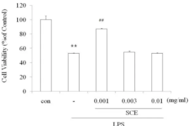
2. SCE가 LPS로 유도된 Raw264.7 cell의 세포생존율에 미치는 영향
SCE가 LPS로 유도된 Raw cell의 NO 생성을 감소시 킨 것이, SCE의 세포독성에 기인한 것인지를 관찰하기 위하여, SCE를 0.001, 0.003, 0.010 mg/ml로 처리 하고 24시간 후 MTT assay를 실시하여 세포생존율을 측정하 였다. 실험결과 LPS는 cell viability를 50%로 세포 독성
Fig. 2. Effects of SCE on the cell viability in LPS-stimulated Raw264.7 cells
Raw 264.7 cells were treated with 0.001, 0.003, 0.010 mg/ml of SCE for 1 h prior to the addition of LPS (2 μg/ml), and the cells were further incubated for 24 h. Data represent the mean ± S.D. with eight separate experiments (* : significant as compared to control. ** : p<0.01. # : significant as compared to LPS alone. ## : p<0.01).
을 나타냈었다. 그러나 SCE 0.001 mg/ml에서는 LPS에 의한 세포사멸을 유의하게 억제 하였다(Fig. 2). 이것은 LPS에 의해 증가된 NO의 독성을 SCE가 세포로부터 보 호 할 수 있는 가능성을 나타낸다.
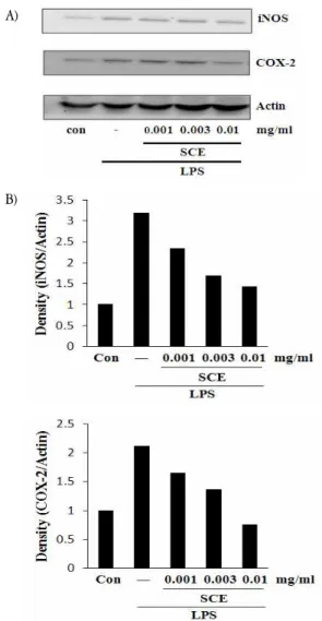
3. SCE가 iNOS 및 COX-2 발현에 미치는 영향
NO 생성 억제에 관련된 iNOS 단백질의 관련성과 prostaglandin합성을 증가시켜 염증반응의 중추적 역할을 하는 COX-2와의 연관성을 조사하기 위하여 immunoblot analysis를 이용하여, 세포질 내에서의 단백질 발현을 조 사하였다. LPS를 처리한 군에서는 각 단백질의 발현이 증가하였으나 SCE 0.010 mg/ml에서 발현의 감소를 보 였다(Fig. 3).
A)
B)
Fig. 3. Effects of SCE on the expressions of iNOS and COX-2 induced by LPS
The levels of iNOS and COX-2 proteins were monitored 18 h after treatment of cells with LPS (2 μg/ml) with or without SCE pretreatment (i.e. 1 h before LPS). The actin was used as a loading control (A). The relative density levels of protein bands were measured by scanning densitometry (B).
4. SCE가 LPS로 유도된 Raw264.7 cell의 TNF-α 생성에 미치는 영향
TNF-α는 LPS반응의 주요 매개체로서18,19), 염증성 병 변과정에서는 발현이 증가되는데 macrophage와 mast cell에서 분비되는 TNF-α는 tumor cell에 세포독성을 나 타낸다20). 본 연구에서의 TNF-α는 LPS에 의해 증가 되 었으며 SCE는 실험에 사용된 전농도에서 유의성을 나타 내지 않았다(Fig.4).
Fig. 4. Effects of SCE on LPS-inducible TNF-α production Production of TNF-α was measured in the medium of Raw 264.7 cells cultured with LPS (2 μg/ml) in the presence or absence of SCE for 18 h.
The concentrations of TNF-α in culture medium were monitored as described in the methods section. Data represent the mean ± S.D. with three separate experiments (* : significant as compared to control. ** : p<0.01).
5. SCE가 LPS로 유도된 Raw264.7 cell의 IL-1β 생성에 미치는 영향
IL-1β는 pro-inflammatory cytokine으로서 여러 면역 학적 작용들과 연관되어 있는데 T-cell의 활성화, B-cell 의 성숙 등에20) 관련하는 것으로 알려져 있다. 본 실험에 서 LPS는 IL-1β의 분비를 유의성 있게 증가시켰으며, SCE 0.001, 0.003, 0.010 mg/ml은 LPS로 유도된 IL-1β 를 SCE 농도 의존적으로 감소시켰다(Fig.5).
Fig. 5. Effects of SCE on LPS-inducible IL-1β production
Production of IL-1β was measured in the medium of Raw 264.7 cells cultured with LPS (2 μg/ml) in the presence or absence of SCE for 18 h.
The concentrations of IL-1β in culture medium were monitored as described in the methods section. Data represent the mean ± S.D. with three separate experiments (* : significant as compared to control. ** : p<0.01. # : significant as compared to LPS alone. ## : p<0.01).
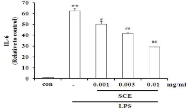
6. SCE가 LPS로 유도된 Raw264.7 cell의 IL-6 생성에 미치는 영향
IL-6는 B cell이 plasma cell로 분화되는 마지막 단계 를 활성화시키고, 염증병변에서 증가하는 것으로 알려져 있다20). LPS는 IL-6의 분비를 유의성 있게 증가시켰으 며, SCE 0.001, 0.003, 0.010 mg/ml 농도에서 감소효과를 확인하였다(Fig. 6).
Fig. 6. Effects of SCE on LPS-inducible IL-6 production.
Production of IL-6 was measured in the medium of Raw 264.7 cells cultured with LPS (2 μg/ml) in the presence or absence of SCE for 18 h. The concentrations of IL-6 in culture medium were monitored as described in the methods section. Data represent the mean ± S.D. with three separate experiments (* : significant as compared to control. ** : p<0.01. # : significant as compared to LPS alone. # : p<0.05. ## : p<0.01).
고 찰
계혈등(
Spatholobi
Caulis)은 콩과식물인 밀화두(Spatholobus suberectus
Dunn)의 줄기를 말린 것으로, 혈액순환을 이 롭게 하고 혈을 보하며 어혈을 없애는 효과가 있어 혈액 순환 개선, 월경불순, 빈혈, 중풍 등의 치료제로 사용되어 왔다. 이 외에도 계혈등은 관상동맥과 말초혈관을 확장하 는데 효과가 있는 약재로서, 혈류 순환을 원활하게 하여 혈압을 하강시키고 혈중지질농도를 낮춰 동맥경화, 고혈 압, 협심증 등에도 처방되어 왔다11). 현재까지 계혈등에 대한 연구는 류마티스성 관절염12,13)에 대한 연구가 대부 분이고 항산화효과21,22), 지질대사 조절16) 및 면역조절작 용17)에 대한 효과도 알려져 있다. 관절염에 대한 연구로 서 최 등12)은 계혈등이 collagen으로 감작시킨 관절염 발 병 실험동물에서 혈청내 류마토이드 인자인 IgG3, IgM, IgG2b의 함량을 감소시키고 IL-10 분비량, NK세포수를 증가시킴으로써 관절염 발병을 억제하는 효과가 있음을 발 표하였다. 또한 차 등은 DPPH법, Ferric-Thiocyanate법, Rancimat법 등의 항산화능 측정기법을 실시하여 ethylacetate로 추출한 계혈 등 분획물에 대하여 우수한 항산 화 효과를 규명하였다21).
본 실험에서는 계혈등 메탄올추출물(SCE)이 LPS에 의해 활성화된 Raw264.7 cell에서 염증성 매개 인자들에 대한 영향을 확인함으로써 계혈등의 항염증 효과를 살펴
보고자 하였다. SCE을 0.001, 0.003, 0.010 mg/ml의 농도 로 처치하여 NO의 생성 저해 정도를 측정한 결과, 18 h, 24 h 처치 후에 NO가 유의하게 감소함을 확인하였고, 24 h에서 MTT assay를 통해 0.001 mg/ml에서는 LPS 에 의해 유도된 세포 독성억제 효과를 관찰하였다. 이것 은 LPS로 인해 증가된 NO가 SCE의 세포독성으로 말미 암은 것이 아니라 SCE에 항염증작용이 있음을 의미한다.
NO는 면역반응, 세포독성, 신경전달계 및 혈관이완 등 생물학적인 과정에 관여하는 것으로 알려져 있으며 농도 에 따라서는 세포 기능유지에 작용을 하기도 하지만 고 농도에서는 세포독성을 유발 한다23,24). 일반적으로 iNOS 는 lipopolysaccharide (LPS), interferon-γ (IFN-γ), interleukin-1 (IL-1) 및 tumor necrosis factor-α (TNF-α) 등의 자극에 의해 활성화되며, 대식세포, 내피 세포, 심근세포 등에서 다량의 NO를 생성하는 것으로 알려져 있다. iNOS에 의한 NO의 생성이 과량일 경우 염증을 유발시켜 조직의 손상, 유전자 변이 및 신경 손상 등을 일으킨다25,26). 염증반응과 관련된 조직 손상에서 NO와 iNOS의 발현이 증가되어 있음이 보고되어 있다27). iNOS의 장시간 유도는 다량의 NO를 생성하며, 생성된 NO는 병리적인 혈관확장, 세포독성, 조직손상 등과 같은 생체에 유해한 작용을 나타낸다. 그리고 염증상태에서 iNOS에 의해 생성된 NO는 혈관투과성, 부종 등의 염증 반응을 촉진시킬 뿐만 아니라 염증매개체의 생합성을 촉 진하여 염증을 심화시키는 것으로 알려져 있다27,28). 그런 까닭에 NO 생성 저해제는 염증반응조절제로의 가능성 때문에 연구가 활발하게 이루어지고 있다.
NO 생성 억제와 iNOS 단백질의 관련성을 immunoblot analysis를 이용하여 세포질 내에서의 iNOS 단백질의 발 현을 조사한 결과 LPS 처치 시에는 iNOS가 강하게 발 현되었으나, SCE에 의해 단백질 발현의 감소를 확인하였 다. COX-2는 대식세포와 활막 세포와 같은 염증세포에서 상처 감염, 스트레스에 의해 유도되는데 prostaglandin synthesis의 생성을 증가시켜 염증상태로 만드는 과정에 있어 중추적 역할을 한다29-32). Immunoblot analysis를 이용하여 COX-2 단백질의 발현을 조사한 결과 LPS 처 치 시에는 COX-2 단백질이 강하게 발현되었으나, 계혈 등을 처치한 실험군에서 발현 감소를 관찰하였다.
Tumor mecrosis factor-α (TNF-α)는 Macrophage와 mast cell에서 분비되는 것으로 tumor cell에 영향을 미 치는 숙주 방어인자 및 염증 매개물질로 알려져 있고 만 성염증성 반응과 관련 된다20). 본 실험에서 LPS는 TNF-α의 분비를 촉진시켰으나, SCE는 증가된 TNF-α 의 생성을 억제하지 못하였다.
IL-1β는 발열작용을 일으킬 뿐만 아니라 면역계, 신경내 분비계, 그리고 신경면역계를 자극하는 pro-inflammatory cytokine으로서 기능 조절 및 종양침습에도 관여한다고 알려져 있다5,33). 결과에서 LPS는 IL-1β의 분비를 유의 성 있게 증가시켰으며, SCE는 농도의존적으로 LPS로 유
도된 IL-1β를 감소시켰다.
IL-6는 TNF-α처럼 염증반응을 조절하는 물질로서 내 인성 발열인자(pyrogen)로 작용하며 면역 체계와 조혈 등에 영향을 끼친다34). 본 실험에서 LPS는 IL-6를 유의 성 있게 증가시켰으며 SCE는 IL-6 생성을 감소시켰다.
계혈등은 微澁微甘溫하고 心脾 2經에 入하여 주로 活 血去瘀, 生新血, 流利經脈, 暖腰膝, 補中調胃의 효능을 가 진다. 본 연구는 계혈등이 LPS로 활성화된 macrophage cell line에서 염증매개물질을 억제하는 결과를 밝힌 것으 로, 계혈등은 gram-negative의 감염이나, 과량의 염증매 개물질로 인한 염증성 질환의 치료에 이용될 수 있는 근 거를 제시하였다.
결 론
본 연구에서는 계혈등이 대식세포 계열인 Raw 264.7 세포에서 LPS로 유도된 NO와 pro-inflammatory mediator 의 생성억제효과, iNOS, COX-2단백질 발현 억제효과를 확인하였다. 계혈 등은 NO의 생성을 억제하고, iNOS 및 COX-2의 발현을 억제하였다. 또 계혈등은 농도의존적으 로 IL-1β, IL-6 cytokine 생성을 억제하였다. 이러한 결 과는 계혈등이 항염증 물질로서 과량의 NO 및 과량의 pro-inflammatory cytokine 생성과 관련된 염증성 질환 의 예방 및 면역치료의 이용, 약물의 개발에 이용될 수 있음을 의미한다.
감사의 글
본 연구는 지식경제부 지역혁신센터사업(대구한의대학 교 한방생명자원연구센터)의 지원에 의하여 이루어졌으 며 이에 감사드린다.
참고문헌
1. Zamora R, Vodovotz Y, Billiar TR. Inducible nitric oxide synthase and inflamamtory diseases. Molecular medicine. 2000 ; 6 : 347-73.
2. Yun HJ, Heo SK, Lee YT, Park WH, Park SD.
Anti-inflammatory Effect of Evodia Officinalis DODE in Mouse Macrophage and Human Vascular Endotherial Cells. Kor J Herbology. 2008 ; 23(1) : 29-38.
3. Willeaume V, Kruys V, Mijatovic T, Huez G.
Tumor necrosis factor-alpha production induced by viruses and by lipopolysaccharides in macrophages:
similarities and differences. J Inflamm. 1995-1996 ; 46 : 1-12.
4. McDaniel ML, Kwon G, Hill JR, Marshall CA, Corbett JA. Cytokines and nitric oxide in islet inflammation and diabetes. Proc Soc Exp Biol Med. 1996 ; 211 : 24-32.
5. Moncada S, Palmer RM, Higgs EA. Nitric oxide:
physiology, pathophysiology, and pharmacology.
Pharmacol Rev. 1991 ; 43 : 109-42.
6. Yun HY, Dawson VL, Dawson TM. Neurobiology of nitric oxide. Crit Rev Neurobiol. 1996 ; 10(3-4) : 291-316.
7. Moncada S, Palmer RM, Higgs EA. Nitric oxide:
physiology, pathophysiology, and pharmacology.
Pharmacol Rev. 1991 ; 43 : 109-42.
8. Ryu JH, Ahn H, Kim JY, Kim YK. Inhibitory activity of plant extracts on nitric oxide synthesis in LPS-activated macrophage. Phytother Res. 2003 ; 17(5) : 485-9.
9. Mu MM, Chakravortty D, Sugiyama T, Koide N, Takahashi K, Mori I, Yoshida T, Yokochi T. The inhibitory action of quercetin on lipopolysaccharide - induced nitric oxide production in Raw264.7 macrophage cells. J Endotoxin Res. 2001 ; 7(6) : 431-8.
10. Kim JY, Jung KS, Jeong HG. Suppressive effects of the kahweol and cafestol on cyclooxygenase - 2 expression in macrophages. FEBS Lett. 2004 ; 569(1-3) : 321-6.
11. Kim JG, Cho BG. Traditional drugs of the east.
Young Lim Sa. 1995 : 150-1.
12. Choi JS, Song TW, Kim DH. A Study on the effect Spatholobus suberectus Dunn on the inhibition of arthritis by collagen on the mouse.
Kor J Herbology. 2003 ; 18 : 79-88.
13. So HG, Oh MS, Kim DH. Immunity Responses of the Spatholobus suberectus Dunn to Synovial Cells Isolated from Patients with Rheumatoid Arthritis.
Korean J Oriental Physiol Pathol. 2003 ; 17 : 780-6.
14. Li RW, Lin GD, Myers SP, Leach DN.
Anti-inflammatory activity of Chinese medicinal vine plants. Journal of Etnopharmacol. 2003 ; 85 : 61-7.
15. Takahashi T, Takasuka N, Iigo M, Baba M, Nishino H, Tsuda H, Okuyama T. Isoliquiritigenin, a flavonoid from licorice, reduces prostaglandin E2 and nitric oxide, causes apoptosis, and suppresses aberrant crypt foci development. Cancer Sci. 2004 ; 95(5) : 448-53.
16. Wang W, Wang J, Zhao D, Liu H, Zhou W, Chen K. Comparison of Spatholobus suberectus Dunn
Euonymus alatus (Thunb) Sied and Eupolyphage sinensis Walkeron regulation of plasma lipid. Zhongguo Zhoug Yao Za Zhi. 1991 ; 16 : 299-301, 320.
17. Lam TL, Lam ML, Au TK, Ip DT, Ng TB, Fong WP, Wan DC. A comparison of human imminodeficiency virus type-1 protease inhibition activities by the aqueous and methanol extracts of Chinese medicinal herbs. Life Sci. 2000 ; 67(23) : 2889-96.
18. Lee AK, Sung SH, Kim YC, Kim SG. Inhibition of lipopolysaccharide - inducible nitric oxide synthase, TNF-α and COX-2 expression by sauchinone effects on I-κBα phosphorylation, C/EBP and AP-1 activation.
British journal of pharmacology. 2003 ; 139 : 11-20.
19. Dinarello CA. Pro-inflammatory cytokines. Chest.
2000 ; 118 : 503-508.
20. Delgado AV, McManus AT, Chambers JP. Production of tumor necrosis factor-alpha, interleukin 1-beta, interleukin 2, and interleukin 6 by rat leukocyte subpopulations after exposure to substance P.
Neuropeptides. 2003 ; 37(6) : 355-61.
21. Cha BC, Lee EH, Noh MA. Antioxidant activity of Spatholobus suberectus Dunn. Kor J Pharmacogn.
2005 ; 36(1) : 50-5.
22. Sim GS, Kim JH, Lee DH, Park SM, Pyo HB, Zhang YH, Lee BC. Effects of the Spatholobi caulis extract on antioxidation and inhibition of matrix metalloproteinase in human skin fibroblasts.
Kor J Biotechnol Bioeng. 2005 ; 20(1) : 40-5.
23. Nathan C, Xie QW. Nitric oxide synthases: roles, tolls and controls. Cell. 1994 ; 78 : 915-8.
24. Kubes P. Inducible nitric oxide synthase; a little bit og good in all of us. Gut. 2000 ; 47 : 6-9.
25. McCartney-Francis N, Allen JB, Mizel DE, Albina JE, Xie QW, Nathan CF, Wahl SM. Suppression of arthritis by an inhibitor of nitric oxide synthase. J Exp Med. 1993 ; 178(2) : 749-754.
26. Weisz A, Cicatiello L, Esumi H. Regulation of the mouse inducible-type nitric oxide synthase gene
promoter by interferon-gamma, bacterial lipopolysaccharide and NG-monomethyl-L-arginine. Biochem J. 1996 ; 316 : 209-15.
27. Tezuka Y, Irikawa S, Kaneko T, Banskota AH, Nagaoka T, Xiong Q, Hase K, Kadota S. Screening of Chinese herbal drug extracts for inhibitory activity on nitric oxide production and identification of an active compound of Zanthoxylum bugeanum.
J Ethnopharmacol. 2001 ; 77 : 209-17.
28. Kim RG, Shin KM, Chun SK, Ji SY, Seo SH, Park HJ, Choi JW, Lee KT. In vitro antiinflammatory activity of the essential oil from Ligularia fischeri var. spiciformis in murine macrophage Raw264.7 cells. Yakhak Hoeji. 2002 ; 46 : 343-7.
29. Needleman P, Isakson, PC. The discovery and function of COX-2, J. Rheumatol Suppl. 1997 ; 49 : 6-8.
30. Surh YJ. Anti-tumor promoting potent of selected spice ingredients with antioxidative and anti- inflammatory activities: a short review. Food Chem Toxicol. 2002 ; 40(8) : 1091-7.
31. Surh YJ, Chun KS, Cha HH, Han SS, Keum YS, Park KK, Lee SS. Molecular mechanisms underlying chemopreventive activities of anti-inflammatory phytochemicals: down-regulation of COX-2 and iNOS through suppression of NF-kappa B activation.
Mutat Res. 2001 ; 480-1, 243-68.
32. Seybold VS, Jia YP, Abrahams LG. Cyclooxygenase-2 contributes to central sensitization in rats with peripheral inflammation. Pain. 2003 ; 105(1-2) : 47-55.
33. Maes M. The cytokine hypothesis of depression:
inflammation, oxidative & nitrosative stress (IO &
NS) and leaky gut as new targets for adjunctive treatments in depression. Neuro Endocrinol Lett.
2008 ; 29(3) : 287-91.
34. Van Snick J. IL-6: an overview. Annu. Rev.
Immunol. 1990 ; 8 : 253-78.