ment as most typical photosynthetic dinoflagellates con- tain, whereas the latter two genera Karenia and Karlodinium contain fucoxanthin or its derivatives as their major carote- noids. Later, new fucoxanthin-containing genus Takayama de Salas, Bolch, Botes & Hallegraeff with a sigmoid apical groove was erected and three unarmored dinoflagellates described by Larsen (1994, 1996), Gymnodinium pulchelum Larsen, Gyrodinium acrotrochum Larsen, and G. cladochroma Larsen, having a sigmoid apical groove and fucoxanthin, were transferred to the genus (de Salas et al. 2003). Those three fucoxanthin-containing genera formed a well-suppor- ted monophyletic clade and constituted a separate evolu- tionary lineage and the new family Kareniaceae comprising those genera was proposed (Bergholtz et al. 2005).
Some species in the family
Kareniaceasuch as Karenia
https://doi.org/10.11626/KJEB.2021.39.2.236
INTRODUCTION
The unarmored dinoflagellate genus Gymnodinium F.
Stein has been recognized to be assemblage of unrelated species for many years. Based on combination of LSU rDNA sequences, chloroplast pigment composition, and outline of apical groove, Daugbjerg et al. (2000) proposed that the heterogeneous genus Gymnodinium sensu lato divi - ded into four genera: Gymnodinium sensu stricto with a horse- shoe-shaped apical groove, Akashiwo G.Hansen & Moe- strup with a clockwise spiral apical groove, Karenia G.Han- sen & Moestrup with a straight apical groove, and Karlodi- nium J.Larsen with a short straight apical groove and a vent- ral pore. The former two genera Gymnodinium and Akashiwo possess peridinin in the chloroplast as a main accessory pig-
Original article
Newly recorded unarmored dinoflagellates in the family Kareniaceae (Gymnodiniales, Dinophyceae) in brackish and coastal waters of Korea
Minji Cho1, Hojoon Choi1, Seung Won Nam2 and Sunju Kim1,3,*
1Division of Earth Environmental System Science, Pukyong National University, Busan 48513, Republic of Kroea
2Protist Research Team, Nakdonggang National Institute of Biological Resources, Sangju 37242, Republic of Korea
3Department of Oceanography, Pukyong National University, Busan 48513, Republic of Korea
Korean J. Environ. Biol.
39(2) : 236-244(2021) ISSN 1226-9999(print) ISSN 2287-7851(online)
* Corresponding author Sunju Kim
Tel. 051-629-6577
E-mail. [email protected]
Received: 14 June 2021 Revised: 18 June 2021
Revision accepted: 21 June 2021
Abstract: Unarmored dinoflagellates, in the family Kareniaceae, include harmful or toxic bloom-forming species, which are associated with massive fish kills and mor- talities of marine organisms worldwide. The occurrence and distribution of the toxigenic species in the family Kareniaceae were investigated in the brackish and coastal waters of Korea between July 2018 and October 2020. During the survey, we collected seven newly recorded species; Karenia papilionacea, Karlodinium digitatum, Karl. veneficum, Karl.
zhouanum, Takayama acrotrocha, T. helix, and T. tasmanica. A total of fifteen strains of the seven taxa were successfully established as clonal cultures and examined using LM, SEM, and molecular phylogeny inferred from LSU rDNA sequences. Herein, we present the taxonomic information, morphological features, and molecular phylogenetic positions of the unrecorded dinoflagellate species collected from Korean coastal waters.
Keywords: first record, Kareniaceae, LSU rDNA, molecular phylogeny, morphology
brevis, K. mikimotoi, and Takayama pulchella have been rep- orted to form harmful algal blooms, causing massive fish kills and fisheries damages worldwide (Steidinger et al.
1998; Pierce and Henry 2008; Brand et al. 2012). In Asia, harmful red tides by Karlodinium australe and Karl. digi- tatum were reported from Johor Strait between Malaysia and Singapore in 2014 and 2015 and from China in 2019, respectively (Lim et al. 2014; Leong et al. 2015; Cen et al.
2019; Sakamoto et al. 2021). In Korea, two Karenia species have been reported to cause massive harmful blooms, lead- ing to economic losses: one by K. mikimotoi from Jinhae Bay in 1992 with economic loss of 5.5 million USD and the other by Karenia sp. (as Gymnodinium sp.) from Tongyeong in August 1992 with damage of approx. 1.82 M USD, res- pectively (Sakamoto et al. 2021).
In this study, we reported unrecorded dinoflagellate spe- cies in the family Kareniaceae from brackish and coastal
waters of Korea and provided their morphological features and molecular phylogenetic relationships inferred from LSU rDNA sequences.
MATERIALS AND METHODS
1. Sampling and culture collectionPlankton samples were collected by vertical and hori- zontal sampling using a 20 μm-mesh plankton net in brack- ish and coastal waters of Korea from July 2018 to October 2020 (Table 1). Water temperature and salinity were mea- sured in situ using a YSI instrument (YSI Inc., OH, USA).
The collected samples were kept at 20°C until microscopic observation. Single cells were individually isolated with a capillary pipette under an inverted microscope (Axio Vert.
A1; Zeiss, Hallbergmoos, Germany) and washed several
Table 1. List of newly recorded unarmored dinoflagellates in the family Kareniaceae in brackish and coastal waters of Korea
Species Strain Date Locality
(Latitude, Longitude) Temp.
(°C) Sal. GenBank
accession Karenia papilionacea KpLomme01 Jul 24, 2018 Yongho, Busan
(35°08′00″N, 129°06′55″E) 26.1 32.8 MZ358888 Karlodinium digitatum KdLomme01 Oct 14, 2020 Yongho, Busan
(35°08′00″N, 129°06′55″E) 20.7 31.2 MZ358887 Karlodinium digitatum KdLomme02 Oct 16, 2020 Yongho, Busan
(35°08′00″N, 129°06′55″E) 20.0 31.6 MZ358883 Karlodinium digitatum KdLomme03 Oct 16, 2020 Yongho, Busan
(35°08′00″N, 129°06′55″E) 20.0 31.6 MZ358886 Karlodinium digitatum KdLomme04 Oct 16, 2020 Yongho, Busan
(35°08′00″N, 129°06′55″E) 20.0 31.6 MZ358884 Karlodinium digitatum KdLomme05 Oct 16, 2020 Yongho, Busan
(35°08′00″N, 129°06′55″E) 20.0 31.6 MZ358885 Karlodinium veneficum KvLomme01 May 01, 2020 Jangchun, Changwon
(35°07′41″N, 128°41′54″E) 17.0 32.8 MZ358877 Karlodinium veneficum KvLomme02 May 01, 2020 Jindong, Gosung
(35°11′19″N, 128°33′58″E) 18.8 32.7 MZ358875 Karlodinium veneficum KvLomme03 May 13, 2020 Yongho, Busan
(35°08′00″N, 129°06′55″E) 15.3 36.0 MZ358876 Karlodinium veneficum KvLomme04 June 10, 2020 Hakri, Gijang
(35°15′30″N 129°14′48″E) 19.1 34.1 MZ358874 Karlodinium zhouanum KzLomme01 Oct 25, 2019 Yongho, Busan
(35°08′00″N, 129°06′55″E) 21.1 31.8 MZ358878 Takayama achrotrocha TaLomme01 Oct 14, 2020 Yongho, Busan
(35°08′00″N, 129°06′55″E) 20.7 31.2 MZ358881 Takayama helix ThLomme01 Sep 23, 2020 Yongho, Busan
(35°08′00″N, 129°06′55″E) 21.5 31.0 MZ358882 Takayama tasmanica TtLomme01 Sep 12, 2020 Jangchun, Changwon
(35°07′41″N, 128°41′54″E) 24.3 26.1 MZ358880 Takayama tasmanica TtLomme02 Oct 14, 2020 Yongho, Busan
(35°08′00″N, 129°06′55″E) 20.7 31.2 MZ358879
times in a series of drops of 0.2 μm filtered and sterilized seawater. The specimen was transferred to a 96-well plate filled with ambient filtered seawater. Clonal cultures were maintained in F/2 medium (Guillard and Ryther 1962) at 20°C, with a 14 : 10 light-dark cycle under 100-120 μmol m
-2s
-1. A pair of gelatin-embedded slides prepared from each clonal culture strain after final conc. 1% glutaralde- hyde fixation were deposited at the Nakdonggang Nation- al Institute of Biological Resources, Korea (NNIBRPR 17563-NNIBRPR17576).
2. Light microscopy
Live cells were observed with Axio Imager A2 (Zeiss) light microscope, equipped with differential interference il- lumination. Light micrographs were taken at
×1000 using a AxioCam HRc (Zeiss) photomicrographic system equip- ped with the microscope. Glutaraldehyde-fixed cells (1%
final concentration) were examined to determine the shape and location of nuclei after staining with 4ʹ-6-diamidino- 2-phenylindole (DAPI: 0.1 μg mL
-1final concentration) under epifluorescence microscope with ultraviolet light (excitation of 360 nm and emission of 460 nm).
3. Scanning electron microscopy
For scanning electron microscopy, 2 mL of culture was fixed with an equal volume of glutaraldehyde (2% final con- centration) in 0.2 M cacodylate buffer at pH 7.4 at 4°C for 1 h. Fixed cells were rinsed twice in distilled water for 1 h and dehydrated using ethanol concentration gradient (30, 50, 70, 80, 95, and two changes of 100% ethanol) soaking for 12 min at each step. Dehydrated samples were critical point dried in liquid CO
2using an HCP-2 (Hitachi, Tokyo, Japan). Finally, the samples were coated with gold-palla- dium for 3 min and examined under a MIRA3 FE-SEM (Tescan Korea, Seoul, Korea).
4. DNA extraction, PCR, Sequencing
Genomic DNA was extracted from 1 mL of exponentially growing culture strains using Chelex extraction method (Kim and Park 2014). The purity and quantity were deter- mined with a NanoDrop spectrophotometer (Thermo Fisher Scientific, DE, USA). Approximately 1050 bp of the LSU rDNA were amplified using the primers D1R and D3B
(Nunn et al. 1996). PCR was conducted using a C1000 Touch thermal cycler with a commercially available PCR premix (Accu-Power PCR PreMix; BIONEER, Daejeon,
Korea). The thermal cycle condition of PCR was as follows:
initial 95°C for 3 min, followed by 39 cycles of denaturation at 95°C for 45 s, annealing at 52°C for 45 s, and extension at 72°C for 1 min, with a final extension at 72°C for 7 min. The PCR products were purified using ExoSAP-IT
TMExpress according to manufacturer’s instructions, confirmed by 1% agarose gel electrophoresis. The purified PCR products were sequenced in an ABI model 3730xl DNA Analyzer (Applied Biosystems; Foster City, CA), using the same primers used for PCR in conjunction with a Big-Dye Ter- minator v3.1 Cycle Sequencing kit (Applied Biosystems).
ContigExpress (Vector NTI v. 10.1; Invitrogen, Grand Island, NY) was used to edited out low quality and to assem- ble the sequence reads and the complete sequences were deposited in GenBank (Table 1).
5. Alignments and phylogenetic analyses
A total of 76 sequences of
Kareniaceanspecies including Korean strains and two genera Gymnodinium and Gyrodi- nium species as outgroup were aligned with MEGA (Kumar et al. 2018) and unambiguously aligned regions (1002 positions) were applied for further phylogenetic analyses.
Maximum likelihood analysis was performed with RAxML 8.0 (Stamatakis 2014) using the GTRGAMMA evolution model and rapid bootstrapping of 2,000 replicates. Bayes- ian analysis was conducted using MrBayes 3.1.1 (Ronquist and Huelsenbeck 2003) running four simultaneous Monte Carlo Markov Chains for 2,000,000 generations and sam- pling every 100 generations, following a burn in of 100,000 generations.
RESULTS AND DISCUSSION
1. Taxonomic summaryThree genera and seven species in the family Kareniaceae were newly recorded in brackish and coastal waters of Korea
(Table 1). The newly recorded species Karenia papiliona- cea, Karlodinium digitatum, Karl. veneficum, Karl. zhouanum, Takayama acrotrocha, T. helix, and T. tasmanica were descri- bed based on their morphological characteristics and the LSU rDNA sequences obtained to perform molecular phy- logenetic analyses.
Class Dinophyceae Fritsch Order Gymnodiniales Apstein
Family Kareniaceae Bergholtz, Daugbjerg, Moestrup &
Fernández-Tejedor
Genus Karenia G.Hansen & Moestrup
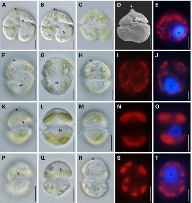
Karenia papilionacea A.J.Haywood & K.A.Steidinger (Fig. 1A-E)
Synonym: Gymnodinium breve C.C.Davis (Haywood et al.
1996).
Reference: Haywood et al. 2004 (Figs. 2e-h and 3a-f).
Specimen examined. Table 1.
Cells are 38.5-44.9 μm long and 22.4-22.9 μm wide and dorsally convex and ventrally concave. The epithca has a prominent apical carina and a short straight apical groove with extended one third of the dorsal epitheca. The cingu- lum is displaced by the cingulum width. The nucleus is sphe- rical and located in the left hyposome. Chloroplasts are few and large or several and small, located peripherally, yellow- green in color.
Distribution. Hawke’s Bay in New Zealand (Haywood et al.
2004), Uranouchi inlet, Nomi Inlet, Sukumo Bay, Saiki Bay, Yatsushiro Sea in western Japan (Yamaguchi et al. 2016).
Site of collection. Specimens were collected from Yongho Bay of Busan, Republic of Korea (35°08ʹ00ʺN, 129°06ʹ55ʺ E) on July 24, 2018.
Voucher slide. NNIBRPR17563-NNIBRPR17564.
Genus Karlodinium J.Larsen
Karlodinium digitatum Gu, Chan & Lu(Fig. 1F-J)
Basionym: Karenia digitata Z.B.Yang, H.
Takayama, K.Mat-
suoka & I.J.Hodgkiss.
Reference: Cen et al. 2019 (Figs. 2 and 3).
Specimen examined. Table 1.
Cells are 16.1-22.7 μm long and 14.5-20.2 μm wide and globular or oval in shape. A straight apical groove extends from the dorsal apex to the ventral epicone. The structure of the curve knot is present in hypocone. The sulcus invades the epicone slightly as a small finger-like intrusion. The cingulum is descended with a displacement of approx. 25%
of cell length. The nucleus is spherical and located in the posterior. Chloroplast is spherical in shape and distributed irregularly in cells.
Distribution. Western coastal waters of Japan (Yang et al.
2000), Silver Mine Bay in Hong Kong (Lee et al. 2011), Fu- jian Province in China (Cen et al. 2019).
Site of collection. Specimens were collected from Yongho Bay of Busan, Republic of Korea (35°08ʹ00ʺN, 129°06ʹ55ʺ E) on October 16, 2020.
Voucher slide. NNIBRPR17565-NNIBRPR17566.
Karlodinium veneficum(Ballantine) J.Larsen(Fig. 1K-O)
Basionym: Gymnodinium veneficum (Ballantine 1956) p.
Synonym: Karlodinium micrum 469. (Leadbeater & Dodge) J.Larsen, Gymnodinium galatheanum Braarud sensu Kite
& Dodge, Gymnodinium micrum (Leadbeater & Dodge) Loeblich III, Gyrodinium galatheanum (Braarud) Taylor sensu Taylor.
References: Bergholtz et al. 2005 (Figs. 13 and 14).
Specimen examined. Table 1.
Cells are 10.6-14.7 μm long and 6.4-10.6 μm wide and ovoid in shape. The cingulum is displaced about two cingu- lum width. The sulcus extends to the left epicone. A short straight apical groove is visible. The nucleus is spherical and located centrally or in the posterior. Four chloroplasts are located peripherally and two in epicone and two in the hypocone.
Distribution. Australia, New Caledonia, New Zealand, Asia (China and Japan, and Qatar), Europe (England, France, Germany, Italy, and Norway), and USA.
Site of collection. Specimens were collected at Jangchun of Changwon (35°07ʹ41ʺN, 128°41ʹ54ʺE) and Jindong of Gosung-gun (35°11ʹ19ʺN, 128°33ʹ58ʺE) on May 01, 2020 and Yongho Bay of Busan (35°08ʹ00ʺN, 129°06ʹ55ʺE) on May 13, 2020 and Hakri harbor of Gijang-gun (35°15ʹ30ʺ N, 129°14ʹ48ʺE) on June 10, 2020, respectively.
Voucher slide. NNIBRPR17567-NNIBRPR17568.
Karlodinium zhouanum Z.Luo & H.Gu(Fig. 1P-T)
References: Luo et al. 2018 (Figs. 1-4).
Specimen examined. Table 1.
Cells are 10.4-14.9 μm long and 7.7-11.9 μm wide and ovoid with a conical epicone and hemispherical hypocone in shape. The cingulum is descended with a displacement of 25% of total cell length. The sulcus intruded into the epi- one as a fingerlike projection. A straight apical groove ext- ends from above the sulcal intrusion to the dorsal epicone.
The nucleus is spherical and situated in the epicone. Six
chloroplasts are peripherally located in the cell with internal
lenticular pyrenoids.
Distribution. South China Sea, Yellow Sea of China (Luo et al. 2018).
Site of collection. The species was observed from Yongho Bay of Busan in Korea (35°08ʹ00ʺN, 129°06ʹ55ʺE) on Oct- ober 25, 2019.
Voucher slide. NNIBRPR17569-NNIBRPR17570.
Genus Takayama de Salas, Bolch, Botes & Hallegraeff
Takayama acrotrocha(Larsen) de Salas, Bolch and Hallegraeff(Fig. 2A-E)Basionym: Gyrodinium acrotrochum Larsen.
References: Larsen 1996 (Figs. 2-4 and 35).
Fig. 1. Light and scanning electron micrographs of four species in the two genera Karenia and Karlodinium. (A-E) Karenia papilionacea. (F-J) Karlodinium digitatum. (K-O) Karlodinium veneficum. (P-T) Karlodinium zhouanum. Cells showing sulcal intrusion into epicone(arrow); apical groove(arrowhead); structure of curve knot(double arrowheads); nucleus(N); pyrenoid(P). Scale bars represent 10μm in (A-E) and 5μm in (F-T), respectively.
A
F
K
P
B
G
L
Q
C
H
M
R
D
I
N
S
E
J
O
T
Specimen examined. Table 1.
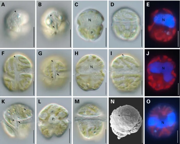
Cells are 14.9-21.7 μm wide and 12.9-20.2 μm wide and spherical in shape. The cingulum is descended with a dis- placement of one fourth of the cell length. A sigmoid apical groove extends from the proximal end of girdle around the apex. The nucleus occupies much of the episome. Several disc-shaped chloroplasts are present.
Distribution. Hobsons Bay in Australia (Larsen 1996).
Site of collection. The species was observed from Yongho Bay of Busan in Korea (35°08ʹ00ʺN, 129°06ʹ55ʺE) on Oct- ober 14, 2020.
Voucher slide. NNIBRPR17571-NNIBRPR17572.
Takayama helix de Salas, Bolch, Botes and Hallegraeff (Fig. 2F-J)
Synonym: Gymnodinium sp. 6 (Takayama 1998, plate 6,
Figs. 8 and 9).
References: de Salas et al. 2003 (Figs. 7-10, 13C and D).
Specimen examined. Table 1.
Cells are 21.0-32.4 μm long and 17.8-31.9 μm wide and rhomboidal to spherical in shape with a conical epicone and truncated hypocone. The cingulum is deeply excava- ted with a displacement of approximately 25% of total cell length. A shallow sigmoidal apical groove extends from below the right of the sulcus, passes to left of the cell apex, to about one third of the way down the dorsal epicone. The nucleus is large, variable in shape and located in the epicone or centrally. The sulcus extends into the epicone as a finger- like protrusion. Chloroplasts are thin, elongated and arran- ged in spiraling bands.
Distribution. East coast of Tasmania (Australia), Port Lin- coln (South Australia), Port Phillip Bay (Victoria, Austra-
Fig. 2. Light and scanning electron micrographs of three species in the genus Takayama. (A-E) Takayama acrotrocha. (F-J) Takayama helix.
(K-O) Takayama tasmanica. Cells showing sulcal intrusion into epicone(arrow); apical groove(arrowhead); nucleus(N); pyrenoid(P). Scale bars=10μm.
F
K
B
G
L
C
H
M
D
I
N
E
J
O
A
lia), South Africa, and Japan. (de Salas et al. 2003).
Site of collection. Specimens were collected from Yongho Bay of Busan in Korea (35°08ʹ00ʺN, 129°06ʹ55ʺE) on Sep-
tember 23, 2020.
Voucher slide. NNIBRPR17573-NNIBRPR17574.
Fig. 3. Phylogenetic tree inferred from LSU rRNA gene sequences including the family Kareniaceae and two genera of Gymnodinium and Gyrodinium as outgroup. Numbers above the nodes represent ML bootstrap supports(left, LBS) and Bayesian posterior probabilities(right, BPP) higher than 60% and 0.7, respectively. Robust statistical supports(100 of LBS or 1.0 of BPP) are shown as an asterisk(*).
Takayama tasmanica de Salas, Bolch & Hallegraeff (Fig. 2K-O)
References: de Salas et al. 2003 (Figs. 2-4, 13A and B).
Specimen examined. Table 1.
Cells are 20.9-33.2 μm long and 17.5-30.1 μm wide and obovate to spherical in shape with a hemispherical epicone and truncated hypocone. The sulcus is wide and extends shortly into the epicone as a finger-like projection. The cin- gulum is wide and displaced approximately one fourth of the total cell length. A sigmoid apical groove extends from below the right of the sulcal extension, detours around the cell apex, to two third of the way down the dorsal epicone.
The nucleus is cup-shaped and located centrally. Chloro- plasts radiate from a central pyrenoid, through the nucleus, and branch peripherally.
Distribution. North- and southeastern Tasmania (Austra- lia) (de Salas et al. 2003).
Site of collection. The species was observed from Yongho Bay of Busan and Jangchun harbor of Masan in Republic of Korea (35°08ʹ00ʺN, 129°06ʹ55ʺE) on September 12 and October 14, 2020, respectively.
Voucher slide. NNIBRPR17575-NNIBRPR17576.
2. Molecular phylogeny
ML tree inferred from LSU rDNA sequences (D1-D3 re- gions, 1002 aligned positions) showed that all newly recor- ded seven species of three genera Karenia, Karlodinium, and Takayama obtained from Korea nested within the family Kareniaceae, forming a monophyletic group with strong bootstrap supports and Bayesian posterior probability
(LBP/BPP
=100/1.00) (Fig. 3).
All sequences of Korean Karl. veneficum strains were identical based on LSU rDNA region (988 bp) and tightly clustered with the Karl. veneficum strains from USA, China, and France (EF036540, MG737363, and KJ508381). The sequence of Korean Karl. zhouanum (Kz-Lomme01) strain was identical with that from South China Sea (MG737358) and formed a highly supported clade. All sequences of the Korean Karl. digitatum strains were identical and formed a clade with the isolates from China (Cen et al. 2019) and a sister group of the clade of Karl. azanzae and Karl.
australe. However, the sequence of Karl. digitatum HK1 (MG737365) from Hongkong (Luo et al. 2018) was dis- tantly related from the clade of other Karl. digitatum strains and formed a clade with Karl. decipiens strains from Austra-
lia and Southern Ocean, suggesting that Karl. digitatum strain HK1 may be misidentified. Additionally, this clade consisting of Karl. decipiens and Karl. digitatum HK1 was more closely related to Takayama species with moderate sta- tistical supports (LBP/BPP
=90/1.00).
Sequences of our two Korean strains of T. tasmanica were identical and formed a clade with T. tasmanica (AY284948) from Australia and two sequences of Takayama sp. from France. Takayama tuberculata (EF469230) from the South- ern Ocean branched as a sister lineage for the clade. Seq- uence of Korean T. helix strain was identical with T. helix
(AY284950) from Australia. The Korean T. acrotrocha strain was closely related with Takayama cf. pulchella from New Zealand (U92254) and T. acrotrocha GT15 from Singapore (DQ656116).
All LSU rDNA sequences of Karenia papilionacea strains including the Korean strain KpLomme 01 formed a mono- phyletic clade with moderate statistical supports (LBS/BPP of 93/1.00). Recent report revealed that LSU rDNA seq- uences of K. papilionacea exhibited some degree of diver- gence as original K. papilionacea phylotype and its novel sis- ter phylotype I (Yamaguch et al. 2016). The sequence of the Korean strain K. papilionacea (Kp-Lomme01) clustered with the original K. papilionacea phylotype from Australia.
ACKNOWLEDGEMENTS
This research was funded by the Nakdonggang national Institute of Biological Resources (NNIBR202101102).
REFERENCES
Ballantine D. 1956. Two new marine species of Gymnodinium isolated from the Plymouth area. J. Mar. Biol. Assoc. 35:467- 474.
Bergholtz T, N Daugbjerg, Ø Moestrup and M Fernández-Tejedor.
2005. On the identity of Karlodinium veneficum and descrip- tion of Karlodinium armiger sp. nov.(Dinophyceae), based on light and electron microscopy, nuclear-encoded LSU rDNA, and pigment composition. J. Phycol. 42:170-193.
Brand LE, L Campbell and E Bresnan. 2012. Karenia: The biology and ecology of a toxic genus. Harmful Algae 14:156-178.
Cen JY, JY Wang, LF Huang, GM Ding, YZ Qi, RB Cao, L Cui and SH Lü. 2019. Who is the “murderer” of the bloom in coastal waters of Fujian, China, in 2019? J. Oceanol. Limnol. 38:722- 732.
Daugbjerg N, G Hansen, J Larsen and Ø Moestrup. 2000. Phylog- eny of some of the major genera of dinoflagellates based on ultrastructure and partial LSU rDNA sequence data, including the erection of three new genera of unarmoured dinoflagel- lates. Phycologia 39:302-317.
De Salas MF, CJS Bolch, L Botes, G Nash, SW Wright and GM Hallegraeff. 2003. Takayama gen. nov.(Gymnodiniales, Dino- phyceae), a new genus of unarmoured dinoflagellates with sigmoid apical grooves, including the description of two new species. J. Phycol. 39:1233-1246.
Guillard RR and JH Ryther. 1962. Studies of marine planktonic diatoms. I. Cyclotella nana Hustedt, and Detonula confervacea (Cleve) Gran. Can. J. Microbiol. 8:229-239.
Gu H, N Zeng, T Liu, W Yang, A Mu and B Krock. 2013. Morphol- ogy, toxicity, and phylogeny of Alexandrium(Dinophyceae) species along the coast of China. Harmful Algae 27:68-81.
Haywood AJ, KA Steidinger, EW Truby, PR Bergquist, PL Bergq- uist, Adamson J and L MacKenzie. 2004. Comparative mor- phology and molecular phylogenetic analysis of three new species of the genus Karenia(Dinophyceae) from New Zea- land. J. Phycol. 40:165-179.
Haywood AJ, L MacKenzie, I Garthwaite and N Towers. 1996.
Gymnodinium breve ‘‘look-alikes’’: three Gymnodinium iso
lates from New Zealand. pp. 227-30. In: Harmful and Toxic Al- gal Blooms(Yasumoto T, Y Oshima and Y Fukuyo, eds.), Pro- ceedings of the Seventh International Conference on Toxic Phytoplankton. Intergovernmental Oceanographic Commis- sion of UNESCO. Sendai, Japan.
Kim S and MG Park. 2014. Amoebophrya spp. from the bloom forming dinoflagellate Cochlodinium polykrikoides: parasites not nested in the “Amoebophrya ceratii complex”. J. Eu- karyot. Microbiol. 61:173-181.
Kumar S, G Stecher, M Li, C Knyaz and K Tamura. 2018. MEGA X:
molecular evolutionary genetics analysis across computing platforms. Mol. Biol. Evol. 35:1547-1549.
Larsen J. 1994. Unarmoured dinoflagellates from Australian waters I. The genus Gymnodinium(Gymnodiniales, Dinophy- ceae). Phycologia 33:24-33.
Larsen J. 1996. Unarmoured dinoflagellates from Australian waters II. Genus Gyrodinium(Gymnodiniales, Dinophyceae).
Phycologia 35:342-349.
Lee FW, KC Ho, YL Mak and CL Lo. 2011. Authentication of the proteins expression profiles(PEPs) identification methodo- logy in a bloom of Karenia digitata, the most damaging harm- ful algal bloom causative agent in the history of Hong Kong.
Harmful Algae 12:1-10.
Leong SCY, LP Lim, SM Chew, JWK Kok and SLM Teo. 2015.
Three new records of dinoflagellates in Singapore’s coastal waters, with observations on environmental conditions associ- ated with microalgal growth in the Johor Straits. Raffles Bull.
Zool. Suppl. 31:24-36.
Lim HC, CP Leaw, TH Tan and NF Kon. 2014. A bloom of Karlodi
nium australe(Gymnodiniales, Dinophyceae) associated with mass mortality of cage-cultured fishes in West Johor Strait, Malaysia. Harmful Algae 40:51-62.
Luo Z, L Wang, L Chan, S Lu and H Gu. 2018. Karlodinium zhoua
num, a new dinoflagellate species from China, and molecular phylogeny of Karenia digitata and Karenia longicanalis(Gym- nodiniales, Dinophyceae). Phycologia 57:401-412.
Nunn GB, BF Theisen, B Christensen and P Arctander. 1996. Sim- plicity-correlated size growth of the nuclear 28S ribosomal RNA D3 expansion segment in the crustacean order Isopoda.
J. Mol. Evol. 42:211-223.
Pierce RH and MS Henry. 2008. Harmful algal toxins of the Florida red tide(Karenia brevis): Natural chemical stressors in South Florida coastal ecosystems. Ecotoxicology 17:623-631.
Ronquist F and JP Huelsenbeck. 2003. MRBAYES 3: Bayesian phylogenetic inference under mixed models. Bioinformatics 19:1572-1574.
Sakamoto S, WA Lim, D Lu, X Dai, T Orlova and M Iwataki. 2021.
Harmful algal blooms and associated fisheries damage in East Asia: current status and trends in China, Japan, Korea and Russia. Harmful Algae 102:101787.
Stamatakis A. 2014. RAxML version 8: a tool for phylogenetic analysis and post-analysis of large phylogenies. Bioinforma- tics 30:1312-1313.
Steidinger KA, GA Vargo, PA Tester and CR Tomas. 1998. Bloom dynamics and physiology of Gymnodinium breve with empha- sis on the Gulf of Mexico. pp. 133-153. In: Physiological Ecol- ogy of Harmful Algal Blooms(Anderson DM, AD Cembella and GM Hallegraeff, eds.). Springer Verlag. Berlin.
Takayama H. 1998. Morphological and taxonomical studies on the free-living unarmored dinoflagellates occurring in the Seto Inland Sea and adjacent waters. Ph.D. Thesis. The University of Tokyo. Tokyo.
Yamaguchi H, T Hirano, T Yoshimatsu, Y Tanimoto, T Matsumoto, S Suzuki, Y Hayashi, A Urabe, K Miyamura, S Sakamoto, M Ya- maguchi and Y Tomaru. 2016. Occurrence of Karenia papilion
acea(Dinophyceae) and its novel sister phylotype in Japanese coastal waters. Harmful Algae 57:59-68.
Yang ZB, H Takayama, K Matsuoka and IJ Hodgkiss. 2000. Karenia digitata sp. nov.(Gymnodiniales, Dinophyceae), a new harmful algal bloom species from the coastal water of west Japan and Hong Kong. Phycolgia 39:463-470.