Swyer-James (MacLeod) 증후군에 병발한 기관지원성 편평 상피세포암 1예
Bronchogenic Squamous Cell Carcinoma in Patient with Swyer-James Syndrome
-One Case Report-
Swyer-James syndrome is a rare disease with patients presenting with unilateral hyperlucent lungs and hypoperfusion due to hypoplasia of the pulmonary artery and bronchiolitis obliterans. A unilateral hyperlucent lung generally develops after a lower respiratory tract infection during early childhood. In extremely rare cases, an association of bronchogenic carcinoma with Swyer-James syndrome has been reported. We report a case of bronchogenic squamous cell carcinoma associated with Swyer-James syndrome that performed right upper lobectomy and lymph node dissection with a relevant literature review.
(Korean J Thorac Cardiovasc Surg 2003;36:784-788) Key words
:1. Swyer-James-Macleod's syndrome
2. Carcinoma, bronchogenic
3. Bronchial neoplasms
Fig. 1. The pre-operative chest PA shows consolidation at right upper lobe & relatively increased right hilum and the hyper- lucent, decreased hilum & peripheral vascularity at left lung.
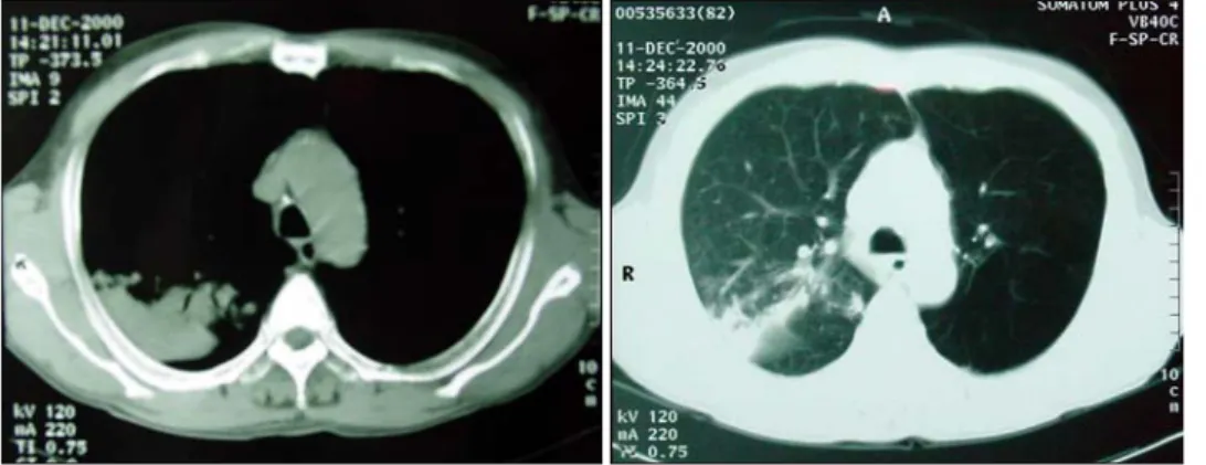
Fig. 2. The pre-operative chest CT shows the emphysematous change of the entire left lung and revealed the consolidation &
mild endobronchial stenosis be- low the right upper lobe bron- chus and the right lower para- tracheal lymph node is enlarged.
Fig. 3. The pre-operative bronchoscopic finding revealed a protruding mass on the right upper lobe bronchus.
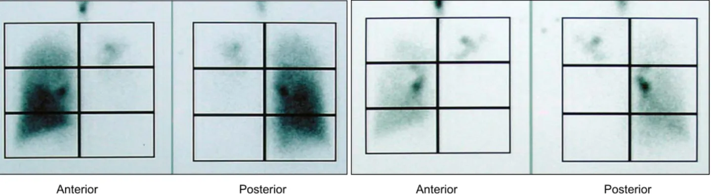
Fig. 4. The ventilation-perfusion scan shows a unilateral matched ventilation and perfusion defect on left lung and right upper lung.
Anterior Posterior Anterior Posterior
Fig. 5. The Histopahthologic finding of an endobronchial mass. A: Well differentiated keratinizing squamous cell carcinoma with an infiltrative growth around the bronchial carcinoma (H&E, ×100). B: Well differentiated squamous cell carcinoma showing a keratin pearl and a central dyscohesion by keratinizing cells (H&E, ×400).
A B
=국문 초록=
중심 단어