207
©The Korean Society of Food Science and Technology
목향 헥산추출물이 대장암세포인 HT-29 세포의 증식에 미치는 영향
김은지
1·박희숙·임순성
1·김정상
2·신현경
1·윤정한
1∗
한림대학교 식품영양학과, 1한림대학교 식의약품의 효능평가 및 기능성소재개발센터, 2경북대학교 동물공학과
Effect of the Hexane Extract of Saussurea lappa on the Growth of
HT-29 Human Colon Cancer Cells
Eun Ji Kim1
, Heesook Park, Soon Sung Lim1
, Jong-Sang Kim2
, Hyun-Kyung Shin1 , and Jung Han Yoon Park1∗
Department of Food Science and Nutrition, 1
Center for Efficacy Assessment and Development of Functional Foods and Drugs, Hallym University
2
Department of Animal Science and Biotechnology, Kyungpook National University
Abstract In Asia Saussurea lappa (SL) has been used as a traditional herbal medicine to treat abdominal pain and
tenesmus. Recently, in vitro cell culture studies have shown that SL has anti-ulcer, anti-inflammatory, and anti-tumor properties. To explore its potential chemopreventive and chemotherapeutic effects in colon cancer, we examined whether the hexane extract of SL (HESL) could inhibit the growth of HT-29 human colon cancer cells, and investigated the mechanisms for this effect. The cells were cultured with various concentrations (0-5 µg/mL) of HESL. The results indicated that HESL markedly decreased the numbers of viable HT-29 cells; whereas at the concentration of 5 µg/mL, HESL slightly decreased the viable cell numbers of CCD 1108Sk human skin normal fibroblasts at 72 hr. HESL substantially increased the numbers of cells in the sub G1 phase, and dose-dependently increased apoptotic cell numbers. Western blot analysis of the total cell lysates revealed that HESL increased Bax protein levels, but did not affect Bcl-2 levels. HESL induced the cleavage of poly (ADP-ribose) polymerase and caspases 8, 9, 7, and 3. This study demonstrated that HESL inhibits cell growth and induces apoptosis in HT-29 cells, which may be mediated by its ability to increase Bax levels and activate the caspase pathway. These findings may lead to the development of new therapeutic strategies for colon cancer treatment.
Key words: Saussurea lappa, colon cancer, apoptosis, Bax, caspase
서
론
미국 등 서구사회에서 여러 암 중 발병률과 이로 인한 사망률 에서 2위를 차지하고 있는 대장암은(1) 우리나라에서도 식생활이 서구화됨에 따라 급증하여 심각한 건강 문제가 되고 있다. 대장 암은 발견 당시 병이 많이 진행 되어서 수술로 완치가 불가능한 경우가 많다(2). 이러한 경우 방사선치료 또는 항암치료를 병행 하여 실시하고 있다(3). 현재 5-fluorouracil 등 여러 종류의 항암 제가 대장암 치료를 위해 사용되고 있으나 효과가 좋지 않으며 (3), 정상세포에 독성 및 다양한 부작용을 야기하고 있다(4,5). 따 라서 독성과 부작용은 적고 암세포의 증식은 효과적으로 억제하 는 항암제의 개발이 요구되고 있다. 예로부터 식용 또는 약용으 로 사용되고 있는 천연물은 어느 정도 안전성이 입증되었기 때 문에 이들 천연물을 이용하여 독성과 부작용이 없으면서 효과적 인 항암제를 개발하기 위해 세계적으로 활발히 연구가 진행되고 있다.목향은 국화과의 다년생 식물인 Saussurea lappa Clarke의 말 린 뿌리로 현재 한의학에서 구토, 설사, 및 호흡기 질병 치료 등 에 널리 사용되고 있는 한약재이다(6). 목향은 많은 종류의
sesquiterpene과 sesquiterpene lactone을 함유하고 있으며(7) 이외에 polyene alcohols(8), triterpene, lignan, amino acid-sesquiterpene adduct, alkaloid 및 tannin 등을 함유하고 있는 것으로 보고되고
있다(9). 목향 추출물 및 목향에 함유되어 있는 각각의 성분들은 다양한 생리활성을 나타내고 있음이 알져지고 있다. 목향과 목향 에서 유래한 각각의 성분들은 항궤양(10), 항바이러스(11), 항균 (12), 항염증(13,14), 혈관생성억제(15) 효과가 있음이 보고되었다. 또한 목향과 목향에서 유래한 다양한 sesquiterpene lactone이 위 암(16,17), 유방암(18,19), 혈액암(20) 세포의 증식을 억제함으로써 항암 작용이 있음이 보고되었다. 그러나 현재까지 목향이 대장암 세포의 증식에 미치는 영향 및 그 작용 기전에 대해서는 연구된 바가 없다. 세포사멸(apoptosis)은 세포가 정상적인 상태 또는 병리학적인 요인에 노출된 후에 죽음에 이르게 되는 생리적인 과정으로 정 상세포의 기능 유지에 필수적인 과정이다(21). 세포사멸은 세포 내·외적인 신호에 의해 시작되어 Bcl-2 family 단백질(22)과
*Corresponding author: Jung Han Yoon Park, Department of Food Science and Nutrition, Hallym University, 39 Hallymdaehak-gil, Chuncheon 200-702, Korea
Tel: 82-33-248-2134 Fax: 82-33-256-0199 E-mail: [email protected]
caspases(23) 등의 다양한 세포사멸 조절인자에 의해 조절된다. 대 장 상피는 계속적으로 빠르게 세포교체(cell turnover)가 이루어지 는 곳으로 대장 상피의 형태 및 기능 유지를 위해 세포사멸은 필수적인 과정이다. 대장에서 세포사멸이 적절하게 이루어지지 않고 감소한 경우 대장 상피세포의 과증식이 이루어지고 결국 에는 대장암이 발생된다(24). 또한 세포사멸에 장애가 생기면 암 은 더욱 진전되어 악화되고 치료가 불가능해진다(25). 그러므로 세포사멸을 적절하게 촉진하는 것은 대장암의 발생과 진전을 억 제하고 암을 치료할 수 있는 좋은 방법이라 할 수 있다. 실제로 현재 사용되고 있는 많은 항암제는 암세포의 세포사멸을 유도 하여 항암제로 작용하는 것으로 알려지고 있다(26-28). 그러므로 세포사멸의 주요한 조절인자인 Bcl-2 family 단백질의 발현이나 caspase들의 활성을 조절하여 세포사멸을 유도하는 성분은 대장 암의 암예방제 및 암치료제로 개발될 수 있는 가능성이 크다고 할 수 있다. 본 연구에서는 목향의 대장암 예방제 및 치료제로의 개발 가 능성을 조사하기 위해 목향 헥산추출물이 인간의 대장에서 유래 한 암세포인 HT-29 세포의 증식에 미치는 영향을 조사하였다. 또 한 목향 헥산추출물의 HT-29 세포 증식 억제 작용 기전을 규명 하고자 하였다.
재료
및
방법
재료 인간의 대장에서 유래한 암세포인 HT-29 세포, 인간의 대장에 서 유래한 정상 상피세포인 FHC 세포와 인간의 피부에서 유래 한 정상 섬유아세포인 CCD 1108Sk 세포는 American TypeCulture Collection(Rockville, MD, USA)에서 구입하였다. 세포배
양에 사용한 Dulbecco's Modified Eagle's Medium:Nutrient Mixture
Ham's F12(DMEM/F12)는 Gibco/BRL(Gaitherburg, MD, USA)에
서 구입하였다. Fetal bovine serum(FBS), penicillin-streptomycin,
Trypsin-EDTA 등은 Cambrex Bio Technology(Walkersville, MD, USA)에서 구입하였다. Bovine serum albumin(BSA), 3-(4,5-dimethylthiazol-2-yl)-2,5-diphenyltetrazolium bromide(MTT), Hoechst H 33258, propidium iodide, anti-β-actin antibody와 본 연구에 사
용한 일반적인 시약은 Sigma Chemical Co.(St. Louis, MO, USA) 에서 구입하였다. Phycoerythrin(PE)-conjugated Annexin V와
7-amino-actinomycin D은 BD Pharmingen(Franklin Lakes, NJ,
USA)에서 구입하여 사용하였다. Bcl-2, Bax antibody는 Santa
Cruz Biotechnology Inc.(Santa Cruz, CA, USA)에서 구입하였고, cleaved caspase-3, cleaved caspase-7, cleaved caspase-8, cleaved caspase-9, cleaved poly(ADP-ribose) polymerase(PARP)에 대한 antibody는 Cell Signaling(Beverly, MA, USA)에서 구입하여 사용
하였다. Horse radish peroxidase-linked anti-rabbit IgG와 horse
radish peroxidase-linked anti-mouse IgG는 Amersham (Bukinghamshire, England)에서 구입하여 사용하였다. 목향 헥산추출물의 제조 실험에 사용한 목향(Saussurea lappa)은 중국에서 수입한 것을 한약재료상에서 구입하여 사용하였다. 분쇄하여 가루로 만든 목 향 100 g에 2 L의 헥산을 가하여 환류 조건하에서 70o C의 조건 으로 3시간 추출하였다. 추출을 2회 반복 실시한 후 추출물을 여 과지로 여과하여 감압 농축하여 3 g의 추출물을 얻어 목향 헥산 추출물로 사용하였다. 세포 배양
세포는 DMEM/F12 배지에 10% FBS, 100 units/mL penicillin 과 100 µg streptomycin을 첨가한 세포 배양액을 사용하여 37 습 윤한 CO2 incubator(5% CO2/95% air)에서 배양하였다. 세포가 배 양 접시의 80% 정도 차면 phosphate-buffered saline(PBS, pH 7.4)으로 세포의 단층을 씻어낸 후 0.25% trypsin-2.65 mM EDTA 로 처리하여 계대 배양하였고 배지는 2일마다 교환하였다. 세포 증식 측정 목향 헥산추출물이 HT-29 세포, FHC 세포와 CCD 1108Sk 세 포의 증식에 미치는 영향을 측정하기 위해 세포를 10% FBS가 첨가된 배지로 희석하여 50,000 cells/well의 밀도로 24 well plate 에 분주하였다. 24시간이 지난 후 1% FBS가 포함되어 있는 배 지로 교환하여 혈청에 들어있는 여러 성분들의 효과를 최소화하 였다. 세포를 1% FBS가 포함되어 있는 배지에서 24시간 배양한 후 1% FBS가 포함되어 있는 배지에 여러 농도의 목향 헥산추출 물(0, 1, 3, 5 µg/mL)를 첨가한 배지로 교환하여 세포를 배양하 였다. 추출물을 첨가하여 0, 1, 2, 3일 동안 배양한 후 MTT assay 방법(29)을 이용하여 살아있는 세포수를 측정하였다. 세포주기 측정
HT-29 세포를 50,000 cells/well의 밀도로 24 well plate에 분주
한 후 위와 동일한 방법으로 0 또는 5 µg/mL 농도로 목향 헥산 추출물을 첨가하여 세포를 2시간 또는 24시간 배양하였다. 세포 에 trypsin-EDTA를 처리하여 세포를 수집한 후 Kim 등 (30)과 같 은 방법으로 세포를 propidium iodide로 염색하였다. Propidium
iodide에 의해 염색된 세포를 FACScanTM(Becton Dickinson,
Franklin Lake, NJ, USA)을 사용하여 flow cytometry 방법에 의해
세포주기를 측정하였고, 얻은 결과는 Modfit version 1.2 software
(Becton Dickinson)를 사용하여 분석하였다. Hoechst H 33258 염색 HT-29 세포를 chamber slide에 분주한 후 위와 동일한 방법으 로 0 또는 5 µg/mL 농도로 목향 헥산추출물을 처리하였다. 목향 헥산추출물을 처리하여 세포를 48시간 배양한 후 차가운 PBS로 세포단층을 씻어낸 후 4% formaldehyde로 세포를 고정하였다. 세 포에 10 µg/mL Hoechst H 33258 용액을 넣어 어두운 곳에서 1 시간 염색하였다. 세포를 PBS로 충분히 헹군 후 형광현미경으로 관찰하였다(31). 세포사멸 세포수 측정 (Annexin V 염색) 세포를 24 well plate에 분주한 후 위와 동일한 방법으로 0, 1, 3, 5 µg/mL 농도로 목향 헥산추출물을 세포 배양액에 첨가하여 세포를 48시간 동안 배양하였다. 세포 단층을 PBS로 씻어낸 후 trypsin-EDTA으로 처리하여 세포를 수집한 후 Kim 등(32)과 같 은 방법으로 세포를 phycoerythrin(PE)-conjugated Annexin V와
7-amino-actinomycin D로 염색한 후 Annexin V 또는 7-amino-actinomycin D에 의해 염색된 세포를 FACScanTM
(Becton Dickinson)을 사용하여 flow cytometry 방법에 의해 측정하였다.
Western blot analysis
세포를 100 mm dish에 분주하여 위와 동일한 방법으로 세포 배양액에 목향 헥산추출물을 첨가하여 48시간 배양한 후 Kim 등
(31,33)과 같은 방법으로 cell lysate를 만들었다. Cell lysate의 단
를 사용하여 측정하였다. Cell lysate(50 µg 또는 100 µg protein) 를 4-20% 또는 10-20% sodium dodecyl sulfate polyacrylamide
gel electrophoresis(SDS-PAGE)로 분리한 후 polyvinylidene difluoride membrane(Millipore, Bedford, MA, USA)에 이동시켰다. Membrane
은 5% skim milk-TBST(20 mM TrisHCl, pH 7.5, 150 mM
NaCl, 0.1% Tween 20)에서 1시간 동안 blocking하고, anti-bcl-2 antibody(1:1,000 희석), bax antibody(1:1,000 희석), anti-cleaved caspase-3 antibody(1:750 희석), anti-cleaved caspase-7 antibody(1:750 희석), anti-cleaved caspase-8 antibody(1:1,000 희석), anti-cleaved caspase-9 antibody(1:1,000 희석), anti-cleaved PARP (1:1,000 희석), anti-β-actin antibody(1:2,000 희석) 등 측정하고자
하는 antibody를 각각 첨가하여 4o
C에서 16시간 또는 상온에서 1
시간 동안 교반하였다. 그 후 horse radish peroxidase-linked
anti-rabbit IgG 또는 horse radish peroxidase-linked anti-mouse IgG를
첨가하여 1시간 교반하였다. 각 protein band는 SuperSignal® West Dura Extended Duration Substrate(Pierce)을 사용하여 enhanced chemiluminescence 방법으로 가시화하였다. 각 단백질 밴드의 강
도는 Bioprofile Bio-1D application(Vilber-Lourmat, Marine la
Vallee, France)을 사용하여 측정하였다.
통계 처리
모든 분석 수치는 mean ± SEM 으로 나타내었다. 수집된 결과 는 SAS(Statistical Analysis System) Windows v. 8.12 프로그램
(SAS Institute, Cary, NC, USA)을 이용하여 통계 분석하였으며,
각 실험군들의 평균치간의 유의성은 α=0.05 수준에서 analysis of
variance와 Duncan's multiple range test에 의해 분석하였다.
결과
및
고찰
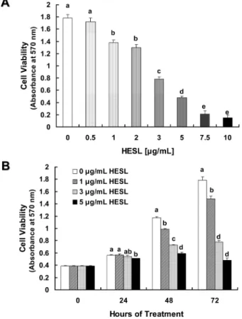
목향 헥산추출물이 HT-29 세포의증식에 미치는 영향 목향 헥산추출물이 인간의 대장에서 유래한 암세포인 HT-29 세포의 증식에 미치는 영향을 조사하기 위해 세포 배양액에 다 양한 농도(0, 0.5, 1, 2, 3, 5, 7.5, 10 µg/mL)의 목향 헥산추출물 을 첨가하여 세포를 72시간 배양한 후 MTT assay(29)를 실시하 여 살아있는 세포수를 측정하였다. HT-29 세포의 증식은 0.5 µg/ mL 농도로 목향 헥산추출물을 처리한 경우 추출물을 처리하지 않은 대조군과 유의적인 차이를 나타내지 않았고, 1 µg/mL 이상 의 농도로 처리한 경우 세포 증식이 현저히 감소하였다. 또한 살 아있는 세포수는 목향 헥산추출물을 7.5 µg/mL 또는 10 µg/mL 농도로 처리한 경우 추출물을 처리하지 않은 대조군에 비해 각 각 88%, 92% 감소하였다(Fig. 1A). 이 결과를 통해 7.5 µg/mL 이상의 농도에서는 목향 헥산추출물이 HT-29 세포에 세포 독성 을 야기하는 것으로 생각되어 추후 실험에서는 목향 헥산추출물 처리 최고농도를 5 µg/mL로 하였다. 목향 헥산추출물의 처리 시 간에 따른 차이를 조사하기 위해 세포배양액에 0, 1, 3, 5 µg/mL 농도로 목향 헥산추출물을 처리하여 세포를 24, 48, 72시간 동안 배양하였다. 목향 헥산추출물을 첨가하여 세포를 24시간 배양한 경우 5 µg/mL 농도에서만 세포 증식이 유의적으로 감소하였다. 목향 헥산추출물을 48시간, 72시간 처리한 경우 처리 농도에 따 라 유의적으로 세포 증식이 감소하였으며, 5 µg/mL 농도로 처리 하여 72시간 배양한 경우 목향 헥산추출물을 처리하지 않은 대 조군 (0 µg/mL)에 비해 73% 세포 증식이 감소하였다(Fig. 1B). 목향은 많은 종류의 sesquiterpene과 sesquiterpene lactone을 함유 하고 있고(7) 그 중 주성분은 sesquiterpene lactone계 화합물인costunolide와 dehydrocostus lactone으로 알려지고 있다(9).
Costunolide를 주성분으로 함유하고 있는 목향 에탄올추출물은 위 암세포인 AGS 세포의 세포주기를 지연하고 세포사멸을 유도하 여 세포 증식을 억제하였고(17), costunolide는 유방암(18,19)과 혈 액암(20) 세포의 증식을 억제하였다. 본 연구에서는 목향 헥산추 출물에서 HT-29 세포의 증식을 현저히 억제한 유효성분 동정에 대한 연구는 수행하지 않았으나, 목향 헥산추출물에 함유된 costunolide가 HT-29 세포의 세포주기를 지연하고 세포사멸을 유 도하여 세포 증식을 억제함을 유추할 수 있다. 그러나 목향 헥산 추출물의 유효성분과 그 성분의 항암기전을 알기 위해서는 추가 연구가 필요하다. HT-29 세포의 증식을 현저히 억제한 목향 헥산추출물이 정상 세포에 미치는 영향을 인간의 대장에서 유래한 정상 상피세포인 FHC 세포와 인간의 정상 섬유아세포인 CCD 1108Sk 세포를 사 용하여 관찰하였다. FHC 세포 배양액에 목향 헥산추출물을 처리 하여 72시간 배양한 후 MTT assay(29)을 실시하여 얻은 결과를 Fig. 2A에 나타내었다. 목향 헥산추출물 처리에 의해 FHC 세포 의 증식은 변화하지 않았다(Fig. 2A). CCD 1108Sk 세포 배양액 에 0-5 µg/mL의 목향 헥산추출물을 처리하여 세포를 24시간, 48 시간 배양한 경우 살아있는 세포수에는 유의적인 차이를 나타내
Fig. 1. Effect of the hexane extract of Saussurea lappa (HESL) on viable cell numbers of HT-29 human colon cancer cells. HT-29
cells were plated at a density of 50,000 cells/well in 24-well plates with DMEM/F12 supplemented with 10% FBS. One day after plating, the monolayers were serum-deprived with DMEM/F12 supplemented with 1% FBS serum-deprivation medium for 24 hr. After serum deprivation, cells were incubated in serum-deprivation medium in the absence or presence of various concentrations of HESL. Cell numbers were estimated by the MTT assay. Each bar represents the mean±SEM (n = 6). Means with different lowercase letters are significantly different ( p < 0.05).
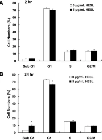
지 않았다. 목향 헥산추출물을 처리하여 72시간이 경과한 경우 5 µg/mL 농도에서만 CCD 1108Sk 세포수가 유의적으로(p<0.05) 감 소하였다(Fig. 2B). Ko 등(17)은 목향 에탄올추출물이 위암세포인 AGS 세포의 증식은 현저히 억제한 반면 쥐의 장에서 유래한 정 상 상피세포인 RIE1 세포의 증식은 고농도로 처리시에만 억제하 였다고 보고하였다. 이는 목향 헥산추출물이 정상세포에는 독성 과 부작용이 적고 암세포 증식은 효과적으로 억제함을 나타내 며, 차후 안전하고 효과적인 항암제 개발의 좋은 소재가 될 수 있는 가능성을 제시한다. 목향 헥산추출물이 세포주기 진행과 세포사멸에 미치는영향 암세포의 증식 억제는 세포주기 진행의 지연과 세포사멸 유도 에 의해 이루어진다(34). 목향 헥산추출물에 의한 HT-29 세포 증 식 억제가 세포주기 진행 지연에 의한 것인지 조사하기 위해 목 향 헥산추출물을 처리하여 세포를 배양한 후 핵을 propidium
iodide로 염색하여 flow cytometry를 실시하였다. 목향 헥산추출물
을 5 µg/mL 농도로 세포배양액에 첨가하여 세포를 2시간 배양 한 경우 세포주기 진행에 유의적인 변화는 나타나지 않았다(Fig. 3A). 반면 5 µg/mL 목향 헥산추출물을 24시간 처리한 경우 sub G1기에 머무른 세포수는 유의적으로(p<0.05) 증가하였고 G1기에 머무른 세포수는 유의적으로(p<0.05) 감소하였으며 S기와 G2/M 기에 머무른 세포수는 유의적인 차이를 나타내지 않았다(Fig. 3B). 일반적으로 sub G1기에 머무른 세포수의 증가는 세포사멸의 특 징 중의 하나로 생각되고 있다(34). 본 연구에서 목향 헥산추출 물이 sub G1기에 머무른 세포수를 증가시킴을 관찰하였으며, 이 는 목향 헥산추출물이 HT-29 세포의 세포주기 진행을 지연시키 기 보다는 세포사멸을 유도하여 세포 증식을 억제함을 시사한다. 목향 헥산추출물이 HT-29 세포에 세포사멸를 유도하였는지 확 인하기 위해 목향 헥산추출물을 5 µg/mL 농도로 세포 배양액에 첨가하여 세포를 48시간 배양한 후 세포를 Hoechst H 33258로 염색하여 세포의 형태학적인 변화를 관찰하였다. Fig. 4A에서 보 는 바와 같이 목향 헥산추출물을 5 µg/mL 농도로 처리한 경우 핵이 응축되고 분절되어 나타나는 apoptotic bodies가 뚜렷하게 나 타났다. 목향 헥산추출물에 의해 유도된 세포사멸를 정량하기 위 하여 세포배양액에 목향 헥산추출물을 처리하여 48시간 세포를 배양한 후 Annexin V로 세포를 염색하여 flow cytometry 방법으 로 세포사멸 세포수를 측정한 결과를 Fig. 4B에 나타내었다. 목 향 헥산추출물의 처리 농도가 증가할수록 살아있는 세포수는 현 저히 감소하였고, 세포사멸 세포수는 유의적으로 증가하였다(Fig.
4B). 이상의 결과는 목향 헥산추출물이 HT-29 세포의 세포사멸
를 유도하여 세포의 증식을 억제함을 나타낸다.
Fig. 2. Effect of the hexane extract of Saussurea lappa (HESL) on viable cell numbers of FHC human normal colonic epithelial cells(A) and CCD 1108Sk human skin normal fibroblasts(B).
FHC cells and CCD 1108Sk cells were plated and treated with HESL as described in Fig. 1. Cell numbers were estimated by the MTT assay. Each bar represents the mean±SEM (n = 6). Means with different lowercase letters are significantly different ( p < 0.05).
Fig. 3. Effect of the hexane extract of Saussurea lappa (HESL) on the cell cycle progression in HT-29 cells. HT-29 cells were
treated with 0 or 5 µg/mL HESL for 2 hr (A) or 24 hr (B) as described in Fig. 1. Cells were trypsinzed, fixed, and treated RNase. Cellular DNA was then stained with propidium iodide. The percentage of cells in sub G1, G1, S, and G2-M phase of the cell cycle was analyzed by flow cytometry. Each bar represents the mean±SEM (n = 6). *
Significantly different from 0 µg/mL HESL at p < 0.05.
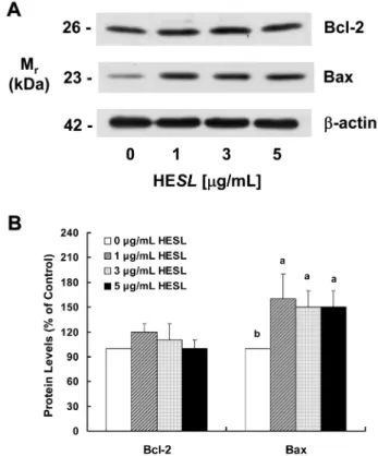
목향 헥산추출물이 Bcl-2와 Bax 단백질 발현에 미치는 영향
Bcl-2 family 단백질은 미토콘드리아의 막 투과성과 cytochrome
c의 방출을 제어하여 세포사멸을 조절하는 것으로 알려져 있다.
Bcl-2 family 단백질은 기능과 아미노산 서열의 유사성에 따라 anti-apoptotic 단백질과 pro-apoptotic 단백질, 그리고 Bcl-2 homology domain(BH)-3 only 단백질로 구분된다(35).
Pro-apoptotic 단백질은 미토콘드리아의 막 투과성을 증가시키고,
cytochrome c의 방출을 증가시킴으로서 세포사멸을 유도한다. 반
면, anti-apoptotic 단백질은 pro-apoptotic 단백질과 heterodimer를 형성하여 pro-apoptotic 단백질의 활성을 억제하여 세포사멸을 억 제한다(35). BH-3 only 단백질은 anti-apoptotic 단백질에 결합하여 anti-apoptotic 단백질에 길항적으로 작용하여 세포사멸을 증가시 킨다(36). HT-29 세포에서 세포사멸을 현저히 유도한 목향 헥산 추출물이 세포사멸의 중요한 조절인자인 Bcl-2 family 단백질 수 준에 미치는 영향을 조사하기 위해 목향 헥산추출물을 세포 배 양액에 첨가하여 세포를 48시간 동안 세포를 배양한 후 세포를 수거하고 cell lysate를 만들어 Western blot를 실시하였다. 대표적 인 anti-apoptotic Bcl-2 family 단백질인 Bcl-2 단백질 수준은 목 향 헥산추출물 처리에 의해 변화하지 않았다. 반면, pro-apoptotic Bcl-2 family 단백질인 Bax 단백질 수준은 목향 헥산추출물 처리 에 의해 유의적으로(p<0.05) 증가하였으나 처리 농도에 따른 차 이는 나타나지 않았다(Fig. 5). 세포의 생존과 사멸을 결정하는 중 요한 요소인 Bcl-2/Bax 비율(37)은 목향 헥산추출물을 0, 1, 3, 5 µg/mL 농도로 처리 농도를 증가하여 처리한 경우 1, 1.3, 1.4, 1.5 로 증가하였다. 이 결과는 목향 헥산추출물에 의한 Bax 단백질 수준과 Bcl-2/Bax 비율 증가가 목향 헥산추출물에 의해 유도된 HT-29 세포의 세포사멸의 기전 중의 하나임을 제시한다. 그러나 목향 헥산추출물에 의한 Bax 단백질 수준과 Bcl-2/Bax 비율 증 가는 목향 헥산추출물에 의해 유도된 세포사멸에 비해 미미하였 다. Kim 등(38)은 차가버섯 물추출물이 Bax와 Bcl-2 단백질 수준 에 영향을 미치지 않고 caspase 경로의 활성을 증가시킴으로서 대장암세포인 HT-29 세포의 세포사멸을 유도함을 보고하였다. 또 한 HT-29 세포에서 보라우무 메탄올추출물(33)과 복분자 물추출 물(39)은 caspase 경로를 활성화하여 세포사멸을 유도하였다고 보 고되었다. 이는 목향 헥산추출물에 의해 유도된 세포사멸은 Bax 단백질 수준과 Bcl-2/Bax 비율 증가에 의하기 보다는 caspase 활 성 증가 등의 다른 경로에 의해 주로 이루어짐을 제시한다. 목향 헥산 추출물이 caspase 활성에 미치는 영향
Cysteine aspartic acid proteases인 caspases는 세포사멸의 중요한
Fig. 4. Effect of the hexane extract of Saussurea lappa (HESL) on apoptosis of HT-29 cells. Cells were plated and treated with
HESL for 48 hr as described in Fig. 1. (A) Hoechst H 33258 stain. Cell were fixed and stained with a DNA specific dye, Hoechst H 33258. Images were obtained using a fluorescence microscope. Microphotographs are representative of three independent experi-ments. Magnification, X 200. (B) Fluorescence-activated cell sorting analysis. Cells were trypsinzed, stained with 7-amino-actinomycin D and Annexin V, and then analyzed by flow cytometry. The number of living cells and early apoptotic cells is expressed as a percentage of total cell number. Each bar represents the mean±SEM (n = 6). Values with different letters above each bar are significantly different (p < 0.05) between concentrations of HESL.
Fig. 5. Effect of the hexane extract of Saussurea lappa (HESL) on the protein levels of Bcl-2 and Bax in HT-29 cells. Cells were
treated with various concentration of HESL for 48 hr as described in Fig, 1. Cell lysates were analyzed by immunoblotting with an antibody against Bcl-2, Bax or β-actin. (A) Photograph of chemilumi-nescent detection of the blots, which were representative of 3 independent experiments, are shown. (B) Quantitative analysis of Western blots. The relative abundance of each band to their own β-actin was quantified and the control levels were set at 100. The adjusted mean±SEM (n = 3), is shown above each blot. Means with different lowercase letters are significantly different (p < 0.05).
조절 인자로 세포 내 단백질의 분절을 유도하여 세포사멸을 유 도한다. Caspases는 세포내에서 불활성화된 proenzyme의 형태로 존재하여 세포사멸 신호에 의해 스스로 또는 다른 caspases에 의 해 분절되어 활성화된다. Caspases는 여러 종류가 존재하며 구조, 기능, 및 세포사멸 신호와의 근접성에 의해 initiator caspase와
effector caspase로 구분된다. Initiator caspase인 caspase-8과 caspase-9은 death-inducing 신호에 의해 활성화되어 effector caspase들을 활성화시키며, caspase-8과 caspase-9은 서로 다른 경
로를 통해 활성화된다. 세포막에 존재하는 cell death receptor에
cell death ligands가 결합하면 caspase-8이 활성화되는 반면 caspase-9은 미토콘드리아 막의 투과성이 증가되어 세포질로 방
출된 cycochrome c에 의해 활성화된다. 활성화된 caspase-8 또는
caspase-9은 effector caspase인 caspase-7 또는 caspase-3을 활성화
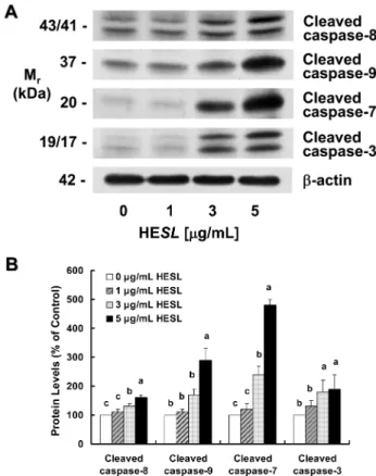
하고, 활성화된 effector caspase은 apoptosis의 형태학적 특징에 관 여하는 lamin A, α-fodrin, DNA fragmentation factor(DFF), PARP 등의 단백질을 분해하여 세포사멸을 유도한다(23). 여러 연구에 서 암세포의 세포사멸을 유도한 항암 성분들은 caspase의 활성을 조절하였다(31,33,34)고 보고된 바 있다. 본 연구에서는 HT-29 세 포에서 세포사멸을 현저히 유도한 목향 헥산추출물이 caspases의 활성에 미치는 영향을 조사하기 위해 caspases 활성 형태인 cleaved caspases들의 단백질 수준을 관찰하였다. 목향 헥산추출물 처리에 의해 cleaved caspase-8, -9, -7, -3 단백질 수준이 유의적으로 증 가하였으며, 목향 헥산추출물을 처리하지 않은 대조군에 비해 5 µg/mL 목향 헥산추출물을 처리한 경우 cleaved caspase-8, -9, -7, -3의 단백질 수준이 각각 1.6배, 2.9배, 4.8배, 1.9배 증가하였다 (Fig 6).
세포사멸 신호에 의해 활성화된 caspase-8은 effector caspase인
caspase-3을 활성하거나 또는 BH-3 only Bcl-2 family 단백질로 세
포질에 존재하는 Bid를 분절한다. 분절된 Bid(truncated-Bid)는 미 토콘드리아로 이동하여 Bax와 결합하고, Bax의 구조적 변화를 초 래한다. 이로 인해 미토콘드리아의 투과성을 증가하여 세포사멸 이 유도된다(36). 목향 헥산추출물에 의해 활성화된 caspase-8이
Bid의 분절을 유도했는지를 조사하였다. Western blot에 의해
truncated-Bid는 검출되지 않았고, 또한 목향 헥산추출물 처리에 의해 Bid 단백질 수준도 변화하지 않았다(결과 미제시). 이는 목 향 헥산추출물이 8의 활성을 증가시키고, 활성화된 caspase-8은 Bid 분절에는 관여하지 않고 caspase-3를 활성화시켜 세포사 멸을 유도함을 제시한다. Caspase-9은 미토콘드리아에서 방출된 cytochrome c에 의해 활 성화되고, cytochrome c의 방출은 Bcl-2 family 단백질의 변화에 의해 미토콘드리아 막 투과성이 증가됨으로서 증가된다(35). 본 연구에서 목향 헥산추출물에 의해 pro-apoptotic Bcl-2 family 단 백질인 Bax 단백질의 수준이 증가하였고(Fig. 5) cleaved
caspase-9 단백질 수준이 증가함(Fig. 6)을 관찰하였다. 이 결과는 목향 헥 산추출물에 의해 증가한 Bax가 미토콘드리아의 막 투과성을 증 가하여 cytochrome c의 방출을 유도하여 caspase-9을 활성화하였 음을 제시한다. 그러나 향후 미토콘드리아 막 투과성과 미토콘드 리아에서 세포질로 방출된 cytochrome c를 확인해야 할 것으로 생각된다. 이상의 결과를 통해 HT-29 세포에서 목향 헥산추출물이 initiator
caspase인 caspase-8과 -9의 활성을 증가시키고, 활성화된 caspas-8과 -9에 의해 effector caspase인 caspase-3과 -7을 활성화하여 세
포사멸을 유도한 것임을 알 수 있다. 이는 caspases 활성 증가가 목향 헥산추출물에 의한 세포사멸 과정에 중요한 조절인자로 작 용했음을 제시한다.
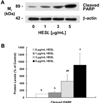
목향 헥산추출물이 poly(ADP-ribose) polymerase (PARP)의
불활성에 미치는 영향
세포의 핵 안에 존재하고, nicotinamide adenine dinucleotide
(NAD+)에서 poly(ADP-ribose)의 생성을 촉매하는 효소인 PARP
는 DNA 수선에 관여하여 세포의 생존 유지에 중요한 역할을 담 당한다. PARP는 caspase-3의 주요한 표적 단백질 중의 하나로
caspase-3에 의해 분절되어 불활성화 되면 세포의 생존을 유지할
수 없어 세포사멸에 기여한다(40). 목향 헥산추출물에 의해 cleaved
caspase-3가 증가(Fig. 6)하였기 때문에 목향 헥산추출물이 PARP
를 분절하여 세포사멸을 유도할 것으로 생각되어, 목향 헥산추출 물이 PARP의 불활성형인 cleaved PARP 단백질 수준에 미치는 영향을 조사하였다. Fig. 7에 나타난 바와 같이 목향 헥산추출물 처리 농도가 증가할수록 cleaved PARP 단백질 수준은 현저히 증 가하였으며, 추출물을 처리하지 않은 대조군에 비해 5 µg/mL 농 도로 처리한 경우 cleaved PARP 단백질 수준이 9.1배 증가하였 다. Lee 등(17)은 AGS 위암세포에 목향 에탄올추출물을 처리한 경우 cleaved caspase-3와 cleaved PARP 단백질 수준이 증가하였 고, 이를 통해 세포사멸이 증가되었다고 보고하였다. 암세포의 세 포사멸을 유도하는 천연물 유래 성분들이 caspase-3 활성과 PARP
Fig. 6. Effect of the hexane extract of Saussurea lappa (HESL) on the protein levels of various cleaved caspases in HT-29 cells.
Cells were treated with various concentration of HESL for 48 hr as described in Fig, 1. Cell lysates were analyzed by immunoblotting with an antibody against cleaved caspase-3, cleaved caspase-7, cleaved casapse-8, cleaved caspase-9, or β-actin. (A) Photograph of chemiluminescent detection of the blots, which were representative of 3 independent experiments, are shown. (B) Quantitative analysis of Western blots. The relative abundance of each band to their own
β-actin was quantified and the control levels were set at 100. The
adjusted mean±SEM (n = 3), is shown above each blot. Means with different lowercase letters are significantly different ( p < 0.05).
의 불활성이 증가됨이 보고되었다(33,38,39). 이 결과들은 목향 헥 산추출물에 의해 활성화된 caspase-3에 의해 PARP가 분절되어 불 활성화 되어 HT-29 세포에 세포사멸을 유도했음을 나타낸다.
요
약
목향(Saussurea lappa)은 항암효과를 비롯하여 다양한 생리활성 을 나타내는 것으로 알려지고 있으나 목향이 대장암에 미치는 영 향에 대해 자세히 연구된 바가 없다. 본 연구에는 목향 헥산추출 물이 인간의 대장에서 유래한 암세포인 HT-29 세포의 증식에 미 치는 영향과 그 작용 기전에 대해 연구하였다. 헥산으로 목향을 추출하여 얻은 목향 헥산추출물을 HT-29 세포의 세포 배양액에 0, 1, 3, 5 µg/mL로 첨가하여 세포를 배양하였다. HT-29 세포의 증식은 목향 헥산추출물 처리 농도가 증가할수록 현저히 감소하 였다. 인간의 대장에서 유래한 정상 상피세포인 FHC 세포의 증 식은 목향 헥산추출물에 의해 변화하지 않았고, 인간의 피부에서 유래한 정상 섬유아세포인 CCD 1108Sk 세포 증식은 목향을 5 µg/mL 농도로 72 시간 배양한 경우에만 감소하였다. 목향 헥산 추출물 처리에 의해 HT-29 세포의 세포주기 진행 중 sub G1기 에 머무른 세포수가 증가하였고, 세포사멸 세포수가 현저히 증가 하였다. 세포사멸의 주요한 조절인자인 Bcl-2 family 단백질 중 Bcl-2은 목향 헥산추출물에 의해 변화하지 않았으나 Bax는 유의 적으로 증가하였다. Bcl-2 family 단백질과 더불어 세포사멸 조절 에 중요한 역할을 하는 caspases의 활성형인 cleaved caspase-8,-9, -7, -3가 목향 헥산추출물에 의해 증가하였다. 또한 목향 헥산 추출물 처리 농도가 증가할수록 cleaved PARP 단백질 수준도 현 저히 증가하였다. 이 결과는 목향 헥산추출물이 세포사멸을 유도 하여 대장암세포인 HT-29 세포의 증식을 억제함을 나타내며, 목 향 헥산추출물에 의해 HT-29 세포의 세포사멸은 Bax 증가와 caspases의 활성 증가에 의한 것임을 나타낸다. 본 연구는 목향을 독성과 부작용이 적은 암예방제나 항암제로 개발할 수 있는 가 능성을 제시한다. 그러나 목향을 이용하여 제품 개발을 위해서는 유효 성분 동정 및 동물시험 등의 연구 수행이 필요할 것으로 사 료된다.
감사의
글
이 연구는 2005년도 정부(교육인적자원부)의 재원으로 한국학 술진흥재단의 지원(MOEHRD, KRF-2005-F00055), 농촌진흥청 바 이오그린21사업(20070301034039)과 2007년도 산업자원부 지역혁 신센터사업(한림대 식의약품의 효능평가 및 기능성소재개발센터) 의 지원에 의해 수행되었으며, 지원에 감사드립니다.문
헌
1. Jemal A, Siegel R, Ward E, Murray T, Xu J, Thun MJ. Cancer statistics, 2007. CA Cancer J. Clin. 57: 43-66 (2007)
2. Lee KH, Yu CS, Kim HC, Kim JR, Kim YM, Kim JS, Kim JC. Outcome of curative resection in patients with completely obstructing colorectal cancer. J. Korean Surg. Soc. 66: 199-204 (2004)
3. Kim WS, Lee RA, Hwang DY, Hong YJ, Hong SI. Histoculture drug response assay in colorectal cancer specimen. J. Korean Surg. Soc. 66: 109-115 (2004)
4. Maroun JA, Anthony LB, Blais N, Burkes R, Dowden SD, Dran-itsaris G, Samson B, Shah A, Thirlwell MP, Vincent MD, Wong R. Prevention and management of chemotherapy-induced diarrhea in patients with colorectal cancer: A consensus statement by the canadian working group on chemotherapy-induced diarrhea. Curr. Oncol. 14(1): 13-20 (2007)
5. de Gramount A, Figer A, Seymour M, Homerin M, Hmissi A, Cassidy J, Boni C, Cortes-Funes H, Cervantes A, Freyer G, Papamichael D, Le Bail N, Louvet C, Hendler D, de Braud F, Wilson C, Morvan F, Bonetti A. Leucovorin and fluorouracil with or without oxaliplatin as first-line treatmet in advanced colorectal cancer. J. Clin. Oncol. 18: 2938-2947 (2000)
6. Jeon YJ, Lee HS, Yeon SW, Ko JH, An KM, Yu SW, Kang JH, Hwang BY, Kim TY. Inhibitory effects of dehydrocostuslactone isolated from Saussureae Radix on CDK2 activity. Korean J. Pharmacogn. 36(2): 97-101 (2005)
7. Jung JH, Ha JY, Min KR, Shibata F, Nakagawa H, Kang SS, Chang IM, Kim YS. Reynosin from Saussurea lappa as inhibitor on CINC-1 induction in LPS-stimulated NRK-52E cells. Planta Med. 64: 454-456 (1998)
8. Jung JH, Kim YS, Lee CO, Kang SS, Park JH, Im KS. Cyto-toxic constituents of Saussurea lappa. Arch. Pharm. Res. 21: 153-156 (1998)
9. Kang SS, Kim JS, Chi HJ, Chang SY, Ha KW. Isolation and quantitative determination of costunolide from Saussurea root. Korean J. Pharmacogn. 30: 48-53 (1999)
10. Yoshikawa M, Hatakeyama S, Inoue Y, Yamahara J. Saussuream-ines A, B, C, D, and E, new anti-ulcer principles from ChSaussuream-inese
Saussureae. Chem. Pharm. Bull. (Tokyo) 41: 214-216 (1993)
11. Chen C, Chou CK, Lee SD, Wang JC, Yeh SF. Active com-pounds from Saussurea lappa Clarks that suppress hepatitis B virus surface antigen gene expression in human hepatoma cells. Antivir. Res. 27: 99-109 (1995)
12. Wedge DE, Galindo JC, Marcias FA. Fungidal activity of natural and synthetic sesquiterpene lactone analogs. Phytochemistry 53:
Fig. 7. Effect of the hexane extract of Saussurea lappa (HESL) on the protein levels of cleaved PARP in HT-29 cells. Cells were
treated with various concentration of HESL for 48 hr as described in Fig, 1. Cell lysates were analyzed by immunoblotting with an antibody against cleaved PARP or β-actin. (A) Photograph of chemiluminescent detection of the blots, which were representative of 3 independent experiments, are shown. (B) Quantitative analysis of Western blots. The relative abundance of each band to their own β-actin was quantified and the control levels were set at 100. The adjusted mean ±SEM (n=3), is shown above each blot. Means with different lowercase letters are significantly different ( p < 0.05).
747-757 (2000)
13. Cho JY, Baik KU, Jung JH, Park MH. In vitro anti-inflammatory effects of cynaropicrin, a sesquiterpene lactone, from Saussurea
lappa. Eur. J. Pharmacol. 398: 399-407 (2000)
14. Matsuda H, Toguchida I, Ninomiya K, Kageura T, Morikawa T, Yoshikawa M. Effects of sesquiterpenes and amino acid-sesquiter-pene conjugates from the roots of Saussurea lappa on inducible nitric oxide synthase and heat shock protein in lipopolysaccha-ride-activated macrophages. Bioorg. Med. Chem. Lett. 11: 709-715 (2003)
15. Jeong SJ, Itokawa T, Shibuya M, Kuwano M, Ono M, Higuchi R, Miyamoto T. Costunoide, a sesquiterpene lactone from
Saus-surea lappa, inhibits the VEGFR KDR/Flk-1 signaling pathway.
Cancer Lett. 187: 129-133 (2002)
16. Ko SG, Koh SH, Jun CY, Nam CG, Bae HS, Shin MK. Induction of apoptosis Saussurea lappa by and Pharbits nil on AGS gastric cancer cells. Biol. Pharm. Bull. 27: 1604-1610 (2004)
17. Ko SG, Kim HP, Jin DH, Bae HS, Kim SH, Park CH, Lee JW.
Saussurea lappa induces G2-growth arrest and apoptosis in AGS
gastric cancer cells. Cancer Lett. 220: 11-19 (2005)
18. Bocca C, Gabriel L, Bozzo F, Miglietta A. A sesquiterpene lac-tone, costunolide, interacts with microtubule protein and inhibits the growth of MCF-7 cells. Chem. Biol. Interact. 147: 79-86 (2004)
19. Choi SH, Im E, Kang HK, Lee JH, Kwak HS, Bae YT, Park HJ, Kim ND. Inhibitory effects of costunolide on the telomerase activity in human breast carcinoma cells. Cancer Lett. 227: 153-162 (2005)
20. Choi JH, Seo BR, Seo SH, Lee KT, Park JH, Park HJ, Choi JW, Itoh Y, Miyamoto K. Costunolide induces differentiation of human leukemia HL-60 cells. Arch. Pharm. Res. 25: 480-484 (2002) 21. Hengartner MO. The biochemistry of apoptosis. Nature 407:
770-777 (2000)
22. Korsmeyer SJ. Regulators of cell death. Trends Genet. 11: 101-105 (1995)
23. Debatin KM. Apoptosis pathways in cancer and cancer therapy. Cancer Immunol. Immun. 53: 153-159 (2004)
24. Ramachandran A, Madesh M, Balasubramanian KA. Apoptosis in the intestinal epithelium: Its relevance in normal and pathophysi-ology conditions. J. Gastoenterol. Hepatol. 15: 109-120 (2000) 25. Kim DG. Apoptosis and gastrointestinal disease. Korean J.
Gas-troenterol. 39: 79-87 (2002)
26. Haldar S, Chintapalli J, Croce CM. Taxol induces bcl-2 phospho-rylation and death of prostate cancer cells. Cancer Res. 56: 1253-1255 (1996)
27. Lowe SW, Lin AW. Apoptosis in cancer. Carcinogenesis 21: 485-495 (2000)
28. Schmitt CA, Lowe SW. Apoptosis and therapy. J. Pathol. 187: 127-137 (1999)
29. Denizot F, Lang R. Rapid colorimetric assay for cell growth and survival modifications to the tetrazolium dye procedure giving improved sensitivity and reliability. J. Immunol. Methods 89: 271-277 (1986)
30. Kim EJ, Shin HK, Cho JS, Lee SK, Won MH, Kim HW, Park JHY. Trans-10,cis-12 conjugated linoleic acid inhibits the G1-S cell cycle progression in DU145 human prostate carcinoma cells. J. Med. Food 9: 293-299 (2006)
31. Jung JI, Lim SS, Choi HJ, Cho HJ, Shin HK, Kim EJ, Chung WY, Park KK, Park JHY. Isoliquiritigenin induces apoptosis by depolarizing mitochondrial membranes in prostate cancer cells. J. Nutr. Biochem. 17: 689-696 (2006)
32. Kim EJ, Houlthuizen PE, Park HS, Ha YL, Jung KC, Park JHY. Trans-10,cis-12 conjugated linoleic acid inhibits Caco-2 colon cancer cell growth. Am. J. Physiol. Gastrointest. Liver Physiol. 283: G357-G367 (2002)
33. Kim EJ, Park SY, Hong J, Shin M, Lim SS, Shin HK, Park JHY. Inhibitory effect of the methanolic extract of Symphyocladia
latiuscula on the growth of HT-29 human colon cancer cells. J.
Korean Soc. Food Sci. Nutr. 36: 431-438 (2007)
34. Yu Z, Li W. Induction of apoptosis by puerarin in colon cancer HT-29 cells. Cancer Lett. 238: 53-60 (2006)
35. Cory S, Huang DC, Adams JM. The Bcl-2 family: Roles in cell survival and oncogenesis. Oncogene 22: 8590-8607 (2003) 36. Bouillet P, Strasser A. BH3-only proteins-evolutionarily
con-served proapoptotic Bcl-2 family members essential for initiating programmed cell death. J. Cell Sci. 115: 1567-1574 (2002) 37. Oltvai ZN, Milliman CL, Korsmeyer SJ. Bcl-2 heterodimerizes in
vivo with a conserved homolog, Bax, that accelerates pro-grammed cell death. Cell 74: 609-619 (1993)
38. Kim EJ, Lee YJ, Shin HK, Park JHY. A study on the mecha-nisms by which the aqueous extract of Inonotus obliquus induces apoptosis and inhibits proliferation in HT-29 human colon cancer cells. J. Korean Soc. Food. Sci. Nutr. 35: 516-523 (2006)
39. Kim EJ, Lee YJ, Shin HK, Park JHY. Induction of apoptosis by the aqueous extract of Rubus coreanum in HT-29 human colon cancer cells. Nutrition 21: 1141-1148 (2005)
40. Oliver FJ, de la Rubia G, Rolli V, Ruiz-Ruiz MC, de Murcia G, Murcia JM. Importance of poly(ADP-ribose) polymerase and its cleavage in apoptosis. Lesson from an uncleavable mutant. J. Biol. Chem. 273: 33533-33539 (1998)