INTRODUCTION
Cyclooxygenase-2 (COX-2) has been associated with the biology of tumors including carcinogenesis, inhibition of apoptosis, proliferation, promotion of angiogenesis, enhanced invasiveness, immune modulation, and increased mutagen- esis (1, 2). Clinical studies have shown that COX-2 overex- pression might be related to the extent of tumors at diagno- sis and treatment outcomes of several cancers (3-12). In addition, COX-2 overexpression has been reported to short- en the disease-free and overall survival after radiation thera- py (RT) in patients with cervical cancer (7, 8, 10). Recently, COX-2 overexpression has been reported to reduce the tumor response to RT in esophageal cancer, cervical cancer and rec- tal cancer (13-16). Inhibitory effect of COX-2 on apoptosis is thought to be the cause of the reduced response rate in such cases.
In squamous cell carcinoma (SCC) of the uterine cervix, COX-2 overexpression has been reported to have an adverse effect on tumor response in two studies (14, 16); however, the relationship between COX-2 expression and tumor res-
ponse remains to be elucidated. In these studies, the meth- ods for evaluation of the tumor response after RT were not described, and possible confounding factors such as tumor volume were not analyzed. Traditional methods including gynecological examination or computerized tomography scanning have limitations with regard to the evaluation of responses; they cannot exactly determine alterations in the extent of tumors. However, assessment of tumors using mag- netic resonance imaging (MRI) could offer more accurate and objective information compared to clinical evaluations (17).
In this study, we used MRI to evaluate the tumor response to RT, and investigated the correlation between COX-2 ex- pression and tumor response in patients with SCC of the uter- ine cervix treated with RT.
MATERIALS AND METHODS Patient selection
We enrolled 57 patients treated with curative radiotherapy
1170
Min Kyu Kang1, Won Park2, Yoon-La Choi3, Eun Yoon Cho3, Geunghwan Ahn3, HeeRim Nam2, Seung Jae Huh2, Yong Chan Ahn2, Do Hoon Lim2, Dong Ryul Oh2, Duk Soo Bae4, and Byoung Gie Kim4
Department of Radiation Oncology1, Yeungnam University College of Medicine, Daegu; Departments of Radiation Oncology2, Pathology3, and Obstetrics and Gynecology4, Samsung Medical Center, Sungkyunkwan University School of Medicine, Seoul, Korea
Address for correspondence Won Park, M.D.
Department of Radiation Oncology, Samsung Medical Center, 50 Irwon-dong, Gangnam-gu, Seoul 135-710, Korea
Tel : +82.2-3410-2616, Fax : +82.2-3410-2619 E-mail : [email protected]
This work was supported by the Samsung grant, #SBRI C-A4-313-1.
DOI: 10.3346/jkms.2009.24.6.1170
The Effect of Cyclooxygenase-2 Expression on Tumor Volume Response in Patients Treated with Radiotherapy for Uterine Cervical Cancer
We investigated the correlation between Cyclooxygenase-2 (COX-2) expression and the tumor response in patients with cervical cancer that were treated with cura- tive radiotherapy (RT). Fifty-seven patients with squamous cell carcinoma were treat- ed with concurrent radiochemotherapy (CRCT, n=29) or RT alone (n=28). The res- ponse of each patient was evaluated by three serial Magnetic Resonance Imaging examinations: before the start of RT, at four weeks after the start of RT (mid-RT) and at four weeks after the completion of RT (post-RT). Forty-three patients had positive COX-2 expression. The COX-2 negative patients achieved a higher rate of complete response (CR) at mid-RT than did the COX-2 positive patients (28.6%
vs. 7.0%, P=0.054), but not at post-RT (64.3% vs. 69.8%). The initial tumor volume was a significant predictor of CR at mid-RT (P=0.003) and post-RT (P=0.004). The multivariate analysis showed that the initial tumor volume (at mid-RT and post-RT) and CRCT (at post-RT) were significant predictors of CR; however, the COX-2 ex- pression was not. In conclusion, the COX-2 expression status has no significant correlation with the tumor response. Further studies on the changes in COX-2 ex- pression levels during RT may be helpful for determination of its role in the tumor response to treatment and patient prognosis.
Key Words : Uterine Cervical Neoplasms; Cyclooxygenase 2; Radiotherapy; Tumor Burden; Tumor Response
Received : 2 June 2008 Accepted : 17 December 2008
for SCC of the uterine cervix from 1997 to 2004 at Samsung Medical Center. All patients had three serial MRIs performed before, during and after radiotherapy and had an available paraffin block sample for immunohistochemical staining.
The patient’s age range was from 34 to 83 yr with a median age of 61 yr. The number of patients with FIGO stages I, II, III, and IV was 7, 33, 12, and 5, respectively. Lymph node metastasis was suspected in the pelvis in 29 patients and the paraaortic region in five patients. The mean tumor size was 4.8 cm (range 1.2-8.2 cm).
Treatment
All patients were treated with external beam RT plus intra- cavitary brachytherapy (ICR). The external beam RT was performed with a 15-MV photon delivered daily in 1.8 Gy, five fractions per week. Whole-pelvic external irradiation was given up to a dose of 39.6 Gy (for stage IB or IIA) or 45 Gy (for stage IIB, III, and IV), and then midline shielding was used. The total dose delivered to the parametrium ranged from 45 Gy to 66.6 Gy (median 50.4 Gy). Patients with paraaor- tic lymph node metastasis were treated with extended pelvic RT, including the paraaortic lymph node region up to 45 Gy.
High dose rate ICR using an 192Ir source was used to deliver a total dose of 24 Gy at point A with six insertions, two frac- tions per week.
Twenty-nine patients (50.9%) received concurrent radio- chemotherapy (CRCT). Eighteen patients were given con- current and adjuvant chemotherapy including cisplatin (60 mg/m2) plus 5-fluorouracil (1,000 mg/m2 in a continuous infusion over 96 hr) every three weeks; a median of three cycles was administered (range 1-6 cycles). Eleven patients were given weekly cisplatin (30 mg/m2) concurrently with RT.
The median overall treatment time for all patients was 55 days (range 37-85 days).
Response evaluation
The patients underwent three serial MRI examinations with- in four weeks before the start of RT (pre-RT), at four weeks after the start of RT (mid-RT) and at four weeks after the com- pletion of RT (post-RT). The tumor volume of each MRI examination was calculated by multiplying the sum of the areas by the slice thickness of T2-weighted axial or sagittal images using the Picture Archiving Communication System (General Electric Medical Systems, Milwaukee, WI, U.S.A.).
The mid-RT and post-RT responses were classified into four categories: complete response (CR) was the complete disap- pearance of the primary cervical tumor, partial response was a reduction of 50% or more in the pretreatment volume of the primary cervical tumor; stable disease was a reduction of less than 50% or an increase of less than 25%; progressive disease was an increase of 25% or more.
Immunohistochemical staining of COX-2
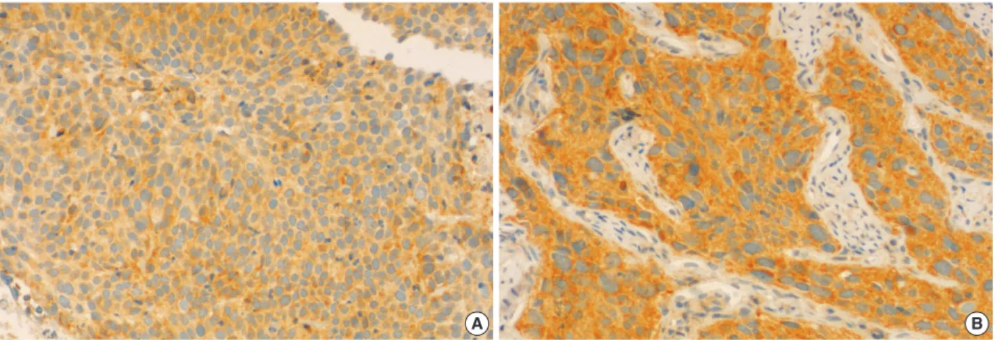
Immunohiostochemical staining was performed with a COX-2 monoclonal antibody (Cayman Chemical, Ann Arbor, MI, U.S.A.). Two pathologists analyzed the immunostaining without knowledge of the patient outcome. They evaluated the slides based on the percentage of tumor cells stained as follows: 0, totally negative; 1, <10% reactive; 2, 10-50%
reactive; 3, >50% reactive. Scores 0 and 1 were regarded as negative and scores 2 and 3 as positive. Cases with discordant results were discussed until a consensus was reached (Fig. 1).
Statistical analysis
The correlations between patient characteristics and COX- 2 expression were evaluated by the Fisher’s exact test. The relationships between variables and tumor response at mid- RT and post-RT were evaluated by the same method. The generalized estimating equation was used to verify the sig-
Fig. 1. Immunohistochemical staining of COX-2. (A) negative (<10% of distribution of immunoreactivity), (B) positive (≥10% of distribu- tion of immunoreactivity) (original magnification ×200).
A B
nificance of the change of the CR rate from mid-RT to post- RT according to the treatment modality. In addition, we used the multiple logistic regression analysis to evaluate signifi- cant predictors of CR at mid-RT and post-RT. P values of less than 0.05 were considered statistically significant. All statistical analyses were performed with the SAS� System (SAS 8.0, SAS Institute Inc., Cary, NC, U.S.A.).
RESULTS Tumor response
The mean tumor volume of all patients at pre-RT, mid-RT and post-RT was 58 mL (range 1.1-166.4 mL), 15.6 mL (range 0-132.6 mL) and 14.2 mL (range 0-96.8 mL), respectively.
Seven patients (12.3%) achieved CR at mid-RT and 39 pati- ents (68.4%) at post-RT.
COX-2 expression
COX-2 expression was positive in 43 patients (75.4%).
COX-2 expression was not related to age, hemoglobin level, initial tumor volume, the FIGO stage or treatment modali- ty (Table 1). Evaluation based on the initial tumor size showed that COX-2 positive cases were more common in patients with larger tumors (>4 cm); however, the difference was mar- ginally significant (P=0.059).
Tumor response according to COX-2 expression
At mid-RT, CR was more common in COX-2 negative patients than in COX-2 positive patients (28.6% vs. 7.0%, respectively) but the correlation was marginally significant (P=0.054). At post-RT, CR was achieved in 64.3% of COX-2 negative patients and in 69.8% of COX-2 positive patients (P=0.747).
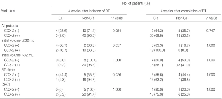
Table 2 shows the relationship between COX-2 expression and tumor response according to the initial tumor volume or treatment modality. For patients with a small pretreatment tumor volume (≤32 mL), the COX-2 negative tumors tend-
Variables No. of patients (%)
P value COX-2 (-) COX-2 (+)
Table 1. Patient characteristics according to COX-2 expression
COX-2, cyclooxygenase-2.
Variables
No. of patients (%) 4 weeks after initiation of RT
CR Non-CR P value
4 weeks after completion of RT
CR Non-CR P value
Table 2. Tumor volume responses during and after radiotherapy according to COX-2 expression
RT, radiation therapy; CR, complete response; COX-2, cyclooxygenase-2; CRCT, concurrent radiochemotherapy.
All patients
COX-2 (-) 4 (28.6) 10 (71.4) 0.054 9 (64.3) 5 (35.7) 0.747
COX-2 (+) 3 (7.0) 40 (93.0) 30 (69.8) 13 (30.2)
Initial volume ≤32 mL
COX-2 (-) 4 (66.7) 2 (33.3) 0.057 5 (83.3) 1 (16.7) 1.000
COX-2 (+) 2 (16.7) 10 (83.3) 12 (100.0) 0 (0.0)
Initial volume >32 mL
COX-2 (-) 0 (0.0) 8 (100.0) 1.000 4 (50.0) 4 (50.0) 1.000
COX-2 (+) 1 (3.2) 30 (96.8) 18 (58.1) 13 (41.9)
RT alone
COX-2 (-) 4 (44.4) 5 (55.6) 0.026 5 (55.6) 4 (44.4) 1.000
COX-2 (+) 1 (5.3) 18 (94.7) 12 (63.2) 7 (36.8)
CRCT
COX-2 (-) 0 (0) 5 (100) 1.000 4 (80.0) 1 (20.0) 1.000
COX-2 (+) 2 (8.3) 22 (91.7) 18 (75.0) 6 (25.0)
Age (yr)
≤60 5 (35.7) 22 (51.2) 0.369
>60 9 (64.3) 21 (48.8)
Hemoglobin
≤10 g/dL 2 (84.6) 12 (71.4) 0.477
>10 g/dL 11 (15.4) 30 (28.6)
Unknown 1 (50.0) 1 (50.0)
Initial tumor size
≤4 cm 8 (57.1) 12 (27.9) 0.059
>4 cm 6 (42.9) 31 (72.1)
Initial tumor volume
≤32 mL 6 (42.9) 12 (27.9) 0.334
>32 mL 8 (57.1) 31 (72.1)
FIGO stage
IB-II 8 (57.1) 32 (74.4) 0.314
III-IVa 6 (42.9) 11 (25.6)
Treatment
Radiotherapy 9 (64.3) 19 (44.2) 0.230
Radiochemotherapy 5 (35.7) 24 (55.8)
ed to achieve a higher rate of CR at mid-RT than COX-2 positive tumors (P=0.057); however, there was no difference in the CR rate at post-RT. In patients who received RT alone, COX-2 negative tumors achieved a significantly higher rate of CR than did COX-2 positive tumors at mid-RT (44.4%
vs. 5.3%, P=0.026); however, this was not observed at post- RT. The tumor response was related to COX-2 expression in neither patients with a large pretreatment tumor volume (>32 mL) nor the CRCT group.
Factors affecting the tumor response
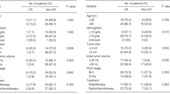
Tables 3, 4 show the relationship between the variables and tumor response during and after RT. Patients with a small pretreatment tumor volume achieved a higher rate of CR both at mid-RT (33.3% for ≤32 mL vs. 2.6% for >32 mL, P=0.003) and at post-RT (94.4% for ≤32 mL vs. 56.4% for
>32 mL, P=0.005). Patients with a small pretreatment tumor size achieved similar results at mid-RT (P=0.006) but not
at post-RT.
According to the treatment modalities, the CR rate was not different at mid-RT and post-RT. However, the CR rate increased more in the CRCT group from 6.9% at mid-RT to 75.9% at post-RT than in the RT alone group from 17.9%
to 60.7%; these findings were marginally significant (P=
0.057).
Factors affecting the tumor response in the multiple logistic regression analysis
The multivariate analysis revealed that the initial tumor volume and concurrent chemotherapy were significant pre- dictors of CR at mid-RT and post-RT (Table 5). Tumors with an initial volume >32 mL had a lower probability of CR at mid-RT (odds ratio [OR]=0.052; 95% confidence interval
[CI], 0.005-0.588) and post-RT (OR=0.037; 95% CI, 0.004- 0.379) compared to those with an initial volume ≤32 mL.
The patients treated with CRCT had a greater probability of
Variable
No. of patients (%)
P value
CR Non-CR
Table 3. Tumor response at 4 weeks after initiation of radiother- apy for all patients
CR, complete response.
Variable No. of patients (%)
P value
CR Non-CR
Table 4. Tumor response at 4 weeks after radiotherapy for all patients
CR, complete response.
Variable 4 weeks after initiation of RT
OR (95% CI) P value
4 weeks after completion of RT
OR (95% CI) P value
Table 5. Multiple logistic regression analysis of the initial tumor volume, COX-2, and concurrent chemotherapy as predictive factors of complete response
RT, radiation therapy; OR, odds ratio; CI, confidence interval; COX-2, cyclooxygenase-2.
Initial tumor volume (>32 mL vs. ≤32 mL) 0.052 (0.005-0.588) 0.017 0.037 (0.004-0.379) 0.006 COX-2 expression (positive vs. negative) 0.188 (0.023-1.504) 0.115 2.042 (0.436-9.569) 0.365 Concurrent chemotherapy (yes vs. no) 1.165 (0.127-10.709) 0.892 4.152 (1.081-15.948) 0.038
Age (yr)
≤60 19 (70.4) 8 (29.6) 0.784
>60 20 (66.7) 10 (33.3)
Hemoglobin
≤10 g/dL 8 (57.1) 6 (42.8) 0.510
>10 g/dL 29 (70.7) 12 (29.3)
Unknown 2 (100) 0 (0)
Tumor size
≤4 cm 15 (75.0) 5 (30.0) 0.555
>4 cm 24 (64.9) 13 (35.1)
Initial tumor volume
≤32 mL 17 (94.4) 1 (5.6) 0.005
>32 mL 22 (56.4) 17 (43.6)
FIGO stage
IB-II 29 (72.5) 11 (27.5) 0.359
III-IVa 10 (58.8) 7 (41.9)
Treatment
Radiotherapy 17 (60.7) 11 (39.3) 0.263
Radiochemotherapy 22 (75.9) 7 (24.1) Age (yr)
≤60 3 (11.1) 24 (88.9) 1.000
>60 4 (13.3) 26 (86.7)
Hemoglobin
≤10 g/dL 1 (7.1) 13 (92.9) 1.000
>10 g/dL 5 (12.2) 36 (87.8)
Unknown 1 (50.0) 1 (50.0)
Tumor size
≤4 cm 6 (30.0) 14 (70.0) 0.006
>4 cm 1 (2.7) 36 (97.3)
Initial tumor volume
≤32 mL 6 (33.3) 12 (66.7) 0.003
>32 mL 1 (2.6) 38 (97.4)
FIGO stage
IB-II 6 (15.0) 34 (85.0) 0.662
III-IVa 1 (5.9) 16 (94.1)
Treatment
Radiotherapy 5 (17.9) 23 (82.1) 0.253
Radiochemotherapy 2 (6.9) 27 (93.1)
CR at post-RT than did those treated with RT alone (OR=
4.152; 95% CI, 1.081-15.948). The COX-2 expression sta- tus was not related to tumor response.
DISCUSSION
The tumor volume prior to RT is a known prognostic fac- tor for cervical cancer, but Gong et al. (18) confirmed an expo- nential relationship between tumor regression rates (TRR) and time regardless of the tumor volume by estimating TRR during external beam RT with serial MRI. They suggested that tumors with a small pretreatment volume have a high- er probability of early disappearance after RT. We also found that small volume tumors achieved a higher rate of CR at mid-RT and post-RT compared to large volume tumors.
Currently, chemotherapy is used concurrently with RT to improve the treatment results for patients with locally ad- vanced cervical cancer (19, 20). Ohara et al. (21) investigat- ed the difference of TRR according to the treatment modal- ity (CRCT vs. RT alone) by fitting an exponential regression equation to the respective curve of each patient. Although TRR was not significantly different between the CRCT group (median, 0.032 per day) and the RT alone group (median, 0.024 per day), rapid TRR (>0.05 per day) was more com- mon in the CRCT group. In addition, TRR for large tumors (>5 cm in diameter) was greater in the CRCT group. In a randomized trial reported by Keys et al. (22), the rate of resid- ual disease in hysterectomy specimens at three to six weeks after treatment was lower in the CRCT group compared to the RT alone group. In our study, the CR rate was not differ- ent according to the use of chemotherapy. However, the ini- tial tumor volume of the CRCT group was significantly larger than that of the RT alone group (average 70.9 mL vs. 44.6 mL, P=0.034). Therefore, we analyzed the extent of change of the CR rate from mid-RT to after-RT, and the increase of the CR rate was prominent in the CRCT group. The CR rate at post-RT was higher in the CRCT group than in the RT alone group where the CR rate was higher at mid-RT. It is therefore reasonable to conclude that chemotherapy acceler- ated the tumor response to RT.
COX-2 overexpression has been accepted as an indepen- dent prognostic factor for recurrence and survival in patients with cervical cancer (7, 8, 10). Kim et al. (8) reported that the 5-yr overall and disease free survival was worse in COX- 2 positive patients than in COX-2 negative patients, and COX-2 overexpression was the only significant prognostic factor in the multivariate analysis. Chen et al. (10) also report- ed similar results. COX-2 overexpression was related to a reduced tumor response to radiotherapy in esophageal and rectal cancer (13, 15). For SCC of the esophagus, COX-2 neg- ative patients achieved a higher CR rate after preoperative radiochemotherapy (60% vs. 0%, P=0.01); however, this result is limited by the small number of patients and confounding
factors affecting tumor response. In rectal cancer, COX-2 over- expression in pretreatment biopsies was related to a poor res- ponse to neoadjuvant radiochemotherapy (P=0.026) (15).
Two studies investigating the correlation between COX- 2 overexpression and complete remission after RT have been reported in patients with uterine cervical cancer (14, 16). Kim et al. (14) reported that all COX-2 negative patients achieved a CR; however, 83% of COX-2 positive patients with SCC and 68% of those with adenocarcinoma achieved a CR (P<
0.001). Ishikawa et al. (16) found that COX-2 overexpres- sion before RT was related to reduced apoptosis after 9 Gy, and tumors without COX-2 overexpression achieved a higher rate of CR after RT although this was not statistically signif- icant (80% vs. 59%, P=0.12). In our study, multiple logistic regression analysis showed that the COX-2 status was not a predictive factor of a CR. Although the COX-2 status seemed to be correlated with tumor response in some selected sub- groups, the tumors that achieved a CR, at mid-RT, were sig- nificantly smaller than the tumors that did not achieve a CR (10.5±14.1 mL vs. 64.6±46.5 mL, P<0.001), Further- more, COX-2 negative tumors, in the small volume group and RT alone group, were significantly smaller than COX- 2 positive tumors in each of the subgroups. This could have caused COX-2 overexpression to be misinterpreted as a sig- nificant predictor of the tumor response. However, we can- not conclude that COX-2 does not have influence on tumor response to radiotherapy on the basis of our study results due to the retrospective study design and potential for selection bias.
Although there is no data on the change of COX-2 protein expression during radiotherapy in patients with cervical can- cer, COX-2 protein expression increased after single or frac- tionated irradiation in PC-3 cells in vitro (23, 24), and after radiotherapy for rectal cancer patients in vivo (25-29). For the patients with rectal cancer treated with preoperative radio- therapy, with or without chemotherapy, the distribution of COX-2 positive tumors or the intensity of COX-2 protein expression increased, and even the tumors without COX-2 expression newly expressed the COX-2 protein. One study reported that the initial COX-2 expression was not correlated with tumor regression (28). However, another study reported that the intensity of COX-2 protein expression, after radio- chemotherapy, was inversely correlated with tumor regres- sion (29). Therefore, COX-2 protein expression in patients with cervical cancer might also increase during radiothera- py. Investigation of the change in COX-2 expression levels during RT, by repeated biopsies, might be indicated to deter- mine the relationship between COX-2 expression and the tumor response.
In conclusion, the results of this study show no correlation between the pretreatment COX-2 expression status and the tumor response. However, the COX-2 expression status is known as an important prognostic factor in patients with cer- vical cancer based on previous studies. Therefore, further stud-
ies are needed to clarify the alteration of COX-2 expression levels during RT to determine its association with the tumor response to treatment and patient prognosis.
REFERENCES
1. Choy H, Milas L. Enhancing radiotherapy with cyclooxygenase-2 enzyme inhibitors: a rational advance? J Natl Cancer Inst 2003; 95:
1440-52.
2. Kanaoka S, Takai T, Yoshida K. Cyclooxygenase-2 and tumor biol- ogy. Adv Clin Chem 2007; 43: 59-78.
3. Fujita T, Matsui M, Takaku K, Uetake H, Ichikawa W, Taketo MM, Sugihara K. Size- and invasion-dependent increase in cyclooxyge- nase 2 levels in human colorectal carcinomas. Cancer Res 1998;
58: 4823-6.
4. Murata H, Kawano S, Tsuji S, Tsuji M, Sawaoka H, Kimura Y, Shi- ozaki H, Hori M. Cyclooxygenase-2 overexpression enhances lym- phatic invasion and metastasis in human gastric carcinoma. Am J Gastroenterol 1999; 94: 451-5.
5. Sheehan KM, Sheahan K, O’Donoghue DP, MacSweeney F, Conroy RM, Fitzgerald DJ, Murray FE. The relationship between cyclooxy- genase-2 expression and colorectal cancer. JAMA 1999; 282: 1254-7.
6. Ryu HS, Chang KH, Yang HW, Kim MS, Kwon HC, Oh KS. High cyclooxygenase-2 expression in stage IB cervical cancer with lymph node metastasis or parametrial invasion. Gynecol Oncol 2000; 76:
320-5.
7. Gaffney DK, Holden J, Zempolich K, Murphy KJ, Dicker AP, Dod- son M. Elevated COX-2 expression in cervical carcinoma: reduced cause-specific survival and pelvic control. Am J Clin Oncol 2001;
24: 443-6.
8. Kim YB, Kim GE, Cho NH, Pyo HR, Shim SJ, Chang SK, Park HC, Suh CO, Park TK, Kim BS. Overexpression of cyclooxygenase-2 is associated with a poor prognosis in patients with squamous cell car- cinoma of the uterine cervix treated with radiation and concurrent chemotherapy. Cancer 2002; 95: 531-9.
9. Nix P, Lind M, Greenman J, Stafford N, Cawkwell L. Expression of Cox-2 protein in radioresistant laryngeal cancer. Ann Oncol 2004;
15: 797-801.
10. Chen HH, Su WC, Chou CY, Guo HR, Ho SY, Que J, Lee WY. In- creased expression of nitric oxide synthase and cyclooxygenase-2 is associated with poor survival in cervical cancer treated with radio- therapy. Int J Radiat Oncol Biol Phys 2005; 63: 1093-100.
11. Khor LY, Bae K, Pollack A, Hammond ME, Grignon DJ, Venkate- san VM, Rosenthal SA, Ritter MA, Sandler HM, Hanks GE, Shipley WU, Dicker AP. COX-2 expression predicts prostate-cancer outcome:
analysis of data from the RTOG 92-02 trial. Lancet Oncol 2007; 8:
912-20.
12. Yoshikawa R, Fujiwara Y, Koishi K, Kojima S, Matsumoto T, Yanagi H, Yamamura T, Hashimoto-Tamaoki T, Nishigami T, Tsujimura T. Cyclooxygenase-2 expression after preoperative chemoradiother- apy correlates with more frequent esophageal cancer recurrence.
World J Gastroenterol 2007; 13: 2283-8.
13. Kulke MH, Odze RD, Mueller JD, Wang H, Redston M, Bertagnol-
li MM. Prognostic significance of vascular endothelial growth fac- tor and cyclooxygenase 2 expression in patients receiving preoper- ative chemoradiation for esophageal cancer. J Thorac Cardiovasc Surg 2004; 127: 1579-86.
14. Kim YB, Kim GE, Pyo HR, Cho NH, Keum KC, Lee CG, Seong J, Suh CO, Park TK. Differential cyclooxygenase-2 expression in squa- mous cell carcinoma and adenocarcinoma of the uterine cervix. Int J Radiat Oncol Biol Phys 2004; 60: 822-9.
15. Smith FM, Reynolds JV, Kay EW, Crotty P, Murphy JO, Hollywood D, Gaffney EF, Stephens RB, Kennedy MJ. COX-2 overexpression in pretreatment biopsies predicts response of rectal cancers to neoad- juvant radiochemotherapy. Int J Radiat Oncol Biol Phys 2006; 64:
466-72.
16. Ishikawa H, Ohno T, Kato S, Wakatsuki M, Iwakawa M, Ohta T, Imai T, Mitsuhashi N, Noda SE, Nakano T, Tsujii H. Cyclooxyge- nase-2 impairs treatment effects of radiotherapy for cervical cancer by inhibition of radiation-induced apoptosis. Int J Radiat Oncol Biol Phys 2006; 66: 1347-55.
17. Narayan K, McKenzie A, Fisher R, Susil B, Jobling T, Bernshaw D.
Estimation of tumor volume in cervical cancer by magnetic resonance imaging. Am J Clin Oncol 2003; 26: e163-8.
18. Gong QY, Tan LT, Romaniuk CS, Jones B, Brunt JN, Roberts N.
Determination of tumour regression rates during radiotherapy for cervical carcinoma by serial MRI: comparison of two measurement techniques and examination of intraobserver and interobserver vari- ability. Br J Radiol 1999; 72: 62-72.
19. Morris M, Eifel PJ, Lu J, Grigsby PW, Levenback C, Stevens RE, Rotman M, Gershenson DM, Mutch DG. Pelvic radiation with con- current chemotherapy compared with pelvic and para-aortic radia- tion for high-risk cervical cancer. N Engl J Med 1999; 340: 1137-43.
20. Rose PG, Bundy BN, Watkins EB, Thigpen JT, Deppe G, Maiman MA, Clarke-Pearson DL, Insalaco S. Concurrent cisplatin-based radiotherapy and chemotherapy for locally advanced cervical can- cer. N Engl J Med 1999; 340: 1144-53.
21. Ohara K, Tanaka YO, Tsunoda H, Oki A, Satoh T, Onishi K, Kagei K, Sugahara S, Hata M, Igaki H, Tokuuye K, Akine Y, Yoshikawa H. Preliminary estimation of treatment effect on uterine cervical squa- mous cell carcinoma in terms of tumor regression rate: comparison between chemoradiotherapy and radiotherapy alone. Radiat Med 2005; 23: 25-9.
22. Keys HM, Bundy BN, Stehman FB, Muderspach LI, Chafe WE, Suggs CL 3rd, Walker JL, Gersell D. Cisplatin, radiation, and adju- vant hysterectomy compared with radiation and adjuvant hysterec- tomy for bulky stage IB cervical carcinoma. N Engl J Med 1999;
340: 1154-61.
23. Steinauer KK, Gibbs I, Ning S, French JN, Armstrong J, Knox SJ.
Radiation induces upregulation of cyclooxygenase-2 (COX-2) pro- tein in PC-3 cells. Int J Radiat Oncol Biol Phys 2000; 48: 325-8.
24. Ohneseit PA, Krebiehl G, Dittmann K, Kehlbach R, Rodemann HP.
Inhibition of cyclooxygenase-2 activity by celecoxib does not lead to radiosensitization of human prostate cancer cells in vitro. Radiother Oncol 2007; 82: 229-38.
25. Watwe V, Javle M, Lawrence D, Groth J, Iyer R, El-Hajjar D, Ger- adts J. Cyclooxygenase-2 (COX-2) levels before and after chemother-
apy: a study in rectal cancer. Am J Clin Oncol 2005; 28: 560-4.
26. Debucquoy A, Goethals L, Geboes K, Roels S, McBride WH, Hau- stermans K. Molecular responses of rectal cancer to preoperative chemoradiation. Radiother Oncol 2006; 80: 172-7.
27. Kobayashi H, Hashiguchi Y, Ueno H, Shinto E, Kajiwara Y, Mochizu- ki H. Absence of cyclooxygenase-2 protein expression is a predictor of tumor regression in rectal cancer treated with preoperative short- term chemoradiotherapy. Dis Colon Rectum 2007; 50: 1354-62.
28. Bouzourene H, Yan P, Sandmeier D, Zouhair A, Matter M, Vuilleu-
mier H, Coucke P. The role of COX-2 in rectal cancer treated with preoperative radiotherapy. Virchows Arch 2008; 452: 499-505.
29. Debucquoy A, Libbrecht L, Roobrouck V, Goethals L, McBride W, Haustermans K. Morphological features and molecular markers in rectal cancer from 95 patients included in the European Organisa- tion for Research and Treatment of Cancer 22921 trial: prognostic value and effects of preoperative radio (chemo) therapy. Eur J Can- cer 2008; 44: 791-7.