http://dx.doi.org/10.5624/isd.2015.45.3.199
Multiple myeloma is a systemic clonal proliferation of plasma cells in the bone marrow that characteristically se- crete a monoclonal immunoglobulin.1 Multiple myeloma is one of the three clinically distinct plasma cell disorders, as well as solitary plasmacytoma of the bone and extra- medullary plasmacytoma.2,3
Multiple bone involvement is common, with conversion to multiple myeloma occurring at least several years after the initial development of solitary plasmacytoma of the bone.4 The most common clinical signs and symptoms of multiple myeloma are bone pain or swelling in localized areas, fatigue, and anemia.5 Radiographic examinations usually reveal generalized bone rarefaction with osteopo- rotic changes, as well as punched-out lesions involving multiple bones.6 Jaw involvement is common and often
occurs in the advanced stages of the disease.7,8
Bisphosphonates are important drugs in the treatment of multiple myeloma.9 They are able to inhibit the progres- sion of the disease and can even prolong survival.10 How- ever, in the case of dental infections, they can lead to bis- phosphonate-related osteonecrosis of the jaw(BRONJ).11 Additionally, tooth extractions have been shown to in- crease the risk of BRONJ in individuals taking bisphos- phonates.12
This report describes the characteristic radiographic features of BRONJ in a patient with multiple myeloma, in whom the administration of bisphosphonates and tooth extraction were likely the etiologic factors of BRONJ.
Case Report
A 59-year-old male visited the Wonkwang Dental Hos- pital complaining of tooth pain around the left maxillary molars. An intraoral examination revealed deep dental caries in the second and third molars and moderate mo-
Bisphosphonate-related osteonecrosis of the jaw in a multiple myeloma patient: A case report with characteristic radiographic features
Byung-Do Lee1,*, Moo-Rim Park2, Kyung-Hwan Kwon3
1Department of Oral and Maxillofacial Radiology and Wonkwang Dental Research Institute, College of Dentistry, Wonkwang University, Iksan, Korea
2Department of Internal Medicine, School of Medicine, Wonkwang University, Iksan, Korea
3Department of Oral and Maxillofacial Surgery, College of Dentistry, Wonkwang University, Iksan, Korea
ABstRACt
A 59-year-old male who had suffered from multiple myeloma for nine years and had been administered bispho- sphonates for seven years visited a dental hospital for pain relief due to extensive caries in his left maxillary molars.
The molars were extracted, leaving an exposed wound for three months. The radiograph showed sequestra formation and irregular bone destruction in the left maxilla. Sudden pain and gingival swelling in the right mandibular molar area occurred six months later. The interseptum of the right lower second molar was observed to be necrotic during surgery. These findings coincided with the features of bisphosphonate-related osteonecrosis of the jaw(BRONJ).
In this case, the long intravenous administration of bisphosphonates and tooth extraction were likely the etiologic factors of BRONJ in a patient with multiple myeloma; moreover, the bilateral occurrence of BRONJ is a character- istic feature.(Imaging Sci Dent 2015; 45: 199-203)
Key woRds: Multiple Myeloma; Bisphosphonate-Associated Osteonecrosis of the Jaw; Tooth Extraction
Copyright ⓒ 2015 by Korean Academy of Oral and Maxillofacial Radiology
This is an Open Access article distributed under the terms of the Creative Commons Attribution Non-Commercial License(http://creativecommons.org/licenses/by-nc/3.0) which permits unrestricted non-commercial use, distribution, and reproduction in any medium, provided the original work is properly cited.
Imaging Science in Dentistry·pISSN 2233-7822 eISSN 2233-7830
*This study was supported by Wonkwang University in 2013.
Received March 14, 2015; Revised May 11, 2015; Accepted May 24, 2015
*Correspondence to : Prof. Byung-Do Lee
Department of Oral and Maxillofacial Radiology, College of Dentistry, Wonkwang University, Iksan-daero 460, Iksan, Jeonbuk 570-749, Korea
Tel) 82-63-859-2912, Fax) 82-63-857-4002, E-mail) [email protected]
bility of the third molar. Panoramic and periapical radio- graphs showed a carious lesion on the teeth extending into the subgingival area, but the radiographs did not show a bony destructive lesion in the left maxillary posterior re- gion(Fig. 1). Since the endodontic treatment of subgingi- val carious lesions cannot be guaranteed to be successful, extraction was considered. However, BRONJ was a con- cern because the patient had been administered pamidro- nate for two years, followed by zoledronate for five years, to treat multiple myeloma. Moreover, the patient did not accept extraction as a treatment option, so tooth extraction was postponed. This patient had been diagnosed with mul- tiple myeloma(IgG kappa type) nine years previously and
had undergone multimodal examinations and treatment at the Wonkwang Medical Hospital. The diagnosis of multi- ple myeloma was based on multiple tests, including serum
Fig. 3. Skull radiographs show a punched-out appearance.
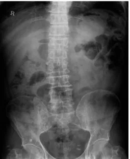
Fig. 2. Compression fracture of T12 and diffuse osteopenia of the pelvic bone.
Fig. 4. At 50 days after extraction, pus discharge from the wound and sequestra formation are observed.
Fig. 1. A panoramic radiograph (A) and periapical radiograph (B) show a carious lesion on the maxillary first, second, and third molars ex- tending into the subgingival area, with a suspected mucosal antral cyst in the left maxillary sinus.
A B
and urine protein electrophoresis, bone marrow biopsy, and radiographs of the bones commonly involved in mul- tiple myeloma. Plain pelvic radiographs showed diffuse decreased bone density and a compression fracture(wedge deformity) of T12(Fig. 2). Skull radiographs showed a characteristic punched-out appearance(Fig. 3). The pa- tient was initially treated with melphalan-based high-dose chemotherapy and autologous stem cell transplantation followed by interferon maintenance therapy. Six years later, he relapsed and was retreated with bortezomib and dexamethasone chemotherapy. Bisphosphonates had been administrated for seven years.
The patient returned to our emergency dental hospital four months later because he suddenly suffered from ext- reme pain due to a vertical fracture of the left maxillary second molar. The left second and third molars were ex- tracted under local anesthesia. Unfortunately, the extrac- tion resulted in a perforation of the left maxillary sinus floor. Ten days after the tooth extraction, the patient com- plained of discomfort and occasional pain around the extraction wound. Approximately 50 days after the ex- traction, pus discharge was observed from the wound (Fig. 4), and the patient was tentatively diagnosed with BRONJ. We continued to administer antibiotics and anal- gesics, in addition to saline rinsing of the wound.
A panoramic radiograph and computed tomograph per-
formed 11 months later showed the pathologic bony frac- ture, sequestra formation, and irregular bone destruction (Fig. 5). Chronic maxillary sinusitis was also observed in the left maxillary sinus. The premolars and first molar were extracted, followed by the surgical removal of nec- rotized bone fragments and alveoloplasty(Fig. 6), and the patient underwent continuous follow-up. After the remov- al of the necrotic bone and the surrounding tissue, the symptoms were relieved. Sudden pain and gingival swell-
Fig. 5. A panoramic radiograph (A) and a computed tomograph (B) reveal the separation of the alveolar ridge from the surrounding maxil- lary bone, sequestra, and irregular bone destruction. Chronic sinusitis is seen in the left maxillary sinus.
A
B
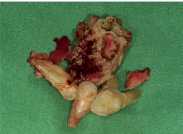
Fig. 6. A photograph shows the extracted premolars, the first mo- lar, and a necrotized bone fragment.
ing occurred in the right mandibular posterior teeth six months later. An intraoral examination showed gingival swelling around the second and third molars. A periapical radiograph showed only moderate periodontitis and prox- imal caries on the distal surface of the first molar, but no specific finding of osteolysis related to osteonecrosis around the root surface was observed(Fig. 7). The inter- septum of the right lower second molar was observed to be necrotic during surgery(Fig. 8). The diagnosis of BRONJ in the left maxilla and right mandible in a multiple my- eloma patient was made based on these clinical and radio- graphic findings.
discussion
The diagnosis of multiple myeloma is based on several criteria, including the identification of an atypical plasma cell population on biopsy, abnormalities of immunoglob- ulin production, and evidence of end-organ damage, such
as osteolytic bone lesions, anemia, hypercalcemia, or renal insufficiency.13 Multiple myeloma is common in patients older than 40 years of age, with a peak incidence rate at 60-70 years, and is rare in people younger than 30 years of age.14 This patient was diagnosed at 50 years of age.
The initial laboratory findings were 3.14g/dL of serum M protein, clonal plasma cells comprising more than 10%
of the bone marrow, and extensive osteolytic bone lesions without anemia, hypercalcemia, or renal insufficiency.
Therefore, the patient was diagnosed with multiple my- eloma of the IgG kappa type at Durie-Salmon stage IIIA.
Multiple myeloma commonly shows numerous punched- out areas of radiolucency on plain radiographs.15 The os- teolytic lesions of multiple myeloma are most commonly observed in the pelvis, spine, ribs, and skull.16 Radiogra- phic evidence of multiple myeloma in the jaw bones usu- ally appears after extensive involvement of other bones, particularly the skull.8,17 The mandible is more commonly affected by multiple myeloma than the maxilla, and the posterior region is more commonly involved than the an- terior region.18 In this case, the characteristic radiographic features of multiple myeloma were the punched-out app- earance of the skull, the absence of detectable jaw lesions, and osteoporotic changes in the spine and pelvis. This presentation was similar to what has been described in other studies.17 Interestingly, the jaw lesion, including the maxilla and mandible, did not show the characteristic bony rarefaction of multiple myeloma despite having existed for nine years.
BRONJ is a severe complication of bisphosphonate therapy.9 Because of the prolonged duration of bisphos- phonate treatment in multiple myeloma patients, BRONJ has frequently been observed in patients with multiple myeloma. With the increasing use of the bisphosphonate
Fig. 8. A photograph shows the necrotic interseptum of the right lower second molar.
Fig. 7. A. An intraoral photograph shows gingival swelling around the lower second and third molars. B. A periapical radiograph shows mod- erate periodontitis and proximal caries on the distal surface of the first molar.
A B
class of drugs, dental professionals are likely to encounter more cases of BRONJ.19
The diagnostic criteria for BRONJ developed by the American Association of Oral and Maxillofacial Surgeon (AAOMS) include a history of bisphosphonate use, no history of radiotherapy, and the presentation of exposed, necrotic jaw bone for more than eight weeks.20 Dental procedures or traumatic injuries are another significant factor contributing to the likelihood of osteonecrosis in the jaw.21 Theoretically, trauma to the mucosa and the ex- posure of bone and surrounding tissues to microbial flora creates an acidic inflammatory milieu.19
In this case, the exposed bone area of the left maxilla was relatively large. The left maxilla had a pathologic bony fracture, sequestra formation, and irregular bone de- struction, which are characteristic features of BRONJ on computed tomography. These radiographic and clinical features corresponded to the AAOMS criteria for BRONJ.
BRONJ of the left maxilla occurred after tooth extraction, suggesting that an invasive dental procedure and the long administration of bisphosphonates were likely the etiolo- gic factors. The right lesion occurred spontaneously, with the most likely etiologic factor being the prolonged ad- ministration of bisphosphonates.
In this case report, the characteristic radiographic fea- tures of multiple myeloma and BRONJ were presented.
Interestingly, the maxillary and mandibular lesion showed no bony rarefaction, which is characteristic of multiple myeloma, but did show an osteonecrotic lesion related to bisphosphonate administration.
References
1. Kyle RA. Multiple myeloma: review of 869 cases. Mayo Clin Proc 1975; 50: 29-40.
2. Batsakis JG. Pathology consultation. Plasma cell tumors of the head and neck. Ann Otol Rhinol Laryngol 1983; 92: 311-3.
3. Wingo PA, Tong T, Bolden S. Cancer statistics, 1995. CA Cancer J Clin 1995; 45: 8-30.
4. Holland J, Trenkner DA, Wasserman TH, Fineberg B. Plasma- cytoma. Treatment results and conversion to myeloma. Cancer 1992; 69: 1513-7.
5. Kyle RA. Diagnostic criteria of multiple myeloma. Hematol Oncol Clin North Am 1992; 6: 347-58.
6. Lee SH, Huang JJ, Pan WL, Chan CP. Gingival mass as the primary manifestation of multiple myeloma: report of two cases. Oral Surg Oral Med Oral Pathol Oral Radiol Endod 1996; 82: 75-9.
7. Epstein JB, Voss NJ, Stevenson-Moore P. Maxillofacial man-
ifestations of multiple myeloma. An unusual case and review of the literature. Oral Surg Oral Med Oral Pathol 1984; 57:
267-71.
8. Lambertenghi-Deliliers G, Bruno E, Cortelezzi A, Fumagalli L, Morosini A. Incidence of jaw lesions in 193 patients with multiple myeloma. Oral Surg Oral Med Oral Pathol 1988; 65:
533-7.
9. Mondello P, Pitini V, Arrigo C, Mondello S, Mian M, Altavilla G. Necrotizing fasciitis as a rare complication of osteonecro- sis of the jaw in a patient with multiple myeloma treated with lenalidomide: case report and review of the literature. Spring- erplus 2014; 3: 123.
10. Coleman R, Gnant M, Morgan G, Clezardin P. Effects of bone-targeted agents on cancer progression and mortality. J Natl Cancer Inst 2012; 104: 1059-67.
11. Lee SH, Chan RC, Chang SS, Tan YL, Chang KH, Lee MC, et al. Use of bisphosphonates and the risk of osteonecrosis among cancer patients: a systemic review and meta-analysis of the observational studies. Support Care Cancer 2014; 22:
553-60.
12. Utreja A, Almas K, Javed F. Dental extraction as a risk factor for bisphosphonate related osteonecrosis of the jaw in cancer patients: an update. Odontostomatol Trop 2013; 36: 38-46.
13. Sukpanichnant S, Cousar JB, Leelasiri A, Graber SE, Greer JP, Collins RD. Diagnostic criteria and histologic grading in multiple myeloma: histologic and immunohistologic analysis of 176 cases with clinical correlation. Hum Pathol 1994; 25:
308-18.
14. Bladé J, Kyle RA, Greipp PR. Multiple myeloma in patients younger than 30 years. Report of 10 cases and review of the literature. Arch Intern Med 1996; 156: 1463-8.
15. Matsumura S, Kishino M, Ishida T, Furukawa S. Radiographic findings for solitary plasmacytoma of the bone in the anterior wall of the maxillary sinus: a case report. Oral Surg Oral Med Oral Pathol Oral Radiol Endod 2000; 89: 651-7.
16. Croucher PI, Apperley JF. Bone disease in multiple myeloma.
Br J Haematol 1998; 103: 902-10.
17. Mozaffari E, Mupparapu M, Otis L. Undiagnosed multiple myeloma causing extensive dental bleeding: report of a case and review. Oral Surg Oral Med Oral Pathol Oral Radiol En- dod 2002; 94: 448-53.
18. Yaegaki K, Kameyama T, Takenaka M, Kimura T, Sujaku C, Tanimura A. Myelomatosis(IgD, lambda) discovered by oral manifestation. Int J Oral Surg 1985; 14: 381-4.
19. Badros A, Weikel D, Salama A, Goloubeva O, Schneider A, Rapoport A. Osteonecrosis of the jaw in multiple myeloma patients: clinical features and risk factors. J Clin Oncol 2006;
24: 945-52.
20. Advisory Task Force on Bisphosphonate-Related Ostenone- crosis of the Jaws, American Association of Oral and Maxil- lofacial Surgeons. American Association of Oral and Maxil- lofacial Surgeons position paper on bisphosphonate-related osteonecrosis of the jaws. J Oral Maxillofac Surg 2007; 65:
369-76.
21. Assael LA. New foundations in understanding osteonecrosis of the jaws. J Oral Maxillofac Surg 2004; 62: 125-6.