Introduction
Injury to the inferior alveolar nerve bundle is a serious complication following the extraction of impacted mandi- bular third molars. Most reports have demonstrated an in- cidence range from 0.5% to 8%.1In order to avoid infe- rior alveolar nerve injury, it is critical to evaluate the proxi- mity of the mandibular canal to the third molar preopera- tively. In the literature, several signs have been defined on panoramic radiographs, which indicate the close pro- ximity of the mandibular canal to the third molar. Nota- bly, four radiographic signs - darkening of the root, inter- ruption of the cortical lines, diversion of the mandibular
canal, and narrowing of the root - are recognized to be associated with inferior alveolar nerve proximity to the mandibular third molar on panoramic radiography.2
Although the incidence of this proximity is low, the complications can be serious, justifying more precise pre- operative evaluation.3 Several aspects of the clinical use- fulness of cone beam computed tomography (CBCT) have been discussed.4,5However, patients are sometimes unne- cessarily exposed to radiation even though conventional evaluation techniques are of high diagnostic value. There- fore, this study was designed to assess the reliability of panoramic radiographs in detecting radiographic signs of inferior alveolar nerve proximity evidenced on CBCT images.
Materials and Methods
This cross-sectional study was performed using panora-
Comparison of panoramic radiography with cone beam CT in predicting the relationship of the mandibular third molar roots to the alveolar canal
Shoaleh Shahidi, Barbod Zamiri*, Pegah Bronoosh**
Biomaterial Research Center, Department of Oral and Maxillofacial Radiology, School of Dentistry, Shiraz University of Medical Sciences, Shiraz, Iran
*Department of Oral and Maxillofacial Surgery, School of Dentistry, Shiraz University of Medical Sciences, Shiraz, Iran
**Department of Oral and Maxillofacial Radiology, School of Dentistry, Shiraz University of Medical Sciences, Shiraz, Iran ABSTRACT
Purpose: Preoperative radiographic assessment of the mandibular third molars is essential to prevent inferior alveolar nerve damage during extraction. The purpose of this study was to assess the reliability of panoramic signs of association between the roots of teeth and the canal, and to compare the panoramic signs with cone beam computed tomography (CBCT) findings.
Materials and Methods: CBCT images of 132 impacted mandibular third molars were evaluated to determine the association of the root to the canal. The CBCT findings were compared with the corresponding panoramic images.
Logistic regression analysis was used to define the diagnostic criteria of the panoramic images.
Results: Among the panoramic signs, loss of the cortical line was the most frequent radiographic sign predicting association (sensitivity: 79.31). Contact of the tooth with the canal was observed in all cases in which the loss of cortical line of the canal or darkening of the roots was found on the panoramic radiographs.
Conclusion: Darkening of the roots and loss of the cortical line on panoramic radiographs might be highly suggestive of the risk of nerve injury. (Imaging Sci Dent 2013; 43: 105-9)
KEY WORDS: Cone-Beam Computed Tomography; Radiography, Panoramic; Molar, Third; Mandibular Nerve
Received September 26, 2012; Revised November 23, 2012; Accepted January 24, 2013 Correspondence to : Dr. Pegah Bronoosh
Department of Oral and Maxillofacial Radiology, School of Dentistry, Shiraz Uni- versity of Medical Sciences, Qasrodasht St., Mehr Cross, Shiraz 7195615878, Iran Tel) 98-711-6263192, Fax) 98-711-6270325, E-mail) [email protected]
Copyright ⓒ 2013 by Korean Academy of Oral and Maxillofacial Radiology
This is an Open Access article distributed under the terms of the Creative Commons Attribution Non-Commercial License (http://creativecommons.org/licenses/by-nc/3.0) which permits unrestricted non-commercial use, distribution, and reproduction in any medium, provided the original work is properly cited.
Imaging Science in Dentistry∙pISSN 2233-7822 eISSN 2233-7830
mic and CBCT images of 96 patients referred by maxillo- facial surgeons whose panoramic features suggested a close relationship between the inferior alveolar canal (IAC) and third molar root. The approval of the Ethical Board of the Institutional Ethics Committee of Shiraz University of Medical Sciences was obtained before conducting this study.
One hundred and thirty-two impacted mandibular third
molars were included in the study. The patient group con- sisted of 50 females and 46 males with a mean age of 25.41 years. The panoramic radiographs were taken using a Pro- line XC unit (Planmeca, Helsinki, Finland) using a 15 cm
×30 cm photostimulable phosphor receptor (Regius 110, Konica Minolta Medical & Graphic, Inc., Tokyo, Japan).
The CBCT images were obtained with a Kodak 9000 (Car- estream Health, Inc., Rochester, NY, USA) at 90 kVp, 6
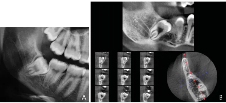
Fig. 1.A. Cropped panoramic radiograph shows the interruption of the superior wall of the mandibular canal due to the root of the left impacted mandibular third molar. B. CBCT image of same tooth demonstrates the direct association of the root and canal.
A B
Fig. 2. A. Cropped panoramic radiograph shows the darkening of both roots of the right impacted mandibular third molar. B. CBCT images of the same tooth show direct contact between the root and canal.
A B
mA, and 10 seconds. A maxillofacial radiologist with more than ten years of experience evaluated the panoramic and CBCT images independently to determine the topogra- phic relationship between the impacted third molar and the mandibular canal.
For the CBCT images, the presence or absence of direct contact between the tooth root and the canal contents was three-dimensionally evaluated, which was used as the diagnostic criterion in predicting neurovascular bundle exposure. Direct contact was considered to be present when loss of canal cortical bone between the two struc- tures was observed on axial, panoramic, and cross-sectio- nal images. Moreover, any root dilacerations, buccal or lingual cortex perforation, and canal diversion were noted for each case.
The presence or absence of direct contact on the panora- mic radiographs was evaluated by the following four fea- tures, all of which have been reported to be suggestive of close contact between the tooth root and the mandibular canal: (a) interruption of the mandibular canal wall, (b) darkening of the root, (c) diversion of the mandibular canal, and (d) root dilaceration.
The sensitivity, specificity, positive predictive value (PPV), and negative predictive value (NPV) of each pan- oramic feature in depicting the association of the root on CBCT images were calculated. The diagnostic criterion for panoramic images was defined using logistic regres- sion analysis to determine the independent predictive value of the four panoramic features listed in the afore- mentioned paragraph.
Results
The study sample consisted of 132 impacted third molars from 96 patients (50 females and 46 males). The most frequent finding by CBCT was a lingual course of the mandibular canal.
Contact of the tooth root with the canal was observed in all cases in which a loss of the cortical line of the canal or darkening of the roots was found on the panoramic radio- graphs (Fig. 1). Loss of the cortical line in the panoramic signs showed the highest sensitivity and was the most fre- quent radiographic sign for predicting association (Table 1).
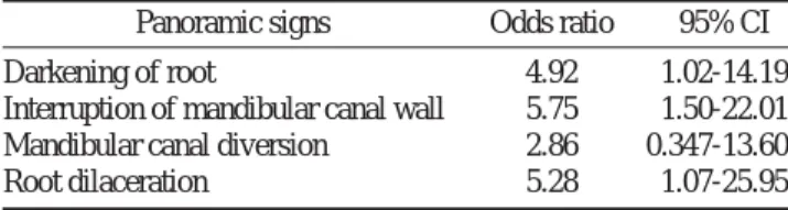
When darkening of the root was seen on a panoramic view, the risk of association in CBCT images was 4.92 times more (Table 2, Fig. 2). However, when darkening of root was detected on panoramic view, the risk of cortex perforation in CBCT images was just 2.8 times greater
(CI==1.18-6.62). When an interrupted border of the man- dibular canal was detected on panoramic radiography, the risk of association in CBCT images was 5.75 times grea- ter. Root dilacerations on panoramic radiography raised the chance of association in CBCT images to 5.28 times greater (Table 2).
Discussion
Although CBCT provides a better evaluation of anato- mical structures and greater intra-operative safety, pano- ramic radiography is still the most widely used technique for assessing the relationship of the third molar root and canal. Several clinical studies have determined the speci- fic radiographic signs detected on panoramic radiographs to be suggestive of a close relationship between the third molars and mandibular canal.2,6,8,11
The results of this study showed that four radiographic signs of proximity on panoramic radiography could be used to predict the association of the canal and root altho- ugh each was of different value.
Interruption of the cortical lines on a panoramic image raised the risk of root and canal contact on CBCT images (odds ratio==5.75) and showed the highest sensitivity among the four signs of association. This result was in accordance with the studies by Nakagawa et al1and Jung et al6 and indicated that the risk of contact between the mandibular canal and the third molar root was enhanced when the white line of the canal was not visible on pano- ramic radiographs.
Darkening of the third molar roots where the mandi-
Table 1.Relationship between signs on panoramic radiography and association of the root to the inferior alveolar nerve
Panoramic signs Sensitivity Specificity PPV NPV Darkening of root 55.17 80.00 96.97 13.33
Interruption of 79.31 60.00 95.83 20.00
mandibular canal wall
Mandibular canal diversion 24.14 80.00 93.33 83.30 Root dilaceration 56.90 80.00 97.06 13.79 PPV: positive predictive value, NPV: negative predictive value
Table 2.Logistic regression analysis of panoramic features Panoramic signs Odds ratio 95% CI
Darkening of root 4.92 1.02-14.19
Interruption of mandibular canal wall 5.75 1.50-22.01 Mandibular canal diversion 2.86 0.347-13.60
Root dilaceration 5.28 1.07-25.95
bular canal was superimposed has been reported to be suggestive of an intimate relationship between the root and canal in several studies.7-10Some others believed that this radiographic sign indicated thinning or perforation of the lingual cortical plate.6,11 The results of the present study showed that the risk of the third molar root contact with the canal in the cases wherein the panoramic sign of the darkening of the roots was observed was higher than in cases of lingual cortical plate perforation (odds ratio== 4.92 and 2.86, respectively). In contrast to the results of Tantanapornkul et al11 and Jung et al,6our study sample consisted mostly of cases in which the canal was located lingual to the roots. This might explain the reason that less lingual cortical plate perforation was detected in the present study. However, it also emphasizes the importance of using an advanced imaging technique to explore whe- ther darkening of the third molar root is related to canal contact or cortical plate perforation.
The probability of molar root association with the mandi- bular canal in the presence of related radiographic signs, known as positive predictive value (PPV), was highest for dilaceration and darkening of the roots in our study. The negative predictive value (NPV) is the probability of no root association with the canal when these radiographic signs are absent; and it was low for all signs except for canal diversion.
Mandibular canal diversion had the least sensitivity to predicting association, which was in contrast with previ- ously published studies.3,8,12This difference could be due to patient selection in our study. Since the referred pati- ents were suspected of a relationship between the canal and root on panoramic radiography, cases which had a sign of a change in the path of the canal might have been less likely to be referred. However, our findings were in agreement with the study of Neves et al13in which man- dibular canal diversion had lower importance in predict- ing the association of the canal and root.
In the present study, root dilaceration showed a higher sensitivity than mandibular canal diversion in predicting the association of the canal and root. This finding sug- gested the possibility of root displacement during the maturation of adjacent anatomic structures rather than the alveolar canal. Detecting root dilaceration might affect surgical planning for the mandibular third molars.
Although the absence of cortical bone in CBCT is highly suggestive of nerve exposure, which has been related with an increased risk of nerve damage, reports indicate a 20% risk of postsurgical paresthesia, with a 70% chance of recovery by one year after surgery.14This recovery rate
showed that anatomical contact was not the sole deter- mining factor for postsurgical prognosis. The association of the inferior alveolar nerve with the root could be clearly visualized using magnetic resonance imaging.15 Further studies are recommended to assess to what extent radio- graphic findings of nerve proximity can predict postsur- gical complications. However, patients with a detected association of the canal to the root should be considered at high risk of nerve damage.
References
1. Nakagawa Y, Ishii H, Nomura Y, Watanabe NY, Hoshiba D, Kobayashi K, et al. Third molar position: reliability of pano- ramic radiography. J Oral Maxillofac Surg 2007; 65: 1303-8.
2. Nakayama K, Nonoyama M, Takaki Y, Kagawa T, Yuasa K, Izumi K, et al. Assessment of the relationship between impact- ed mandibular third molars and inferior alveolar nerve with dental 3-dimensional computed tomography. J Oral Maxillofac Surg 2009; 67: 2587-91.
3. Blaeser BF, August MA, Donoff RB, Kaban LB, Dodson TB.
Panoramic radiographic risk factors for inferior alveolar nerve injury after third molar extraction. J Oral Maxillofac Surg 2003; 61: 417-21.
4. Tyndall DA, Kohltfarber H. Application of cone beam volume- tric tomography in endodontics. Aust Dent J 2012; 57 Suppl 1:
72-81.
5. Ahmad M, Jenny J, Downie M. Application of cone beam computed tomography in oral and maxillofacial surgery. Aust Dent J 2012; 57 Suppl 1: 82-94.
6. Jung YH, Nah KS, Cho BH. Correlation of panoramic radio- graphs and cone beam computed tomography in the assess- ment of a superimposed relationship between the mandibular canal and impacted third molars. Imaging Sci Dent 2012; 42:
121-7.
7. Sedaghatfar M, August MA, Dodson TB. Panoramic radio- graphic findings as predictors of inferior alveolar nerve exposure following third molar extraction. J Oral Maxillofac Surg 2005;
63: 3-7.
8. Monaco G, Montevecchi M, Bonetti GA, Gatto MR, Checchi L. Reliability of panoramic radiography in evaluating the topo- graphic relationship between the mandibular canal and impact- ed third molars. J Am Dent Assoc 2004; 135: 312-8.
9. de Melo Albert DG, Gomes AC, do Egito Vasconcelos BC, de Oliveira e Silva ED, Holanda GZ. Comparison of orthopan- tomographs and conventional tomography images for assess- ing the relationship between impacted lower third molars and the mandibular canal. J Oral Maxillofac Surg 2006; 64: 1030-7.
10. Bell GW. Use of dental panoramic tomographs to predict the relation between mandibular third molar teeth and the inferior alveolar nerve. Radiological and surgical findings, and clinical outcome. Br J Oral Maxillofac Surg 2004; 42: 21-7.
11. Tantanapornkul W, Okochi K, Bhakdinaronk A, Ohbayashi N, Kurabayashi T. Correlation of darkening of impacted man- dibular third molar root on digital panoramic images with cone beam computed tomography findings. Dentomaxillofac Radiol
2009; 38: 11-6.
12. Gomes AC, Vasconcelos BC, Silva ED, Caldas Ade F Jr, Pita Neto IC. Sensitivity and specificity of pantomography to pre- dict inferior alveolar nerve damage during extraction of impact- ed lower third molars. J Oral Maxillofac Surg 2008; 66: 256- 9.
13. Neves FS, Souza TC, Almeida SM, Haiter-Neto F, Freitas DQ, Bóscolo FN. Correlation of panoramic radiography and cone beam CT findings in the assessment of the relationship bet-
ween impacted mandibular third molars and the mandibular canal. Dentomaxillofac Radiol 2012; 41: 553-7.
14. Tay AB, Go WS. Effect of exposed inferior alveolar neurova- scular bundle during surgical removal of impacted lower third molars. J Oral Maxillofac Surg 2004; 62: 592-600.
15. Ferretti F, Malventi M, Malasoma R. Dental magnetic reson- ance imaging: study of impacted mandibular third molars.
Dentomaxillofac Radiol 2009; 38: 387-92.