Efficacy of a Paclitaxel-Eluting Nitinol Stent on the Inhibition of Pseudointimal Hyperplasia in a Transjugular Intrahepatic
Portosystemic Shunt: An Experimental Study in a Swine Model1
Tae-Seok Seo, M.D., PhD2, Joo-Hyeong Oh, M.D., PhD1, Se Hwan Kwon, M.D.1, Young-Koo Park, M.D., PhD3, Ho-Young Song, M.D., PhD 4, Sun-Hong Yuk, PhD5
1Departments of Diagnostic Radiology and 3Pathology, Kyung Hee University Hospital, Seoul, Korea
2 Department of Radiology, Korea University Guro Hospital, Seoul
4Department of Radiology, University of Ulsan, College of Medicine, Seoul
5Department of Polymer Science and Engineering, Hannam University, Taejon
Supported by a grant from the Research Fund, Ministry of Health and Welfare, Republic of Korea Received November 9, 2006 ; Accepted January 2, 2007
Address reprint requests to : Joo-Hyeong Oh, M.D., Department of Diagnostic Radiology, Kyung Hee University Hospital, Hoeki-dong #1, Dongdaemun-gu, Seoul 130-702, Korea
Tel. 82-2-958-8619 Fax. 82-2-968-0787 E-mail: [email protected]
Purpose: To evaluate the efficacy of a paclitaxel-eluting nitinol stent on the inhibition of pseudointimal hyperplasia in a transjugular intrahepatic portosystemic shunt.
Materials and Methods: Twelve pigs were used in this study. Two types of 10-mm di- ameter and 60-mm long nitinol stents were made for a transjugular intrahepatic por- tosystemic shunt by coating them with a polyurethane solution, with and without pa- clitaxel. Each transjugular intrahepatic portosystemic shunt was created successfully in the 12 swine with 7 paclitaxel-eluting stents and 5 polyurethane stents. Five swine in each group were followed-up for 14 days due to the death of 2 swine given the pacli- taxel-eluting stents. The proliferation of the pseudointima was evaluated on both fol- low-up portograms and histopathology examinations. The mean maximum pseudointi- mal hyperplasia is expressed as the percentage of the stent radius.
Results: On the portograms, all the transjugular intrahepatic portosystemic shunts us- ing the paclitaxel-eluting stents maintained patency despite there being a complete oc- clusion of the polyurethane stents in all the animals. The histopathology analysis re- vealed the mean maximum pseudointimal hyperplasia to be 25% and 76% in the pacli- taxel-eluting and control stents, respectively.
Conclusion: A transjugular intrahepatic portosystemic shunt with a paclitaxel-eluting nitinol stent appears to significantly inhibit the formation of pseudointimal hyperpla- sia.
Index words :Interventional procedures, experimental studies Shunts, portosystemic
Stents and prostheses Drugs
A transjugular intrahepatic portosystemic shunt (TIPS) is a safe, effective, and commonly used method for treating the complications of portal hypertension in pa- tients with cirrhosis of the liver. However, the follow-up data on the patency of TIPS has been disappointing be- cause of the high rate of stenosis. Previous studies have reported the 1-year primary patency rates after the cre- ation of a TIPS to range from 22% to 50% (1-4). In addi- tion, it is often associated with the development of pseudointimal hyperplasia within the lumen of the he- patic parenchymal tract.
Restenosis is one of the problems that can occur after placing a stent in a vessel, and is mainly related to an in- flammatory response to an injury from the procedure it- self and a remodeling of the vessel wall that is caused by the sustained force of the stent on the arterial wall (5, 6).
In Several drug-eluting stents with anti-inflammatory and immunosuppressive effects have been developed to overcome this problem, which have been applied clini- cally in vessels such as the coronary arteries. Recently, some studies have reported that a paclitaxel-eluting stent (PES) is effective in reducing the level of myointi- mal proliferation in vessels (7-9).
However, there are few reports on the implantation of drug-eluting stents in local drug delivery in a TIPS and no study using PES in the TIPS tract (10). We hypothe- sized that the paclitaxel released from PES would inhibit the pseudointimal hyperplasia in the TIPS tract and im- prove the patency. The PES was manufactured and an experimental study was performed using them in a swine model. The aim of this study was to evaluate the feasibility and efficacy of a paclitaxel-eluting nitinol stent in reducing the level of pseudointimal hyperplasia in the TIPS tract of a swine model with induced-portal hypertension.
Materials and Methods
Stent Construction
Self-expandable nitinol stents (Hercules vascular stentⓇ, S&G Biotech Inc.; Seongnam, Korea) were used in these experiments. When fully expanded, the stents were 60 mm long and 10 mm in diameter. A PES was created by dipping an expanded bare stent into a solution contain- ing paclitaxel and polyurethane (PU), followed by dry- ing in a clean room. The paclitaxel concentration in the solution was 12%. For the control study, a nitinol stent was coated in a PU solution without paclitaxel using the same method used with the PES. The PES and PU stents
were loaded again in their own introducer sets with an outer diameter of size 8-F .
In-vitro Evaluation of Paclitaxel Released from Paclitaxel- PU membrane
In order to determine the amount of paclitaxel re- leased from the paclitaxel and PU mixture, a membrane was made from the solution, and was soaked in a tube containing a 0.1 M neutral phosphate buffer solution, and placed in a shaking incubator. The tube was contin- uously rotated in an incubator at 37℃. The buffer solu- tion was exchanged every 24 hours, and the amount of paclitaxel released in the buffer solution was measured using high performance liquid chromatography (Rainin Instrument Co.; Wolburn, MA, U.S.A.) for 2 weeks. The wavelength required to detect paclitaxel was 235 nm, which was the peak absorption point of paclitaxel on the preliminary test.
Animal Experiments TIPS
All the experimental procedures were performed un- der the National Institutes of Health guidelines for the humane handling of animals and the committee on ani- mal research at our institution approved this study.
TIPS was performed in 12 domestic swine, each weighing 20 to 25 kg, using 7 paclitaxel stents and 5 PU stents as the control. The technique used for TIPS is de- scribed elsewhere (11, 12). The swine were anesthetized with an intramuscular injection of ketamine hydrochlo- ride (Yuhan Corporation; Seoul, Korea), and was main- tained with intermittent intravenous injections, when- ever needed. Under fluoroscopic guidance, the right ex- ternal jugular vein was cannulated while iodinated con- trast material (UltravistⓇ, Schering Korea; Ansung, Korea) was injected through a vein in the right ear, this was followed by the insertion of a 0.035-inch guide wire (RadiofocusⓇ, Terumo; Tokyo, Japan). The tract was gradually dilated up to 9 Fr using a dilator, and a 9-F Teflon sheath (Transjugular Liver Access SetⓇ; Cook, Bloomington, IN., U.S.A.) was advanced caudally over the guide wire into the inferior vena cava and hepatic vein. The right hepatic vein was selected using a 5-F co- bra catheter (Cook), and a hepatic venogram and wedge portogram was performed to visualize the portal vein.
For the puncture, a sheathed 16-gauge Colapinto needle set (Cook) was placed into the right hepatic vein, and the portal vein was punctured via the intrahepatic route.
The puncture of the portal vein was confirmed by the
aspiration of portal venous blood and the injection of contrast media. A guide wire was then advanced into the portal vein and the needle was removed. A cobra catheter was advanced over the guide wire into the por- tal vein, direct portography was performed, and the por- tal pressure was measured (Fig. 1A). Portal hypotension was induced using a technique described elsewhere (10). A microcatheter (ProgreatⓇ, Terumo; Tokyo, Japan) was inserted into the portal vein through the cobra catheter, and an intraportal injection of N-butyl-2-cyano- acrylate and lipiodol mixture (1:3) was completed in or- der to induce portal hypertension. The mixture was in- jected until the migration of the mixture from the catheter tip to the peripheral portion of the portal vein became sluggish. The total amount of the mixture ranged from 1 to 2 ml. The portal vein was embolozed,
and the occluded portal perfusion on the second por- togram and increased portal pressure were confirmed (Fig. 1B). The parenchymal tract was dilated using a 10- mm diameter, 4-cm long balloon catheter, and a nitinol stent was placed in the tract. The final portogram was taken, and the portal pressure was measured again (Fig.
1C).
Follow-up and Sacrifice
After 2 weeks, the animals were anesthetized using the method employed in the TIPS procedure. In order to perform the follow-up shunt venogram, the portal vein was selected using the same method employed in the TIPS procedure for swine with a patent tract. On the other hand, in those swine with completely occluded TIPS tracts on the hepatic venograms, the portograms
A B
C
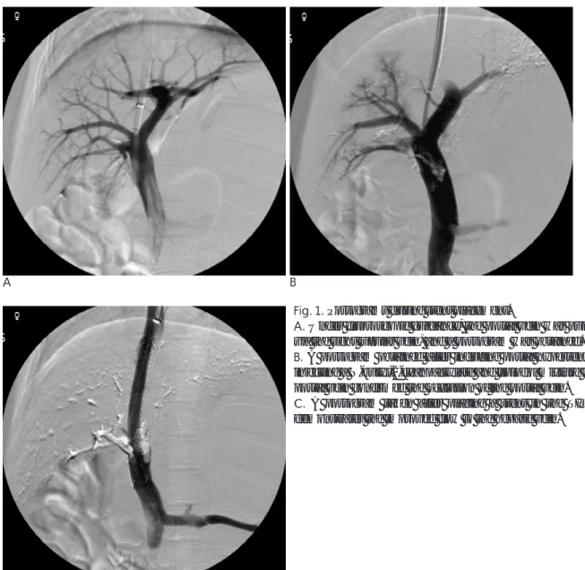
Fig. 1. Portograms during stent placement.
A. Under fluoroscopic guidance, the portal vein was punctured via the right jugular vein, and a portogram was obtained.
B. A portogram obtained after inducing portal hypertension by injecting a N-butyl-2-cyanoacrylate and lipiodol mixture into the portal vein confirmed the occlusion of the portal vein.
C. A portogram taken after placing a stent in the TIPS tract demonstrates the improved flow to the hepatic vein.
were performed by puncturing a tributary of the superi- or mesenteric vein after making an incision in the mid- line of the anterior wall of the abdomen. Each animal was euthanized with an intravenous pentobarbital sodi- um injection (Hanlim Pharm. Co., Ltd.; Seoul, Korea) in order to examine the histology of the tissue around the TIPS. Cross-sections of the TIPS tracts were obtained at the level of the hepatic parenchymal tract; and the speci- mens were evaluated by staining them with hematoxin- eosin and Masson-trichrome. The maximum distance from the stent of pseudointimal hyperplasia in each ani- mal was measured on the histopathology examination.
The level of maximum pseudointimal hyperplasia was calculated by dividing the radius of the stent by the thickness of the pseudointimal hyperplasia not includ- ing the thrombus (10).
Results
In-vitro Evaluation of the Paclitaxel Released from the Stent
The mean increase in stent weight, which was pro- duced by the paclitaxel and PU coating , was 15.2 mil- ligrams. The mean amount of paclitaxel in the PES, which was calculated from the increase in stent weight and the concentration of paclitaxel in the paclitaxel and PU mixture, was 304 μg.
The in-vitro examination showed that (ed note: this should be moved to the methododlogy section.) the
amount of paclitaxel released from the membrane of the paclitaxel and PU mixture during the first three days was 4%, 6%, and 4% of the loading dosage, respectively.
The cumulative amounts of paclitaxel released during the first and second weeks were 31% and 44%, respec- tively.
Animal Experiments TIPS
The TIPS procedures were technically successful in all the swine, and all the stents were deployed accurately without complications. After the stents had been placed, the blood flow to the hepatic vein improved without any disturbances being observed on the final portograms, and the portal pressure was decreased to below the ini- tial pressure. The mean pressure in the portal veins be- fore and after the N-butyl-2-cyanoacrylate injection was 21.0 cmH2O and 33.9 cmH2O, respectively. The latter pressure dropped immediately to 22.2 cmH2O after the TIPS procedure.
Follow-up and Sacrifice
The results of the 2-week follow-up after the TIPS pro- cedure were evaluated in 5 swine from each of the PES and PU stent groups because two swine had died within 3 days of the procedure. On the follow-up portograms in the 5 swine before sacrifice, all of the TIPS tracts that used PES demonstrated luminal patency despite the nar- rowing of the lumen in the intraparenchymal portion
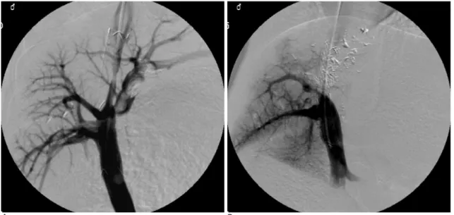
A B
Fig. 2. Follow-up portograms taken 2 weeks after the TIPS procedure.
A. A portogram performed via the hepatic vein in the paclitaxel-eluting stent (PES) group demonstrates a patent TIPS tract, even though a filling defect can be seen within the stent lumen.
B. Portograms in the control group demonstrate the complete occlusion of the TIPS tract.
(Fig. 2A). However, all 5 swine, in which the control stent had been used, had completely occluded lumens of the TIPS tracts (Fig. 2B).
Histopathology Examination
The gross specimens in the PES group showed partial filling of the stent lumen by fibrotic tissue and throm- bus, and the stents were easily separated from the tissue of the TIPS tract. However, those in the PU stent group showed the marked ingrowth of fibrotic tissue within the TIPS tracts, which made separation of the stents from the surrounding tissue difficult (Fig. 3A, B).
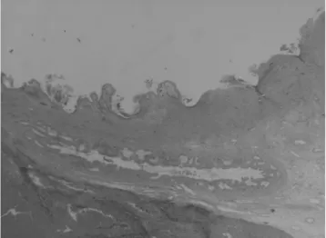
Histopathology analysis revealed the average maxi- mum pseudointimal hyperplasia for the PES and PU stents to be 25.1% and 73.8%, respectively. The micro- scopic findings demonstrated relatively thin and uni- form pseudointimal hyperplasia within the patent TIPS tracts in the PES stents, and complete occlusion of the TIPS tracts by pseudointimal hyperplasia in the PU stents (Fig. 4). The psuedointimal hyperplasia consisted of myofibroblasts, extracellular collagen matrix, and in- flammatory cells infiltrating around the stent wires.
Discussion
Although TIPS is an effective method for portal de- compression in patients with variceal bleeding or in- tractable ascites, it is complicated by portal hyperten- sion, a high rate of restenosis of the tracts and the disap- pointing long-term patency (1-4). The important mech- anisms reported to be responsible for the formation of a restenosis or occlusion of the TIPS tracts are chronic in-
flammatory reactions, inhibition of the endothelializa- tion process, excessive proliferation of pseudointimal granulation tissue, and the ingrowth of hepatocytes into the stented lumens of the hepatic parenchymal tracts (13, 14). The layers of pseudointimal granulation tissue contain mesenchymal cells and collagen covered by a layer of endothelial cells.
Various methods and devices have been used to over- come the problems of restenosis or occlusion, and the low primary patency of the TIPS tract. Several studies of TIPS, which used stent-grafts coated with various mate- rials, have been performed using the swine model (12, 15-17). Although the placement of a stent-graft in a TIPS tract significantly improved the patency after a
A B
Fig. 3. Gross specimen.
A. A stent in the PES group was separated easily from the surrounding tissue and demonstrates fibrotic tissue and thrombus within the lumen.
B. A stent in the control group could not be separated because of the marked ingrowth of fibrotic tissue within the TIPS tract.
Fig. 4. Microscopic specimen (×10) stained with H-E. A.
Microscopic section in the PES group demonstrates relatively thin and uniform pseudointimal hyperplasia within the patent TIPS tract.
short-term follow-up study, they caused a late shunt malfunction in certain cases and did not improve the long-term patency as a result of the increased occur- rence of thrombogenecity and foreign-body reactions.
There have been experimental studies on swine models that used intraluminal irradiation to prevent stenosis of the TIPS tracts. The studies were carried out by apply- ing intraluminal irradiation to the hepatic parenchyma and venous outflow tracts at the time of TIPS place- ment, which was followed by the use of a radionuclide- filled balloon catheter. However, the results were incon- clusive (11, 18, 19). Intraluminal irradiation has several limitations that have to be overcome before it can be used clinically. They include the complexity of the preparation of the radionuclide solution, the difficulty in maintaining the irradiation dose, and the lengthy infla- tion time of the balloon catheter.
Restenosis after the implantation of a stent is a prob- lem that occurs in treated vessels, and many studies have been carried out to determine appropriate preven- tative measures: a drug-eluting stent being one such method. There have been many clinical applications of several drug-eluting stents using various drugs, such as paclitaxel, rapamycin, dexamethasone, and heparin, some with extremely promising results in terms of sup- pressing neointimal hyperplasia and dramatically reduc- ing the rate of restenosis (7, 9, 20-23). In recent experi- mental studies, the application of drug-eluting stents was attempted in non-vascular organs, such as the bile duct and trachea (24, 25). In the TIPS tract, one study used a dexamethasone-eluting stent in a swine model and demonstrated a tendency to inhibit the develop- ment of pseudointimal hyperplasia (10).
Paclitaxel is a cytotoxic, anti-cancer drug that inhibits tumor growth through its anti-proliferative and anti-an- giogenic properties (26, 27). Paclitaxel also inhibits neoinitimal hyperplasia in vessels through persistent fib- rin deposition, inflammatory cell infiltration, and reduc- ing the number of smooth muscle cells, which are asso- ciated with a restenosis after placing the stents (28, 29).
The PES with a topical therapeutic drug within the tar- get tissues is popular in percutaneous coronary interven- tions. The nine-month follow-up results of a prospective randomized study demonstrated that the lumen vol- umes, which were evaluated using serial volumetric in- travascular ultrasound examinations, were larger in the PES group than in the bare-metal stent group, due to a decrease in the neointimal volume (30). In non-vascular organs, it was reported that paclitaxel has a potential an-
ti-proliferative effect in experimental studies using hu- man epithelial gallbladder cells, human fibroblasts, and bladder tissues (27, 31). Shin et al performed an experi- mental application of the PES in non-vascular organs, using a canine urethral model, and reported that is could reduce the level of tissue hyperplasia after placing the stent (32).
In this study, the pseudointima within the patent TIPS tract in the PES group was relatively uniform and thin, which was in contrast to the histopathology findings of the complete occlusion of the TIPS tract in the control stent group. It is believed that those results were the re- sult of a similar mechanism of paclitaxel that is observed in the arteries and other non-vascular organs. Because the pseudointima in the TIPS tract was composed of my- ofibroblasts and inflammatory cells, paclitaxel inhibited the proliferation of fibroblasts and the infiltration of in- flammatory cells, reduced the number of smooth mus- cle cells, and inhibited the proliferation of the pseudoin- tima. However, despite the inhibition of pseudointimal hyperplasia by paclitaxel, the partial thrombosis within the TIPS tract remained a problem, which might in- crease the rate of occlusion of the TIPS tract and de- crease its patency. It was surmised that bile leakage was responsible for the thrombosis within the TIPS tract af- ter placing the PES . Although there is still some contro- versy regarding the issue, the transection of the major bile ducts that occurred during the formation of the TIPS tracts and the leakage of bile from the injured ducts are related to the thrombosis in the TIPS tracts, particularly rapid and early thrombosis (33-35). In this study, a thrombosis within the TIPS tract that is caused by bile leakage might not be suppressed because pacli- taxel does not have an anti-thrombogenic effect.
Therefore, further studies will be needed to determine if the combined use of an anti-thrombotic agent can re- duce the frequency of thrombosis. In addition, an evalu- ation of the relationship between thrombosis and the materials for the PES, such as stent metal or PU, is war- ranted.
There were several limitations in this study . First, there were a small number of animals and the statistical significance was not determined, even though the por- tography and histopathology findings were similar in each of the 5 animals in their respective group. This re- sult was attributed to the effectiveness of paclitaxel.
Second, the concentration of paclitaxel in the blood, which is a parameter for determining the amount of pa- clitaxel released from the PES after its placement in the
TIPS tract, was not evaluated. The release pattern of pa- clitaxel from PES may be different in in vitro and in vivo tests. Furthermore, the cause of the death of the two swine that expired 3 days after PES placement is un- known. This is despite a necropsy being performed. In addition, the possible toxicity of paclitaxel was not de- termined.
In conclusion, the paclitaxel-eluting stent showed a tendency to significantly inhibit the development of pseudointimal hyperplasia in a TIPS tract of a swine model with induced-portal hypertension. Although fur- ther study will be needed, this method has potential to improve the patency of the TIPS tract.
References
1. Haskal ZJ, Pentecost MJ, Soulen MC, Shlansky-Goldberg RD, Baum RA, Cope C. Transjugular intrahepatic portosystemic shunt stenosis and revision: early and midterm results. AJR Am J Roentgenol 1994;163:439-444
2. LaBerge JM, Somberg KA, Lake JR, Gordon RL, Kerlan RK Jr, Ascher NL, et al. Two-year outcome following transjugular intra- hepatic portosystemic shunt for variceal bleeding: results in 90 pa- tients. Gastroenterology 1995;108:1143-1151
3. ter Borg PC, Hollemans M, Van Buuren HR, Vleggaar FP, Groeneweg M, Hop WC, et al. Transjugular intrahepatic portosys- temic shunts: long-term patency and clinical results in a patient co- hort observed for 3-9 years. Radiology 2004;231:537-545
4. Zhuang ZW, Teng GJ, Jeffery RF, Gemery JM, Janne d’Othee B, Bettmann MA. Long-term results and quality of life in patients treated with transjugular intrahepatic portosystemic shunts. AJR Am J Roentgenol 2002;179:1597-1603
5. Duda SH, Poerner TC, Wiesinger B, Rundback JH, Tepe G, Wiskirchen J, et al. Drug-eluting stents: potential applications for peripheral arterial occlusive disease. J Vasc Interv Radiol 2003;14:
291-301
6. Shofti R, Tio F, Beyar R. Neointimal vascularization and intimal thickening in response to self-expanding stents: a swine model. Int J Cardiovasc Intervent 2004;6:61-67
7. Grube E, Silber S, Hauptmann KE, Buellesfeld L, Mueller R, Lim V, et al. Two-year-plus follow-up of a paclitaxel-eluting stent in de novo coronary narrowings (TAXUS I). Am J Cardiol 2005;96:79-82 8. Serruys PW, Sianos G, Abizaid A, Aoki J, den Heijer P, Bonnier H,
et al. The effect of variable dose and release kinetics on neointimal hyperplasia using a novel paclitaxel-eluting stent platform: the Paclitaxel In-Stent Controlled Elution Study (PISCES). J Am Coll Cardiol 2005;46:253-260
9. Werner GS, Krack A, Schwarz G, Prochnau D, Betge S, Figulla HR. Prevention of lesion recurrence in chronic total coronary oc- clusions by paclitaxel-eluting stents. J Am Coll Cardiol 2004;44:
2301-2306
10. Seo TS, Oh JH, Park YK, Song HY, Park SJ, Yuk SH. Efficacy of a Dexamethasone-eluting nitinol stent on inhibition of pseudointi- mal hyperplasia in a transjugular intrahepatic portosystemic shunt: an experimental study in a swine model. Korean J Radiol 2005;6:241-247
11. Lessie T, Yoon HC, Nelson HA, Fillmore DJ, Baldwin GN, Miller FJ. Intraluminal irradiation for TIPS stenosis: preliminary results
in a swine model. J Vasc Interv Radiol 1999;10:899-906
12. Nishimine K, Saxon RR, Kichikawa K, Mendel-Hartvig J, Timmermans HA, Shim HJ, et al. Improved transjugular intrahep- atic portosystemic shunt patency with PTFE-covered stent-grafts:
experimental results in swine. Radiology 1995;196:341-347 13. LaBerge JM, Ferrell LD, Ring EJ, Gordon RL, Lake JR, Roberts JP,
et al. Histopathologic study of transjugular intrahepatic portosys- temic shunts. J Vasc Interv Radio 1991;2:549-556
14. Terayama N, Matsui O, Kadoya M, Yoshikawa J, Gabata T, Miyayama S, et al. Transjugular intrahepatic portosystemic shunt:
histologic and immunohistochemical study of autopsy cases.
Cardiovasc Intervent Radiol 1997;20:457-461
15. Haskal ZJ, Brennecke LJ. Porous and nonporous polycarbonate urethane stent-grafts for TIPS formation: biologic responses. J Vasc Interv Radiol 1999;10:1255-1263
16. Tanihata H, Saxon RR, Kubota Y, Pavcnik D, Uchida BT, Rosch J, et al. Transjugular intrahepatic portosystemic shunt with silicone- covered Wallstents: results in a swine model. Radiology 1997;205:181-184
17. Zhuang ZW, Hoopes PJ, Koutras PC, Ebbighausen WH, Wagner RJ, Bettmann MA. Transjugular intrahepatic portosystemic shunt with an autologous vein-covered stent: results in a swine model. J Vasc Interv Radiol 2001;12:1333-1342
18. Hausegger KA, Portugaller H, Macri NP, Tauss J, Schedlbauer P, Deutschmann J, et al. Covered stents in transjugular portosystemic shunt: healing response to non-porous ePTFE covered stent grafts with and without intraluminal irradiation. Eur Radiol 2003;13:
1549-1558
19. Pokrajac B, Cejna M, Kettenbach J, Schamp S, Fellner C, Seitz W, et al. Intraluminal 192Ir brachytherapy following transjugular in- trahepatic portosystemic shunt revision: long-term results and ra- diotherapy parameters. Cardiovasc Radiat Med 2001;2:133-137 20. Ahn YK, Jeong MH, Kim JW, Kim SH, Cho JH, Cho JG, et al.
Preventive effects of the heparin-coated stent on restenosis in the porcine model. Catheter Cardiovasc Interv 1999;48:324-330 21. Lincoff AM, Furst JG, Ellis SG, Tuch RJ, Topol EJ. Sustained local
delivery of dexamethasone by a novel intravascular eluting stent to prevent restenosis in the porcine coronary injury model. J Am Coll Cardiol 1997;29:808-816
22. Prasad CK, Resmi KR, Krishnan LK, Vaishnav R. Survival of en- dothelial cells in vitro on Paclitaxel-loaded coronary stents. J Biomater Appl 2005;19:271-286
23. Sousa JE, Costa MA, Abizaid AC, Rensing BJ, Abizaid AS, Tnaajura LF, et al. Sustained suppression of neointimal proliferation by sirolimus-eluting stents: one-year angiographic and intravascular follow-up. Circulation 2001;104:2007-2011
24. Lee DK, Kim HS, Kim KS, Lee WJ, Kim HK, Won YH, et al. The effect on porcine bile duct of a metallic stent covered with a pacli- taxel-incorporated membrane. Gastrointest Endosc 2005;61:296-301 25. Shin JH, Song HY, Seo TS, Yuk SH, Kim YH, Cho YM, et al.
Influence of a dexamethasone-eluting covered stent on tissue reac- tion: an experimental study in a canine bronchial model. Eur Radiol 2005;15:1241-1249
26. Dhanikula AB, Panchagnula R. Localized paclitaxel delivery. Int J Pharm 1999;183:85-100
27. Kalinowski M, Alfke H, Kleb B, Durfeld F, Joachim Wagner H.
Paclitaxel inhibits proliferation of cell lines responsible for metal stent obstruction: possible topical application in malignant bile duct obstructions. Invest Radiol 2002;37:399-404
28. Drachman DE, Edelman ER, Seifert P, Groothuis AR, Bornstein DA, Kamath KR, et al. Neointimal thickening after stent delivery of paclitaxel: change in composition and arrest of growth over six
months. J Am Coll Cardiol 2000;36:2325-2332
29. Heldman AW, Cheng L, Jenkins GM, Heller PF, Kim DW, Ware M Jr, et al. Paclitaxel stent coating inhibits neointimal hyperplasia at 4 weeks in a porcine model of coronary restenosis. Circulation 2001;103:2289-2295
30. Weissman NJ, Koglin J, Cox DA, Hermiller J, O’Shaughnessy C, Mann JT, et al. Polymer-based paclitaxel-eluting stents reduce in- stent neointimal tissue proliferation: a serial volumetric intravas- cular ultrasound analysis from the TAXUS-IV trial. J Am Coll Cardiol 2005;45:1201-1205
31. Song D, Wientjes MG, Au JL. Bladder tissue pharmacokinetics of intravesical taxol. Cancer Chemother Pharmacol 1997;40:285-292 32. Shin JH, Song HY, Choi CG, Yuk SH, Kim JS, Kim YM, et al.
Tissue hyperplasia: influence of a paclitaxel-eluting covered
stent—preliminary study in a canine urethral model. Radiology 2005;234:438-444
33. Jalan R, Harrison DJ, Redhead DN, Hayes PC. Transjugular intra- hepatic portosystemic stent-shunt (TIPSS) occlusion and the role of biliary venous fistulae. J Hepatol 1996;24:169-176
34. LaBerge JM, Ferrell LD, Ring EJ, Gordon RL. Histopathologic study of stenotic and occluded transjugular intrahepatic portosys- temic shunts. J Vasc Interv Radiol 1993;4:779-786
35. Saxon RR, Mendel-Hartvig J, Corless CL, Rabkin J, Uchida BT, Nishimine K, et al. Bile duct injury as a major cause of stenosis and occlusion in transjugular intrahepatic portosystemic shunts: com- parative histopathologic analysis in humans and swine. J Vasc Interv Radiol 1996;7:487-497
대한영상의학회지 2007;56:225-232
경경정맥간내문맥정맥단락술 경로내의 가성내막증식 억제를 위한 파클리탁셀방출니티놀스텐트의 효과: 돼지모형에서의 실험연구1
1경희대학교 부속병원 영상의학과, 3병리과
2고려대학교 의과대학 구로병원 영상의학과
4울산대학교 의과대학 서울아산병원 방사선과학교실
5한남대학교 공과대학 화공, 고분자공학부
서태석2・오주형・권세환・박용구3・송호영4・육순홍5
목적: 경경정맥간내문맥정맥단락술 경로 내의 가성내막증식 억제를 위한 파클리탁셀방출나이티놀스텐트의 효과를 평가하고자 하였다.
대상과 방법: 경경정맥간내문맥정맥단락술 경로에 삽입하기 위하여 직경 10 mm, 길이 60 mm의 나이티놀스텐트 를 제작하여 실험군으로 파클리탁셀을 함유한, 대조군으로 함유하지 않은 폴리우레탄용액의 두 가지 형태로 피복 하였다. 경경정맥간내문맥정맥단락술을 12마리의 돼지에서 시행하였고 파클리탁셀방출스텐트 7개, 폴리우레탄피복 스텐트 5개를 삽입하였다. 파클리탁셀방출스텐트를 삽입한 돼지 2마리가 추적과정에서 사망하여 두 가지 스텐트에 서 각각 5마리를 2주간 추적관찰하였다. 희생 전의 문맥조영술과 희생 후 조직병리검사로 가성내막의 증식을 평가 하였다.
결과: 희생 전 문맥조영상에서 파클리탁셀방출스텐트를 삽입한 경경정맥간내문맥정맥단락술 경로는 모두 개통되어 있었지만 폴리우레탄피복스텐트는 모두 폐쇄되어있었다. 조직병리검사에서 가성내막증식이 가장 심한 부위에서 스 텐트반경에 대한 조직두께의 비는 파클리탁셀스텐트가 25%, 폴리우레탄스텐트가 76%였다.
결론: 파클리탁셀방출스텐트는 경경정맥간내문맥정맥단락술 경로에서 가성내막증식을 유의하게 감소시키는 것으로 생각한다.