http://dx.doi.org/10.5090/kjtcs.2013.46.4.274 ISSN: 2233-601X (Print) ISSN: 2093-6516 (Online)
Department of Thoracic and Cardiovascular Surgery, Seoul National University Hospital, Seoul National University College of Medicine Received: November 6, 2012, Revised: February 5, 2013, Accepted: March 7, 2013
Corresponding author: Ki-Bong Kim, Department of Thoracic and Cardiovascular Surgery, Seoul National University Hospital, Seoul National University College of Medicine, 101 Daehak-ro, Jongno-gu, Seoul 110-744, Korea
(Tel) 82-2-2072-3482 (Fax) 82-2-747-5245 (E-mail) [email protected]
C The Korean Society for Thoracic and Cardiovascular Surgery. 2013. All right reserved.
CC This is an open access article distributed under the terms of the Creative Commons Attribution Non-Commercial License (http://creative- commons.org/licenses/by-nc/3.0) which permits unrestricted non-commercial use, distribution, and reproduction in any medium, provided the original work is properly cited.
Anaortic Off-pump Coronary Artery Bypass Grafting in Patients with Takayasu’s Arteritis
Kwon Joong Na, M.D., Kyung-Hak Lee, M.D., Se Jin Oh, M.D., Ho Young Hwang, M.D., Ph.D., Ki-Bong Kim, M.D., Ph.D.
Background: Coronary involvement in Takayasu’s arteritis is a rare but fatal disease. The aim of this study was to evaluate the early and mid-term results of Takayasu’s arteritis patients who underwent coronary artery bypass grafting (CABG). Materials and Methods: Of 2,280 patients who underwent isolated CABG from January 1998 to June 2012, Takayasu’s arteritis was identified in 5 patients. There were 3 female patients, and the mean age was 58±9 years. Takayasu’s arteritis was diagnosed during preoperative evaluation for coronary artery disease in 4 pa- tients, and the initial manifestation was angina pectoris in 4 patients. All of the patients underwent anaortic off-pump CABG (OPCAB) using the in situ left or right internal thoracic arteries (ITA); 3 patients had severe stenosis of the proximal left subclavian artery and the in situ right ITA was used instead. Medical treatment for in- flammatory arteritis during the perioperative and follow-up period was performed if indicated. Early, 1-year, and 5-year angiographic results and clinical outcomes were analyzed. Results: There was no surgical mortality, and all of the patients were discharged without complications on postoperative 8±2 days. Early postoperative (postoperative 2±1 days) angiography demonstrated a graft patency of 100% (12 of 12 distal anastomoses). One-year (13±3 months) angiography was performed in 4 patients, and all of the grafts were patent (100%, 9 of 9 distal anasto- moses). Conclusion: By performing anaortic OPCAB in patients with Takayasu’s arteritis, we were able to avoid complications associated with manipulating an atherosclerotic and severely calcified ascending aorta. The early and mid-term graft patency of OPCAB in Takayasu’s arteritis was maintained when concomitant with medical treatment.
Key words: 1. Takayasu’s arteritis
2. Coronary artery bypass surgery 3. Off-pump
INTRODUCTION
Takayasu’s arteritis (TA) is a vasculitis of the large vessels affecting the aorta and its major branches. The incidence of coronary artery involvement complicating TA has been re- ported as approximately 10% in patients with a clinical diag- nosis of TA [1]. The clinical manifestation of coronary in-
volvement in patients with TA could be lethal because occlu- sion of the ostia and proximal segment of the coronary ar- teries is found frequently [1,2]. Of several therapeutic options for coronary artery disease in TA, surgical revascularization is very often recommended [1,3,4]. Previous studies have re- ported results of coronary artery bypass grafting (CABG) us- ing an aortocoronary bypass technique [3-7]. However, there
Table 2. Preoperative coronary artery angiography findings
Significant lesion No. of patients
Left main coronary artery 3
Ostium of right coronary artery 3
Ostium of LAD or LCx 3
Distal LAD and LCx 3
Distal right coronary artery 2
Diagonal branch 1
LAD, left anterior descending coronary artery; LCx, left cir- cumflex coronary artery.
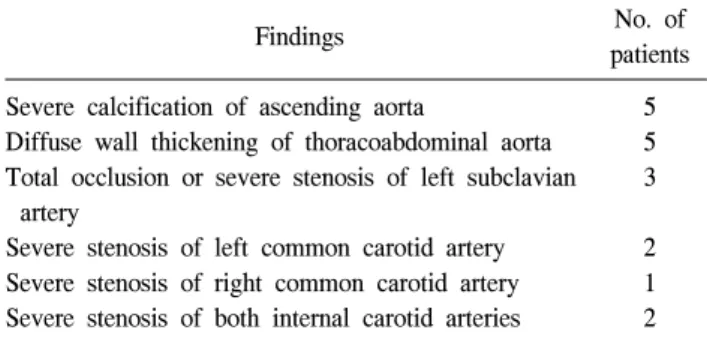
Table 1. Preoperative computed tomographic angiography findings
Findings No. of
patients Severe calcification of ascending aorta
Diffuse wall thickening of thoracoabdominal aorta Total occlusion or severe stenosis of left subclavian
artery
Severe stenosis of left common carotid artery Severe stenosis of right common carotid artery Severe stenosis of both internal carotid arteries
5 5 3
2 1 2
have been only a few studies demonstrating the results of anaortic off-pump CABG (OPCAB) using an in situ arterial graft in this population. The aim of this study was to eval- uate early and mid-term angiographic results of anaortic OPCAB using in situ arterial grafts in patients with TA.
METHODS 1) Patients
Of 2,280 patients who underwent isolated CABG from January 1998 to June 2012, 5 patients (0.2%) had a history of TA. Four patients were diagnosed with TA while they were under evaluation for coronary artery disease, and 1 pa- tient had been diagnosed with TA before diagnosis of coro- nary artery disease. Preoperative computed tomographic (CT) angiography showed severe atherosclerosis of the thoracic and abdominal aorta and severe stenosis of multiple large arteries in all of the patients (Table 1). The diagnosis of TA was es- tablished by the American College of Rheumatology 1990 criteria for the classification of TA (at least 3 of the follow- ing criteria: age ≤40 years at disease onset, claudication of the extremities, decreased brachial artery pressure, blood pres- sure difference >10 mmHg between the arms, bruit over the subclavian arteries or aorta, and abnormal arteriographic re- sults) [8]. There were 3 female (60%) and 2 male (40%) pa- tients, and their mean age was 58±9 years (range, 44 to 65 years). Four patients (80%) had angina as an initial manifes- tation, and one patient was incidentally diagnosed as having severe coronary artery disease during a diagnostic work-up for underlying renal cell carcinoma. Preoperative coronary an- giography revealed that the ostium or proximal part of the
coronary arteries, such as the right coronary artery (RCA), left coronary artery, and left main coronary artery, were in- volved in most cases; lesions of the distal coronary artery system were relatively rare (Table 2).
2) Operative strategies
OPCAB was performed as previously described [9]. Our surgical strategies during the study period were as follows:
(1) performing off-pump revascularization while avoiding aortic manipulation; (2) complete revascularization if possible, using a composite graft anastomosed to the available in situ internal thoracic arteries (ITA). Aortocoronary bypass was not considered in patients with TA because of severe calcification and thickening of the aortic wall, which was identified in preoperative CT angiography and intra-operative examination.
An in situ left ITA was used for revascularization in 2 pa- tients who had a preserved left subclavian artery. In the other 3 patients, an in situ left ITA was not used because the pa- tients showed severe stenosis of the proximal left subclavian artery; an in situ right ITA was used instead. The grafts cre- ated for composite grafting were the right gastroepiploic ar- tery (n=3), saphenous vein (n=2), and left ITA (n=1). The numbers of anastomoses were 1 in 1 patient, 2 in 1 patient, and 3 in 3 patients (mean number of distal anastomoses per patient=2.4±0.9).
The patients received an initial dose of 1.5 mg/kg of hep- arin and periodically received supplemental doses to maintain an activated clotting time of greater than 300 seconds. The distal anastomosis was constructed using a continuous techni- que with 8-0 polypropylene sutures.
All of the patients took aspirin (200 mg/day) until the day
of surgery and resumed it as soon as possible after surgery, usually 1 day postoperatively. Ticlopidine hydrochloride (200 mg/day) was used simultaneously with aspirin for 2 months during the early postoperative period. Antilipid therapy was initiated postoperatively if the patient had a high blood low-density lipoprotein cholesterol level (>100 mg/day).
Perioperative anti-inflammatory therapy was considered by rheumatologists in patients in the active stage of TA. One pa- tient was in the active stage of TA preoperatively, and he un- derwent steroid therapy for 3 months before undergoing OPCAB. All of the patients underwent steroid therapy after the surgery to control the activity of the TA. Two patients showed aggravated clinical symptoms of vascular in- sufficiency and elevation of the erythrocyte sedimentation rate and C-reactive protein levels during follow-up after the sur- gery; azathioprine was thus added to the steroid therapy.
3) Evaluation of clinical outcomes
The patients underwent regular postoperative follow-up with a cardiac surgeon and rheumatologist through the out- patient clinic. Operative mortality was defined as death within 30 days, including death after hospital discharge. Cardiac death was defined as a death related to cardiac events, in- cluding sudden death during follow-up. Major adverse car- diovascular or cerebral events included acute myocardial in- farction, coronary reintervention, death from any cause, and cerebrovascular event.
4) Angiographic evaluation of patency
Patients underwent early, 1-year, and 5-year follow-up cor- onary angiograms regardless of angina symptoms for evalua- tion of the anastomotic sites and patency of the grafts. All of the patients underwent early postoperative (2.0±1.0 days) angiograms. One-year (13±3 months) angiography was per- formed in 4 patients who were followed up for 1 year or more, and 5-year (63±3 months) angiography was performed in 3 of the 4 patients who were followed up for 5 years or more.
RESULTS
The patients’ characteristics and clinical data are described
in Table 3.
1) Early and mid-term clinical outcomes
There was no operative mortality or postoperative morbidities. The patients were discharged on postoperative 8±2 days (range, 6 to 10 days). One patient underwent coro- nary reintervention 5 years after the surgery because of occlu- sion of the right ITA graft anastomosed to the RCA of a moderately stenosed lesion. The other 4 patients had no ma- jor adverse cardiovascular or cerebrovascular events during follow-up. One late sudden death occurred about 3 years after the surgery.
2) Early, 1-year and 5-year angiographic results
The early postoperative angiography demonstrated a pa- tency rate of 100% (12 of 12 distal anastomoses). One-year follow-up angiography demonstrated a patency rate of 100%
(9 of 9 distal anastomoses). Five-year follow-up angiography was performed in 3 patients and the patency rate was 83% (5 of 6 distal anastomoses). Occlusion of a right ITA graft anas- tomosed to the RCA of a moderately stenosed lesion oc- curred in 1 patient. The patient underwent percutaneous coro- nary intervention at postoperative 67 months.
DISCUSSION
The present study demonstrated two major findings. First, anaortic off-pump revascularization using available in situ ar- terial grafts may avoid operative morbidity in patients with TA. Second, early and mid-term results of angiographic graft patency and clinical outcomes showed satisfactory results when followed up with a rheumatologist for control of the in- flammatory disease.
TA is a chronic inflammatory disease of medium and large arteries, and mainly involves the aorta and its major branches, especially their orifices [2]. It predominantly affects young fe- males (almost 90%) with an onset between 10 and 50 years, and a high prevalence is reported in Japan, Southeast Asia, India, and Mexico [1]. Cardiac manifestations of TA are the consequences of the various features related to TA: hyper- tension, aortic regurgitation, coronary artery and pulmonary artery involvement, and direct involvement of the
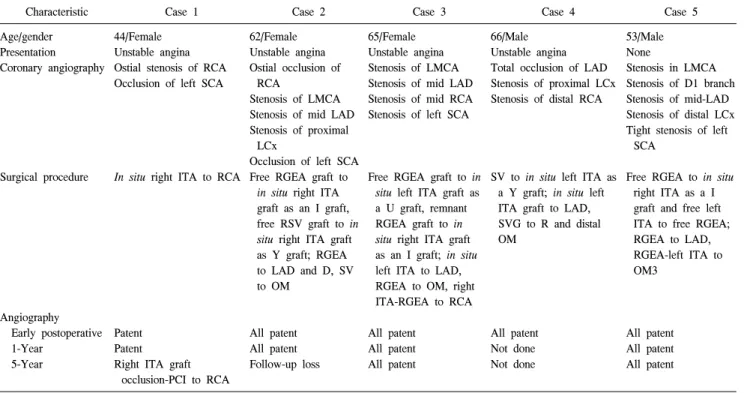
Table 3. Summary of characteristics of five cases
Characteristic Case 1 Case 2 Case 3 Case 4 Case 5
Age/gender Presentation Coronary angiography
Surgical procedure
Angiography Early postoperative 1-Year
5-Year
44/Female Unstable angina Ostial stenosis of RCA Occlusion of left SCA
In situ right ITA to RCA
Patent Patent
Right ITA graft occlusion-PCI to RCA
62/Female Unstable angina Ostial occlusion of
RCA
Stenosis of LMCA Stenosis of mid LAD Stenosis of proximal
LCx
Occlusion of left SCA Free RGEA graft to
in situ right ITA graft as an I graft, free RSV graft to in situ right ITA graft as Y graft; RGEA to LAD and D, SV to OM
All patent All patent Follow-up loss
65/Female Unstable angina Stenosis of LMCA Stenosis of mid LAD Stenosis of mid RCA Stenosis of left SCA
Free RGEA graft to in situ left ITA graft as a U graft, remnant RGEA graft to in situ right ITA graft as an I graft; in situ left ITA to LAD, RGEA to OM, right ITA-RGEA to RCA
All patent All patent All patent
66/Male Unstable angina Total occlusion of LAD Stenosis of proximal LCx Stenosis of distal RCA
SV to in situ left ITA as a Y graft; in situ left ITA graft to LAD, SVG to R and distal OM
All patent Not done Not done
53/Male None
Stenosis in LMCA Stenosis of D1 branch Stenosis of mid-LAD Stenosis of distal LCx Tight stenosis of left
SCA
Free RGEA to in situ right ITA as a I graft and free left ITA to free RGEA;
RGEA to LAD, RGEA-left ITA to OM3
All patent All patent All patent
RCA, right coronary artery; LMCA, left main coronary artery; LAD, left anterior descending coronary artery; SCA, subclavian artery; LCx, left cir- cumflex coronary artery; D, diagonal; ITA, internal thoracic artery; RGEA, right gastroepiploic artery; RSV, reversed saphenous vein; SVG, saphe- nous venous graft; R, Ramus; OM, obtuse marginal; SV, saphenous vein; PCI, percutaneous coronary intervention.
myocardium. Coronary involvement occurs in approximately 10% of TA patients, and causes symptoms by myocardial is- chemia and congestive heart failure [1].
Of the therapeutic options available for coronary artery dis- ease, percutaneous coronary intervention may be performed in TA; however, it has been reported in very limited cases, and the results have not been satisfactory [10]. The presence of chronic inflammation and calcification of the aorta and its branches associated with severe coronary artery disease also pose difficulties for surgeons choosing the optimal surgical option. The inflammatory process usually involves the entire aortic wall, resulting in thickening of the intima, degeneration of the elastic tissue, and proliferation of the connective tissue in the medial layer, which leads to aortic wall thickening and loss of elasticity [11]. Preoperative evaluation of in- flammatory activity is mandatory in TA patients. If the TA is in the active stage of inflammation, treatment for coronary ar- tery disease should be delayed and anti-inflammatory treat- ment for TA must come first for a better outcome [12].
However, in patients with unstable angina or myocardial in- farction, treatment for coronary artery disease is usually re- quired without delay because of the potentially fatal outcome [12].
The incidence of cerebrovascular accident becomes higher when manipulating the aorta with heavy calcification. A re- cent meta-analysis demonstrated that avoiding aortic manipu- lation during CABG decreased the rate of perioperative stroke [13]. For myocardial revascularization, a saphenous vein graft has commonly been used with a method of direct anasto- mosis to the ascending aorta because the subclavian arteries, from which the ITA originate, are commonly affected vessels in TA [3-5,7]. In addition, ITAs have rarely been used for in situ grafting because the arterial grafts can be influenced by the underlying TA itself and graft occlusion may occur later.
However, failure of saphenous vein grafts occurs frequently, mainly at the proximal anastomosis site, due to intimal thick- ening of the aorta. The long-term graft patency of the saphe- nous vein graft in TA was limited to 60% at 4 years after
surgery [14]. In the present study, all 5 patients had severe calcification, atherosclerosis, and thickening of the aortic wall diagnosed by preoperative CT angiography and intra-operative examination. In these patients, aortic cannulation and partial clamp application for direct graft anastomosis may cause crit- ical results such as cerebrovascular accident or aortic wall disruption. We avoided manipulating the thickened and calci- fied ascending aorta by performing anaortic OPCAB, and there were no complications such as cerebrovascular event or other embolic events. Available in situ arterial grafts were used for coronary revascularization instead of aortocoronary bypass. In situ arterial grafts such as ITA or right gastro- epiploic artery were used if there was no significant stenosis or occlusion at the inflow arteries; the inflow artery of the graft was confirmed to be patent in preoperative coronary an- giography and CT angiography. In the present study, 1-year follow-up angiography showed graft patency of 100% in four patients, and 5-year follow-up angiography showed graft pa- tency of 83%. The outcome of intervention or bypass surgery in TA is significantly influenced by the activity of arteritis [15]. Control of disease activity in TA is one of the most im- portant factors for maintaining long-term graft patency [11].
In the present study, 3 patients (60%) were further treated with anti-inflammatory medications during follow-up at the rheumatology outpatient clinic.
There are limitations to the present study that must be recognized. First, our study included only a small number of patients. Second, more follow-up is needed on these patients to evaluate long-term graft patency and clinical outcomes.
CONFLICT OF INTEREST
No potential conflict of interest relevant to this article was reported.
REFERENCES
1. Rav-Acha M, Plot L, Peled N, Amital H. Coronary involve- ment in Takayasu’s arteritis. Autoimmun Rev 2007;6:566-71.
2. Lupi-Herrera E, Sanchez-Torres G, Marcushamer J, Mispireta J, Horwitz S, Vela JE. Takayasu’s arteritis: clinical study of 107 cases. Am Heart J 1977;93:94-103.
3. Young JA, Sengupta A, Khaja FU. Coronary arterial steno- sis, angina pectoris and atypical coarctation of the aorta due to nonspecific arteritis: treatment with aortocoronary by- pass graft. Am J Cardiol 1973;32:356-60.
4. Cipriano PR, Silverman JF, Perlroth MG, Griepp RB, Wexler L. Coronary arterial narrowing in Takayasu’s aor- titis. Am J Cardiol 1977;39:744-50.
5. Abad C. Coronary artery bypass surgery for patients with left main coronary lesions due to Takayasu’s arteritis. Eur J Cardiothorac Surg 1995;9:661-3.
6. Yamaguchi A, Endo H, Adachi H, Kawahito K, Ino T.
Off-pump coronary artery bypass in patients with Takayasu’s disease. Ann Thorac Surg 2004;77:2186-8.
7. Endo M, Tomizawa Y, Nishida H, et al. Angiographic find- ings and surgical treatments of coronary artery involvement in Takayasu arteritis. J Thorac Cardiovasc Surg 2003;125:
570-7.
8. Arend WP, Michel BA, Bloch DA, et al. The American College of Rheumatology 1990 criteria for the classification of Takayasu arteritis. Arthritis Rheum 1990;33:1129-34.
9. Kim KB, Kang CH, Chang WI, et al. Off-pump coronary ar- tery bypass with complete avoidance of aortic manipulation.
Ann Thorac Surg 2002;74:S1377-82.
10. Fava MP, Foradori GB, Garcia CB, et al. Percutaneous transluminal angioplasty in patients with Takayasu arteritis:
five-year experience. J Vasc Interv Radiol 1993;4:649-52.
11. Maksimowicz-McKinnon K, Clark TM, Hoffman GS. Limi- tations of therapy and a guarded prognosis in an American cohort of Takayasu arteritis patients. Arthritis Rheum 2007;
56:1000-9.
12. Lee GY, Jang SY, Ko SM, et al. Cardiovascular manifes- tations of Takayasu arteritis and their relationship to the disease activity: analysis of 204 Korean patients at a single center. Int J Cardiol 2012;159:14-20.
13. Edelman JJ, Yan TD, Bannon PG, Wilson MK, Vallely MP.
Coronary artery bypass grafting with and without manipu- lation of the ascending aorta: a meta-analysis. Heart Lung Circ 2011;20:318-24.
14. Ando M, Sasako Y, Okita Y, et al. Surgical considerations of occlusive lesions associated with Takayasu’s arteritis. Jpn J Thorac Cardiovasc Surg 2000;48:173-9.
15. Amano J, Suzuki A. Coronary artery involvement in Takaya- su’s arteritis: collective review and guideline for surgical treatment. J Thorac Cardiovasc Surg 1991;102:554-60.