thereof. Here, we describe one case that we treated and briefly summarize the available data on E. lenta bacteremia.
CASE REPORT
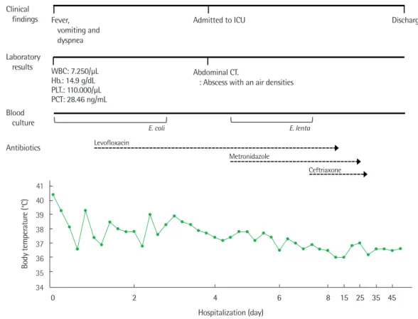
A 73-yr-old male presented with fever, vomiting, and dyspnea.
His medical history included a prior cerebrovascular attack and multiple complications caused by hemiplegia, including recurrent urinary tract infections and pressure sores requiring continuous medical care.
On the day of admission, his body temperature, pulse rate, re- spiratory rate, and blood pressure were 40.4°C, 108/min, 39/min, and 170/90 mmHg, respectively. Abdominal examina-tion re- vealed diffuse tenderness and guarding with absence of bowel sounds. The initial laboratory test data were as follows: Hb, 14.9 g/dL; white blood cell count, 7,250/μL (neutrophils, 78.0%); and platelets, 110,000/μL. His procalcitonin level was elevated to 28.46 ng/mL. Sputum and urine cultures were negative. PCR to detect Mycobacterium tuberculosis was negative. Abdominal and pel- vic contrast computed tomography scans were suggestive of an
INTRODUCTION
Eggerthella lenta is part of the normal human intestinal flora and has been most commonly associated with infections arising from the gastrointestinal tract [1]. Predisposing conditions include gastrointestinal tract disease, malignancies, hepatobiliary disease, immobilization, bedsores, diabetes mellitus, and stroke [2-4]. E.
lenta is an emerging pathogen that has been under-recognized because of difficulties with its laboratory identification [3]. Few re- ports have addressed E. lenta infections or optimal treatments
복강 내 농양을 가진 노인 환자에서 발생한 Eggerthella lenta 균혈증
Bacteremia Caused by Eggerthella lenta in an Elderly Patient with an Intra-abdominal Abscess
엄기원·김솔잎·엄태현·조종래
Ki-Won Eom, M.D., Sollip Kim, M.D., Tae Hyun Um, M.D., Chong Rae Cho, M.D.
인제대학교병원 진단검사의학과
Department of Laboratory Medicine, Inje University School of Medicine, Goyang, Korea http://dx.doi.org/10.3343/lmo.2016.6.2.106
Corresponding author: Chong Rae Cho
Department of Laboratory Medicine, Inje University, Ilsan Paik Hospital, 170 Juhwa-ro, Ilsanseo-gu, Goyang 10380, Korea
Tel: +82-31-910-7282, Fax: +82-31-910-7286, E-mail: [email protected] Received: June 19, 2015
Revision received: October 14, 2015 Accepted: October 14, 2015
This article is available from http://www.labmedonline.org 2016, Laboratory Medicine Online
This is an Open Access article distributed under the terms of the Creative Commons Attribution Non-Commercial License (http://creativecommons.org/licenses/by-nc/3.0/) which permits unrestricted non-commercial use, distribution, and reproduction in any medium, provided the original work is properly cited.
Eggerthella lenta is an anaerobic, non-spore-forming, non-motile, gram-positive bacillus that can be isolated from human feces and a few other clinical specimens. Bacteremia caused by the organism is rare but, when present, is always of clinical significance. E. lenta is an emerging patho- gen that has been under-recognized because of difficulties with its laboratory identification. Few reports on E. lenta infections and the optimal treatment thereof are available. We describe a case of bacteremia caused by E. lenta in an elderly patient with an intra-abdominal abscess. We also review the current literature.
Key Words: Eggerthella lenta, Bacteremia, Identification
abdominal abscess; air appeared to be present at the junction of the distal ileum and cecum. On admission, we ordered blood cul- tures to evaluate the fever. Escherichia coli was isolated, so we prescribed empirical intravenous levofloxacin. Despite 4 days of such treatment, the patient experienced fluctuating high-level fe- ver, tachycardia, and hypotension and required intensive care.
Blood was re-cultured and we additionally prescribed metronida- zole. On hospital day 6, gram-positive rods were evident in an an- aerobic blood culture bottle and were identified using several techniques, including Gram staining, examination of colony mor- phology, and VITEK 2 analysis (bioMérieux, Marcy, l’Etoile, France). The smooth gray colonies of rod-shaped non-sporulating bacteria were gram-positive, and the bacterial cells occurred ei- ther singly or in short chains. The isolate was catalase-positive and urease-negative. Glucose, galactose, maltose, sucrose, and saccharose were not fermented. VITEK MS (bioMérieux)- associated matrix-assisted laser desorption-ionization time-of- flight mass spectrometry identified E. lenta at a confidence level of 99.9%. For 16S rRNA sequencing, bacterial DNA was extracted, amplified, and sequenced with the aid of a 3,730 xL DNA Ana-
lyzer (sequencing service from Macrogen, Seoul, Korea). Compar- ison of the amplicon with database sequences (NCBI BLAST, Ez- Taxon database) revealed that the sequence was 99.9% (702/703 base pairs) identical to that of an authentic E. lenta strain (ATCC 25559). The minimum inhibitory concentrations of four antimicro- bial agents were estimated using the Etest (bioMérieux), following the 2012 Clinical and Laboratory Standards Institute (CLSI) guide- line M100-S22 [5]. The bacteria were grown on brucella agar sup- plemented with 5 µg/mL hemin, 1 µg/mL vitamin K1, and 5% (w/
v) lysed sheep blood cells. The 2012 CLSI breakpoints [5] indi- cated that the isolate was susceptible to clindamycin, metronida- zole, and meropenem, but resistant to ceftriaxone.
The fever and leukocytosis resolved slowly, and the patient was discharged after symptom relief. A nursing home stay was planned and follow-up outpatient visits were scheduled (Fig. 1).
DISCUSSION
E. lenta is an anaerobic, non-spore-forming, gram-positive ba- cillus of the Coriobacteriaceae first described in 1935 by Eggerth
Fig. 1. Schematic diagram of the patient’s clinical course.
Abbreviations: Hb, hemoglobin; PLT, platelet; PCT, procalcitonin.
41 40 39 38 37 36 35 34
Body temperature (°C)
Hospitalization (day)
0 2 4 6 8 15 25 35 45
Clinical findings
Laboratory results
Blood culture Antibiotics
Fever, vomiting and dyspnea
Admitted to ICU Discharge
Abdominal CT.
: Abscess with an air densities WBC: 7.250/µL
Hb.: 14.9 g/dL PLT.: 110.000/µL PCT: 28.46 ng/mL
Levofloxacin
Metronidazole
Ceftriaxone
E. coli E. lenta
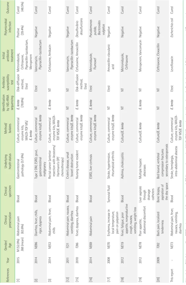
Table 1. Reported cases of Eggerthella lenta infection References YearGender/ AgeClinical presentationClinical specimenUnderlying heath statusMethod/ Isolates Identification by 16S rRNA sequencing Antibiotic susceptibility test Initial antibiotic treatment
Polymicrobial infectionOutcome [1]2015M (52.0%) /68 (mean)Abdominal pain (63.6%)BloodGastrointestinal pathology (57.6%)Culture, commercial enzyme kits, MALDI-TOF MS/ E. lenta
E. lenta (78.8%)Disk-diffusion method, Etest Metronidazole, Ceftriaxone, Piperacillin-tazobactam, Meropenem Positive (39.4%)Cured (48.5%) [2]2014M/86Diarrhea, fever, chills, rigor, dyspneaBloodType 2 DM, ESRD, gout, gastrointestinal malignancy
Culture, commercial enzyme kits/E. lentaNTEtestVancomycin, Piperacillin-tazobactamNegativeCured [3]2014M/53Abdominal pain, fever, chillsBloodRectal cancer (anterior resection with ileostomy/ Hartmann’s OP/ chemotherapy) Culture, commercial enzyme kits, MALDI- TOF MS/E. lenta
E. lentaNTCefotaxime, AmikacinNegativeCured [12]2011F/21Abdominal pain, nausea, vomiting, diarrheaBloodCrohn’s disease, small bowel obstructionCulture, commercial enzyme kits/E. lentaNTNTVancomycin, Piperacillin-tazobactamNegativeCured [15]2010F/86Fever, dyspnea, diarrheaBloodNursing home residentCulture, commercial enzyme kits/E. lentaE. lentaDisk-diffusion method, Etest
Cefuroxime, AmoxicillinDesulfovibrio desulfuricansCured [16]2014M/69Abdominal painBloodESRD, liver cirrhosisCulture, commercial enzyme kits/E. lentaE. lentaEtestMetronidazole, FlumoxefPseudomonas putida, Bacteroides vulgatus Cured [17] 2008M/70Erythema, increase in local temperature, pain on palpation
Synovial fluidStroke, hypertension, rheumatoid arthritisCulture, commercial enzyme kits/E. lentaNTEtestAmoxicillin-clavulanic acidNegativeCured [18]2012M/19Fever, fatigue, poor appetite, nonproductive cough, nausea, vomiting, weight loss
BloodAsthma, cholecystitisCulture/E. lentaNTNTMetronidazole, CeftriaxoneNegativeCured [19]2012M/78Poor appetite, abdominal discomfortLiver aspirate through drainage catheters Multiple hepatic abscessesCulture/E. lentaE. lentaNTMeropenem, VancomycinNegativeCured [20]2009F/82Back pain, localized tendernessBone biopsy, aspiration of the disc space
Bed bound, vertebral compression fractures secondary to osteoporosis Culture/E. lentaNTNTCeftriaxone, CloxacillinNegativeCured This report -M/73Abdominal pain, fever, nausea, vomiting, diarrhea
BloodStroke, hemiplegia, intra-abdominal abscessCulture, commercial enzyme kits, MALDI- TOF MS/E. lenta E. lentaEtestLevofloxacinEscherichia coliCured Abbreviations: DM, diabetes mellitus; ESRD, end-stage renal disease; Hartmann’s OP, Hartmann’s operation; MALDI-TOF MS, matrix-assisted laser desorption/ionization time-of-flight mass spectrometry; NT, not tested.
[6]. The bacterium was previously named Eubacterium lentum, but genetic analysis performed in 1999 showed that the bacterium merited its own genus [7]. Eggerthella and Eubacterium can be differentiated from closely related genera, including Propionibac- terium, Bifidobacterium, Lactobacillus, and Actinomyces, on the basis of acid-production patterns upon growth in peptone/
yeast extract/glucose broth. However, the bacteria are fastidious and slow-growing, and correct identification of E. lenta at the species level is difficult using conventional methods. Not even biochemical or fermentative reactions can reliably identify Eu- bacterium to the subspecies level [8].
Over the last decades, various commercial enzyme-based kits identifying clinically relevant anaerobes have been developed.
These include the RapID-ANA II panel (Thermo Fisher Scientific Inc., Lenexa, KS, USA), the Minitek systems (BBL Microbiology Systems, Cockeysville, MD, USA), the Vitek Anaerobe Identifica- tion (ANI) card (bioMérieux), the BBL Crystal ANR ID kit (BD Di- agnostics, Sparks Glencoe, MD, USA), and the API rapid ID 32A and API 20A systems (bioMérieux) [9-11]. 16S rRNA sequencing is useful for confirming an E. lenta infection and is considered to be the gold standard [12]. Although we used the VITEK 2 ANI card, VITEK MS, and 16S rRNA sequencing, we suggest that the VITEK 2 system alone is adequate; the system is in use by many hospitals.
We reviewed 10 reports of laboratory-documented E. lenta bacteremia occurring from 2008 to 2015 (Table 1). Initially, tradi- tional methods of bacterial identification (Gram staining, culture, and biochemical techniques) were used. Molecular techniques, including real-time PCR or microarray analysis, were secondarily employed to identify the causative organisms (71.4%). Many pa- tients (81.0%) exhibited symptoms of gastroenteritis associated
with diarrhea and/or vomiting. Abdominal pain was also often present. Those infected by E. lenta were already ill, and included patients hospitalized in intensive care units, the elderly, and pa- tients with diabetes and/or chronic kidney failure (86.0%). Cathe- ters (tubes inserted into blood vessels or the gallbladder) and other bodily tubes (e.g., running from the nose to the stomach) were common causes of bacteremia (19.0%). Several antimicrobial susceptibility testing methods (broth and agar dilution methods, disk-diffusion tests, the Etest, and automated testing systems) were used. The isolates were commonly susceptible to both clindamycin and metronidazole (Table 2).
In our case, E. coli was cultured from blood taken on the first day of hospitalization. Although we (empirically) prescribed levo- floxacin, an intermittent fever (over 38.9˚C) persisted for 4 days.
On day 5, E. lenta was detected in the blood. After commence- ment of metronidazole, the fever slowly resolved. Because the isolate was resistant to levofloxacin but susceptible to metronida- zole, we considered that the bacteremia was caused by a mixed infection of E. coli and E. lenta (the latter is not a normal compo- nent of skin flora). At the time of admission, the intra-abdominal abscess was the only obvious source of infection.
Recent reports have shown that several E. lenta infections were associated with the use of intrauterine devices, spondylodiscitis, female genital tract infections, cutaneous abscesses, and bactere- mia (in Crohn’s disease patients) [1-4, 12]. These data emphasize the importance of further investigation for bacterial virulence fac- tors. It is suspected that transfer of E. lenta from an intra-abdomi- nal abscess to the bloodstream may reflect a defect in the gastroin- testinal tract; E. lenta is normally a gut commensal bacterium [13, 14]. Some reports found that E. lenta infection caused morbidity
Table 2. Antimicrobial susceptibility testing results of previously reported Eggerthella lenta infections References Metronidazole Clindamycin Imipenem Amoxicillin-
clavulanic acid Penicillin G Trimethoprim-
sulfamethoxazole Meropenem Chloramphenicol
[1] S (100%)* S (91.0%)* NR S (100%)* S (39.0%)* NR S (100%)* NR
[2] S S NR NR R NR NR NR
[3] S NR S NR NR NR NR NR
[15] S S S S R NR NR R
[16] S NR NR NR R NR S S
[17] S S S S S NR NR NR
[18] S NR S NR NR NR S NR
This report S S S
*Numbers within parentheses represent the proportions of patients responding to antibiotics.
Abbreviations: NR, not reported; R, resistant; S, susceptible.
and mortality associated with septic shock and multiple organ fail- ure. Although the pathogenesis of E. lenta bacteremia has not been well described [15], 10 recent reports indicate that the prog- nosis is rather favorable [1-3, 12, 15-20]. Our patient required inten- sive care for several days but eventually recovered. Few data on E.
lenta infections or the optimal treatment thereof are available.
In summary, we have reported a case of bacteremia caused by E. lenta in an elderly patient with an intra-abdominal abscess. We also reviewed the current literature.
요 약
Eggerthella lenta는 그람 양성이면서 포자는 형성하지 않고 운 동성이 없는 혐기성 간균으로서 주로 사람의 장에서 분리되며 임 상 검체에서는 드물게 발견된다. 특히 균혈증인 경우 심각한 임상 경과를 초래하지만 동정이 쉽지 않고 보고가 적어 E. lenta의 감염 과 치료에 관한 자료를 쉽게 접할 수 없었다. 이에 저자들은 복강 내 농양을 가진 노인 환자에서 발생한 E. lenta 균혈증 1예를 문헌 고찰과 함께 보고하는 바이다.
REFERENCES
1. Gardiner BJ, Tai AY, Kotsanas D, Francis MJ, Roberts SA, Ballard SA, et al. Clinical and microbiological characteristics of Eggerthella lenta bacteremia. J Clin Microbiol 2015;53:626-35.
2. Wong D, Aoki F, Rubinstein E. Bacteremia caused by Eggerthella lenta in an elderly man with a gastrointestinal malignancy: a case re- port. Can J Infect Dis Med Microbiol 2014;25:e85-6.
3. Lee HJ, Hong SK, Choi WS, Kim EC. The first case of Eggerthella lenta bacteremia in Korea. Ann Lab Med 2014;34:177-9.
4. Goupil R, Nadeau-Fredette AC, Tennankore KK, Bargman JM. Perito- nitis caused by Veillonella species and Eggerthella lenta in peritoneal dialysis. Perit Dial Int 2014;34:245-7.
5. Clinical and Laboratory Standards Institute. Performance standards for antimicrobial susceptibility testing; twenty-second informational sup- plement, M100-S22, Wayne, PA: Clinical and laboratory Standards In- stitute, 2012.
6. Eggerth AH. The gram-positive non spore bearing anaerobic bacilli of human feces. J Bacteriol 1935;30:277-99.
7. Wade WG, Downes J, Dymock D, Hiom SJ, Weightman AJ, Dewhirst FE, et al. The family Coriobacteriaceae: reclassification of Eubacte- rium exiguum (Poco et al. 1996) and Peptostreptococcus heliotrinre-
ducens (Lanigan 1976) as Slackia exigua gen. nov., comb. nov. and Slackia heliotrinireducens gen. nov., comb. nov., and Eubacterium lentum (Prevot 1938) as Eggerthella lenta gen. nov., comb. nov. Int J Syst Bacteriol 1999;49:595-600.
8. Kageyama A, Benno Y, Nakase T. Phylogenetic evidence for the trans- fer of Eubacterium lentum to the genus Eggerthella as Eggerthella lenta gen. nov., comb. nov. Int J Syst Bacteriol 1999;49:1725-32.
9. Hansen SL and Stewart BJ. Comparison of API and minitek to Center for Disease Control methods for the biochemical characterization of anaerobes. J Clin Microbiol 1976;4:227-31.
10. Lee EH, Degener JE, Welling GW, Veloo AC. Evaluation of the Vitek 2 ANC card for identification of clinical isolates of anaerobic bacteria. J Clin Microbiol 2011;49:1745-9.
11. Marler LM, Siders JA, Wolters LC, Pettigrew Y, Skitt BL, Allen SD. Eval- uation of the new RapID-ANA II system for the identification of clini- cal anaerobic isolates. J Clin Microbiol 1991;29:874-8.
12. Thota VR, Dacha S, Natarajan A, Nerad J. Eggerthella lenta bactere- mia in a Crohn’s disease patient after ileocecal resection. Future Mi- crobiol 2011;6:595-7.
13. Lee MR, Huang YT, Liao CH, Chuang TY, Wang WJ, Lee SW. Clinical and microbiological characteristics of bacteremia caused by Egger- thella, Paraeggerthella, and Eubacterium species at a university hos- pital in Taiwan from 2001 to 2010. J Clin Microbiol 2012;50:2053-5.
14. Venugopal AA, Szpunar S, Johnson LB. Risk and prognostic factors among patients with bacteremia due to Eggerthella lenta. Anaerobe 2012;18:475-8.
15. Liderot K, Larsson M, Boräng S, Özenci V. Polymicrobial bloodstream infection with Eggerthella lenta and Desulfovibrio desulfuricans. J Clin Microbiol 2010;48:3810-2.
16. Lee J, Cho YG, Kim DS, Lee HS. Eggerthella lenta bacteremia after en- doscopic retrograde cholangiopancreatography in an end-stage renal disease patient. Ann Clin Microbiol 2014;17:128-31.
17. Sánchez FP, Jung PAR, Lamas LM, Barca BA. Septic bursitis due to Eg- gerthella lenta. Reumatol Clin 2008;4:80-4.
18. Salameh A, Klotz SA, Zangeneh TT. Disseminated infection caused by Eggerthella lenta in a previously healthy young man: a case report.
Case Rep Infect Dis 2012:517637.
19. Elias RM, Khoo SY, Pupaibool J, Nienaber JH, Cummins NW. Multiple pyogenic liver abscesses caused by Eggerthella lenta treated with er- tapenem: a case report. Case Rep Med 2012:e718130.
20. Bok CW, Ng YS. Eggerthella lenta as a cause of anaerobic spondylo- discitis. Singapore Med J 2009;50:e393-6.