ISSN 0378-6471 (Print)⋅ISSN 2092-9374 (Online)
https://doi.org/10.3341/jkos.2019.60.6.606
Case Report
정맥동 혈전증에 의한 시신경유두부종
Papilledema with Cerebral Venous Sinus Thrombosis
백민수⋅경성은
Min Su Baek, MD, Sung Eun Kyung, MD, PhD
단국대학교 의과대학 안과학교실
Department of Ophthalmology, Dankook University College of Medicine, Cheonan, Korea
Purpose: We report two patients diagnosed with a sinus thrombosis with papillary edema.
Case summary: Case 1 was a 27-year-old male who presented with complaints of headache and vomiting for 2 months and blur- red vision in both eyes. The best-corrected visual acuity (BCVA) was 1.0 in the right eye and 1.0 in the left eye. A visual field (VF) examination revealed a binocular peripheral VF defect and optical coherence tomography (OCT) and a fundus examination in- dicated optic disc swelling in both eyes. Brain magnetic resonance imaging (MRI) showed no specific finding but magnetic reso- nance venography revealed filling defect signs in the transverse sinus and a cerebrospinal fluid examination indicated elevated intracranial pressure (ICP). Case 2 was a 54-year-old female who came to our hospital with suspicion of bilateral optic disc swelling. The BCVA was 0.9 in the right eye and 1.0 in the left eye. A VF examination revealed an inferior-temporal VF defect and blind spot enlargement in the right eye. OCT and a fundus examination showed optic disc swelling in both eyes. Brain MRI showed no specific finding but magnetic resonance venography revealed a decrease in blood flow in the transverse sinus, sig- moid sinus. A cerebrospinal fluid examination indicated elevated ICP.
Conclusions: In the case of optic disc swelling in both eyes, a secondary cause of ICP elevation and the possibility of optic disc swelling due to sinus thrombosis should be considered, and brain MRI and venography are needed to distinguish these possibilities.
J Korean Ophthalmol Soc 2019;60(6):606-611
Keywords: Optic neuropathy, Papilledema, Venous sinus thrombosis
■Received: 2018. 8. 8. ■ Revised: 2018. 10. 29.
■Accepted: 2019. 5. 17.
■Address reprint requests to Sung Eun Kyung, MD, PhD Department of Ophthalmology, Dankook University Hospital,
#201 Manghyang-ro, Dongnam-gu, Cheonan 31116, Korea Tel: 82-41-550-6497, Fax: 82-41-561-0137
E-mail: [email protected]
*Conflicts of Interest: The authors have no conflicts to disclose.
ⓒ2019 The Korean Ophthalmological Society
This is an Open Access article distributed under the terms of the Creative Commons Attribution Non-Commercial License (http://creativecommons.org/licenses/by-nc/3.0/) which permits unrestricted non-commercial use, distribution, and reproduction in any medium, provided the original work is properly cited.
정맥동 혈전증은 드문 질환으로 매년 성인 백만 명당 3, 4명 의 유병률을 보인다. 전 연령에 걸쳐 발생할 수 있지만 주 로 30대에서 호발하며, 75%는 여성에서 발생한다.1 정맥동 혈전증은 광범위한 임상양상으로 인해 진단하기 어려운 질 환으로 두통이 가장 흔한 임상 증상이나 이차적 전신발작
을 동반하거나 동반하지 않는 부분발작, 편측 혹은 양측마 비, 인지저하나 시각장애와 같은 다른 신경적 증상도 동반 가능한 것으로 알려져 있다.2,3 정맥동 혈전증의 원인은 크 게 감염성과 비감염성으로 나눌 수 있으며,2,4 감염성은 주 변 안면이나 부비동, 치아에서 전파되는 감염에 의하여 발 생하며 비감염성은 외상, 수술이나 시술 후, 자가면역질환, 응고항진질환, 뇌동맥류 등의 원인들에 의하여 발생하는 것으로 보고된 바 있다.4
시신경유두에 발생하는 부종은 양안성인 경우 뇌척수액 압 상승, 독소, 침윤, 악성고혈압 등이 주된 원인이며, 단안 성인 경우 시신경염, 앞허혈시신경병증, 압박, 망막정맥폐 쇄, 당뇨시신경유두병증, 감염 등이 주 원인이다. 이 중에서
A B
C D E
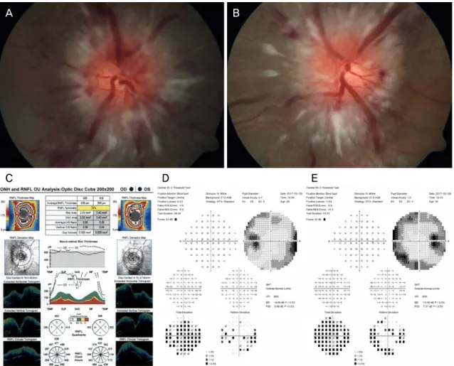
Figure 1. Case 1. The fundus showed the diffuse optic disc swelling and hemorrhage in both eye at the first visit (A) OD, (B) OS.
(C) The Optical coherence tomography showed the increased RNFL thickness in both eyes at the first visit. (D, E) The visual field showed peripheral constriction and enlarged blind spot in both eyes at the first visit. OD = oculus dexter; OS = oculus sinister;
ONH = optic nerve head; RNFL = retinal nerve fiber layer; OU = oculus unitas; S = superior; N = nasal; T = temporal; I = inferior; POS = positive; NEG = negative; RX = prescription; DS = dioptres sphere; DC = dioptres cylinder; GHT = global hemifield test; VFI = visual field index; MD = mean deviation; PSD = pattern standard deviation.
시신경유두부종(papilledema)은 뇌척수액압의 증가에 의하 여 시신경유두에 부종(optic disc swelling)이 발생한 것을 의미한다.5 시신경유두부종은 정맥동 혈전증이 있는 환자 중 28% 정도에서 발생한다.3 저자들은 외상이나 기저질환 의 기왕력 없이 두통 또는 무증상의 양안 시신경유두부종 으로 내원한 2명의 환자에서 정맥동 혈전증을 진단하였기 에 이를 보고하고자 한다.
증례보고
증례1
고혈압 및 당뇨 등 특이기저질환이 없던 26세 남자 환자 가 1주일 전부터 시작된 구토 및 열감, 식은땀을 주소로 내 원하였다. 나안시력 우안 1.0, 좌안 1.0이었고 안압은 우안 22 mmHg, 좌안 17 mmHg, 상대구심성동공운동검사 및 Ishihara
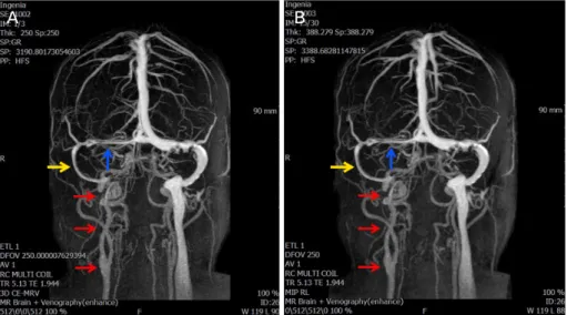
색각검사에서 양안 정상이었다. 안저검사에서 양안 시신경 유두부종 소견이 관찰되었다(Fig. 1A, B). 빛간섭단층촬영 에서 양안 망막신경섬유층의 두께 증가 소견(Fig. 1C)을 보 이고 시야검사(Humphrey visual field test, central 30-2 threshold)에서 양안 주변부 시야감소 및 맹점확대 소견이 보였다(Fig. 1D, E). 신경과 검사에서 뇌척수압은 36 cmH2O 로 증가되어 있었고, 뇌 자기공명영상에서는 정상 소견을 보였으나 뇌 자기공명정맥조영검사에서 횡정맥동에서의 충만결손 소견이 관찰되었다(Fig. 2). 신경과에서 Warfarin 5 mg을 경구투여하였으며 2개월 뒤 마지막 내원 시 나안시 력 우안 1.0, 좌안 0.6, 안압은 우안 18 mmHg, 좌안 20 mmHg, 빛간섭단층촬영 및 안저검사에서 양안 시신경유두부종 호 전 소견 및 출혈 감소 소견을 보였으나 이후 추적관찰에 실 패하였다.
A B
Figure 2. Case 1. Low signal intensity contrast filling defect in superior sagittal sinus (posterior portion, red arrows), great vein of
Galen, straight sinus, right transverse sinus (blue arrow), right sigmoid sinus (yellow arrow), and right IJV (green arrow) (A, B).SE = series; IJV = internal jugular vein; MR = magnetic resonance.
증례2
과거력상 고혈압 및 당뇨 등 특이기저질환이 없던 54세 여자 환자가 정기 안과검진상 양안 유두부종 소견을 주소 로 내원하였다. 최대교정시력 우안 0.9, 좌안 1.0으로 측정 되었으며, 안압은 우안 10 mmHg, 좌안 12 mmHg였고 상 대구심성동공운동검사 및 Ishihara 색각검사에서 양안 정상 이었다. 안저검사에서 양안 시신경유두부종 소견이 관찰되 었다(Fig. 3A, B). 빛간섭단층촬영에서 양안 망막신경섬유 층의 두께 증가 소견(Fig. 3C)을 보이고 시야검사(Humphrey visual field test, central 30-2 threshold)에서 우안 하이측 시 야결손, 맹점확대 소견이 보였다(Fig. 3D, E). 신경과검사에 서 뇌척수압은 22 cmH2O로 증가되어 있었고, 뇌 자기공명 영상은 정상 소견을 보였으나 뇌 자기공명정맥조영검사에 서 우측 횡정맥동 및 구불정맥동에서의 혈류감소 및 우측 속목정맥의 비관류 소견이 관찰되었다(Fig. 4). 신경과에서 Acetazolamide 500 mg을 경구투여하였으며, 3개월 후 시행 한 안저검사와 빛간섭단층촬영검사상 양안 시신경유두부 종 호전 소견을 보였다. 이후 정기적으로 검사한 결과 자동 시야검사에서 시야 결손 등의 변화가 없어 뇌실복강막단락 등의 치료 없이 18개월 동안 경과관찰하였다.
고 찰
유두부종이란 두개내압 상승에 의한 이차적인 시신경유 두부종을 의미한다. 두개내압 상승에 의한 유두부종은 시 신경 주변의 증가된 압력이 사상판전 신경섬유 내에서 축 삭형질흐름을 정체시키고, 정맥혈류의 압박 또는 축삭돌기
팽윤으로 인해 발생한 정맥울혈이 간질성 부종을 일으켜 발생하게 된다.6
정맥동 혈전증은 임신, 신장질환, 구강 피임약 같은 약물, 베체트병, 유육종증 및 전신성 홍반성 루푸스 같은 염증성 질환과 관련이 있는 것으로 알려져 있다. 특히 뇌 정맥동 혈전증은 요추 천자, 경정맥 카테터 삽입, 수술 및 약물 사 용 후, 두부 외상이 있을 경우 발병 위험도가 높다고 알려 져 있다. 65세 이상의 환자에게 가장 흔한 위험 요인은 혈 전증과 악성 종양으로 알려져 있으며, 원인을 알 수 없는 경우도 20-25%로 보고된 바 있다.2 두통이 가장 흔한 임상 증상이고 가장 흔한 시야 변화는 맹점의 확대이며, 하이측 시야소실 및 전반적인 시야감도의 저하가 일반적이다.7-12 임상적으로 정맥동 혈전증이 두개내압 상승을 일으키는 경 우는 흔하지 않지만,13 발생하는 경우 두개내 정맥동에 존 재하는 거미막 과립을 통한 흡수가 감소되어 뇌척수액 압 력이 이차적으로 증가하는 것으로 알려져 있다. 혈전이 상 시상 정맥동에 위치할 때 정맥동 혈전증에 의한 보상 기전 으로 대뇌 정맥의 팽창과 함께 측부혈관이 생성되며, 궁극 적으로 증가한 정맥압으로 인해 모세혈관 관류를 감소시켜 혈관성 부종 또는 세포독성 부종을 일으키고 이로 인해 두 개내압 상승이 발생한다. 임상적으로 정맥동 혈전증 환자 는 국소적인 두개내압 상승 증후군, 국소 증후군, 뇌병증의 특징적이고 전형적인 세 가지 신경학적 증상을 보인다.14
정맥동 혈전증의 징후와 증상이 나타날 때 대개 정맥동 혈전증의 진단은 신경 영상으로 확진되며 뇌 자기공명영상 촬영 또는 전산단층촬영검사에서 특징적인 징후가 나타날 수 있다. Ozsvath et al15은 대뇌 정맥과 경막 정맥동만을 조
A B
Figure 4. Case 2. Smooth margined non visualization of flow signal right IJV (red arrows) and decreased flow signal in right trans-
verse (blue arrow) and sigmoid sinus (yellow arrow) (A, B). SE = series; IJV = internal jugular vein; MR = magnetic resonance.A B
C D E
Figure 3. Case 2. The fundus showed the diffuse optic disc swelling and hemorrhage in both eye at the first visit (A) OD, (B) OS.
(C) The Optical coherence tomography showed the increased RNFL thickness in both eyes at the first visit. (D, E) The visual field showed enlarged blind spot with inferior visual field defect in right eyes at the first visit. OD = oculus dexter; OS = oculus sinister;
ONH = optic nerve head; RNFL = retinal nerve fiber layer; OU = oculus unitas; S = superior; N = nasal; T = temporal; I = inferior; POS = positive; NEG = negative; RX = prescription; DS = dioptres sphere; DC = dioptres cylinder; GHT = global hemifield test; VFI = visual field index; MD = mean deviation; PSD = pattern standard deviation.
영하는 면에 있어서는 전산단층촬영 뇌혈관조영술이 자기 공명영상촬영보다 우수하지만 정맥동 혈전증의 진단에 있 어서는 전산단층촬영과 자기공명영상촬영을 모두 유용하 다고 하였다. 그러나 전산단층촬영은 방사선 노출의 위험 이 있으며 뇌 자기공명영상촬영만으로는 정맥동 혈전증을 진단하기 어렵기 때문에 정맥동 혈전증을 가진 환자를 특 발성 두개강내압 증가 환자로 잘못 진단할 가능성이 있다.
따라서 진단의 민감도는 뇌 자기공명영상과 정맥조영술을 동시에 시행할 때 가장 높으며,정맥동 혈전증이 의심되는 환자에서 뇌 자기공명정맥조영검사를 같이 시행하는 것이 필요한 것으로 알려져 있다.14
정맥동 혈전증으로 인한 대뇌피질과 인접한 백질의 양측 출혈성 경색은 치명적인 결과일 수 있으므로, 조기 진단 및 치료는 정맥동 혈전증 환자의 시각적 기능과 신경학적 기 능을 유지하는 데 중요한 역할을 할 것으로 생각된다.16 본 증례는 기저질환 없이 갑작스럽게 발생한 양안 유두부종 환자들에서 뇌 자기공명정맥조영검사상 정맥 혈전이 발견 되어 유두부종의 원인으로 확인되었다. 정맥동 혈전증은 다양한 신경학적 증상으로 나타날 수 있으며 뇌 자기공명 영상에는 정상 소견을 보이고 뇌척수압이 높은 특발성 두 개강내압 증가의 증상과 비슷하므로 감별 진단에 유의해야 할 것으로 생각된다. 따라서 유두부종이 있는 경우 환자에 서 정맥동 혈전증과 특발성 두개내압 상승과의 감별을 위 해 뇌 자기공명영상과 함께 뇌 자기공명정맥조영검사를 고 려하여야 할 것으로 생각된다.
REFERENCES
1) Einhäupl K, Bousser MG, de Bruijn SF, et al. EFNS guideline on
the treatment of cerebral venous and sinus thrombosis. Eur J Neurol 2006;13:553-9.
2) Ferro JM, Canhão P, Stam J, et al. Prognosis of cerebral vein and dural sinus thrombosis: results of the International Study on Cerebral Vein and Dural Sinus Thrombosis (ISCVT). Stroke 2004;35:664-70.
3) Kimber J. Cerebral Venus Thrombosis. QJM 2002;95:137-42.
4) Keane JR. Cavernous sinus syndrome. Analysis of 151 cases. Arch Neurol 1996;53:967-71.
5) Van Stavern GP. Optic disc edema. Semin Neurol 2007;27:233-43.
6) Singh RJ, Saini J, Varadharajan S, et al. Headache in cerebral venous sinus thrombosis revisited: exploring the role of vascular con- gestion and cortical vein thrombosis. Cephalalagia 2018;38:503-10.
7) Lessell S, Rosman NP. Permanent visual impairment in childhood pseudotumor cerebri. Arch Neurol 1986;43:801-4.
8) Baker RS, Carter D, Hendrick EB, Buncic JR. Visual loss in pseu- dotumor cerebri of childhood. A follow-up study. Arch Ophthalmol 1985;103:1681-6.
9) Corbett JJ, Savino PJ, Thompson HS, et al. Visual loss in pseudotu- mor cerebri. Follow-up of 57 patients from five to 41 years and a profile of 14 patients with permanent severe visual loss. Arch Neurol 1982;39:461-74.
10) Wall M, George D. Visual loss in pseudotumor cerebri. Incidence and defects related to visual field strategy. Arch Neurol 1987;44:170-5.
11) Corbett JJ, Nerad JA, Tse DT, Anderson RL. Results of optic nerve sheath fenestration for pseudotumor cerebri: the lateral orbitotomy approach. Arch Ophthalmol 1988;106:1391-7.
12) Dersh J, Schlezinger NS. Inferior nasal quadrantanopia in pseudo- tumor cerebri. Trans Am Neurol Assoc 1959;84:116-8.
13) Orcutt JC, Page NG, Sanders MD. Factors affecting visual loss in benign intracranial hypertension. Ophthalmology 1984;91:1303-12.
14) O'Rourke TL, Slagle WS, Elkins M, et al. Papilloedema associated with dural venous sinus thrombosis. Clin Exp Optom 2014;97:133-9.
15) Ozsvath RR, Casey SO, Lustrin ES, et al. Cerebral venography:
comparison of CT and MR projection venography. Am J Roentgenol 1997;169:1699-707.
16) Shah S, Saxena D. Bilateral papilledema: a case of cerebral venous sinus thrombosis. Oman J Ophthalmol 2014;7:33-4.
= 국문초록 =
정맥동 혈전증에 의한 시신경유두부종
목적: 양안 시신경유두부종이 관찰된 2명의 환자에서 정맥동 혈전증을 진단하여 이를 보고하고자 한다.
증례요약: 증례 1) 27세 남자 환자가 2달 전부터 발생한 두통 및 구토, 양안 시야 흐림을 주소로 내원하였다. 최대교정시력은 우안 1.0, 좌안 1.0이었으며, 시야검사에서 양안 주변부 시야결손, 빛간섭단층촬영 및 안저검사에서 양안 시신경유두부종이 관찰되었다.
뇌 자기공명영상에서는 정상 소견을 보였으나 뇌 자기공명정맥조영검사에서 횡정맥동 충만결손 소견이 있었고, 뇌척수액검사에서 뇌압상승 소견을 보였다. 증례 2) 54세 여자 환자가 양안 시신경유두부종으로 의뢰되었다. 최대교정시력은 우안 0.9, 좌안 1.0이었으 며, 시야검사에서 우안 하이측 시야결손과 맹점확대 소견, 빛간섭단층촬영 및 안저검사에서 양안 시신경유두부종이 관찰되었다. 뇌 자기공명영상에서는 정상 소견을 보였으나 뇌 자기공명정맥조영검사에서 횡정맥동, 구불정맥동에서의 혈류 감소 소견이 있었으며, 뇌척수액검사에서 뇌압상승 소견을 보였다.
결론: 양안의 시신경유두부종이 있는 경우 정맥동 혈전증에 의한 유두부종의 가능성을 고려하여 뇌 자기공명영상과 함께 뇌 자기공명 정맥조영검사를 고려하여야 할 것으로 생각된다.
<대한안과학회지 2019;60(6):606-611>
백민수 / Min Su Baek
단국대학교 의과대학 안과학교실 Department of Ophthalmology, Dankook
University College of Medicine