Original Article
Synergistic growth inhibition by combination of adenovirus mediated p53 transfer and cisplatin
in ovarian cancer cell lines
Sang Young Ryu1, Kidong Kim1, Woo Sik Lee2, Hee Chung Kwon2, Kee Ho Lee2, Chang Min Kim3, Soon-Beom Kang4
1Department of Obstetrics and Gynecology, 2Laboratory of Molecular Biology, Korea Cancer Center Hospital, Seoul, 3Department of Internal Medicine, National Cancer Center, Goyang, 4Department of Obstetrics and Gynecology, Seoul National University College of Medicine, Seoul, Korea
Objective: This study was to investigate the synergistic growth inhibitory effect by combination of adenovirus mediated p53 gene transfer and cisplatin in ovarian cancer cell lines with different p53 gene mutation patterns.
Methods: Three ovarian cancer cell lines, p53 deleted SKOV3, p53 mutated OVCAR-3, and PA-1 with wild-type p53 were transduced with human adenovirus vectors carrying p53 gene (Ad-p53) and treated with a sublethal concen- tration of cisplatin before and after Ad-p53. The cell number was counted daily for 5 days after Ad-p53 transduction.
Western blotting was used to identify p53 and p21 protein expressions, and flow cytometric analysis was performed to investigate any change of DNA ploidy after Ad-p53 transfer.
Results: Ad-p53 transduced cells successfully expressed p53 and p21 proteins after 48 hours of Ad-p53 transduction.
Synergistic growth inhibition by combination of Ad-p53 and cisplatin was detected only in SKOV3 and OVCAR-3 cells, but not in PA-1 cells. In p53 deleted SKOV3 cells, cisplatin treatment after Ad-p53 showed higher growth inhibition than the treatment before Ad-p53 transduction, and reverse relationship was observed in p53 mutated OVCAR-3 cells. In SKOV3 cells, the fraction of cells at G2/M phase increased after cisplatin treatment, however, it decreased dramatically with Ad-p53 transduction.
Conclusion: The synergistic growth inhibition by combination of Ad-p53 and cisplatin may depend on the p53 status and the temporal sequence of cisplatin treatment, suggesting judicious selective application of this strategy in clinical trials.
Key Words: p53, Adenoviruses, Gene therapy, Cisplatin
Received October 23, 2008, Revised December 12, 2008, Accepted December 13, 2008
Address reprint requests to Soon-Beom Kang
Department of Obstetrics and Gynecology, Seoul National Uni- versity College of Medicine, 28, Yeongeon-dong, Jongno-gu, Seoul 110-744, Korea
Tel: 82-2-2072-3384, Fax: 82-2-762-3599 E-mail: [email protected]
INTRODUCTION
Ovarian cancer is a malignancy with a poor prognosis, and the majority of patients have disease disseminated outside of the ovary at the time of initial diagnosis.1 Despite aggressive surgical debulking and introduction of platinum based che- motherapy regimens, the mortality rate associated with this tumor remains high, and the overall 5-year survival rate is on- ly approximately 11-25%.2 Because of the poor prognosis as- sociated with this cancer, it has become imperative that new treatment modalities continue to be introduced and tested. In
recent years, the use of gene therapy with adenoviral vector constructed to express the protein products of tumor sup- pressor genes has received a great deal of attention. Because of its inherent death-inducing activity, and it's prevalent muta- tion in 40-80% of ovarian tumors, the majority of gene ther- apy research has focused on the p53 tumor suppressor gene.3-6 In the presence of chromosomal alterations, p53 up-regu- lates not only p21-cyclin-cdk pathway that is essential in the transition from G1 to S phase of cell cycle, but also induces apoptotic pathway to programmed cell death.7-9 Restoration of p53 gene function by wild-type p53 gene transfer leads cells to G1 arrest and apoptosis, resulting in cell growth inhibition and death in vitro and in vivo.9-14 However, the gene therapy with the tumor suppressor gene alone has pitfalls. Aside from p53 gene mutations, most human cancer cells are known to have coincidental down-stream gene mutations involved in apoptosis or growth arrest. To overcome such limitations of gene therapy, a combination with conventional treatment mo- dalities, such as chemotherapeutic agents or radiation, is sug-
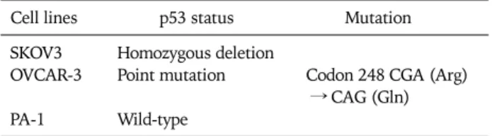
Table 1. The p53 status of three ovarian cancer cell lines
Cell lines p53 status Mutation
SKOV3 Homozygous deletion
OVCAR-3 Point mutation Codon 248 CGA (Arg) → CAG (Gln) PA-1 Wild-type
gested as a possible alternative strategy for human adenovirus vectors carrying p53 gene (Ad-p53) single gene therapy.15-17
Cisplatin, a cell cycle nonspecific DNA damaging agent, is a chemotherapeutic agent widely used in human ovarian cancer. Singularly, cisplatin is one of the most effective che- motherapeutic drugs in advanced ovarian cancers with re- sponse rates of 30-60%.18,19 A combination of Ad-p53 and cis- platin is known to have not only additive cytotoxic effect on human ovarian cancer cells but also a synergistic manner, ren- dering this combination as an effective gene therapy strategy.15-17 However, most of the studies have been per- formed with limited number of ovarian cancer cell lines.
Furthermore, a combination of Ad-p53 and cisplatin theoret- ically may elicit different cytotoxic effect depending on p53 gene status of cancer cells. However, questions of whether the synergistic tumor growth inhibition depends on p53 gene sta- tus in the ovarian cancer cells have not been well studied.
The temporal sequence of adminstration of Ad-p53 and cis- platin is an another critical factor for this combination strategy. In several cancer cell lines including ovarian cancer, sequence of Ad-p53 administration followed by cisplatin is known to inhibit tumor growth more effectively than that of other dosing schedules.17,20 On the other hand, a recent study found that the reverse sequence was more effective in p53 de- leted lung cancer cell lines in inhibiting growth compared to other dosing schedules.21 It appears, therefore, that the differ- ential cytotoxic effect of Ad-p53 and cisplatin combination due to temporal sequence of adminstration depends on the type of cancer cells, and this differential effectiveness is an ex- tremely important factor in clinical settings and should be clearly elucidated.
The mechanism of synergy with combination of Ad-p53 and cisplatin, however, is not well understood. With restoration of p53 function, cell cycle progression of the damaged cancer cells is checked at the G1/S phase and the cells are led to the apoptotic pathway and eventually cell death.7-9,22 However, the p53 gene has recently been shown to be a major factor in the G2/M phase cell cycle check point.23 Therefore, after DNA damage, cell arrest occurs at the transition of not only the G1/S phase but also the G2/M phase of the cell cycle, and the p53 gene in the G2/M phase cell cycle check point inhibits damaged cells from progressing to mitosis, thereby producing aneuploidy or polyploidy cells, which are known to be re- sistant to chemotherapeutic agents.24-27 Therefore, there is a good possibility that DNA ploidy might be altered after p53 gene transfer, and this could be one of the mechanisms in- volved in the synergy of combination of Ad-p53 and cisplatin.
In this study, we investigated the synergistic tumor sup- pression effect of combination of Ad-p53 gene transfer and cisplatin in ovarian cancer cell lines with various p53 gene mutation patterns and also the change of DNA ploidy after Ad-p53 transfer.
MATERIALS AND METHODS 1. Cell lines
SKOV3 cell line with homozygous deleted p53 and PA-1 cell line with wild type p53 were maintained in DMEM (GIBCO, Grand Island, NY, USA) containing 10% fetal bovine serum (FBS), 100 U/ml penicillin and 100 μg/ml streptomycin.
OVCAR-3 cell line with point mutated p53 was maintained in RPMI 1640 supplemented wild 10% FBS and insulin (Table 1).28 The adenovirus stocks were propagated in the human transformed embryonal kidney 293 cell line, as described previously.29
2. Vectors
The replication defective human adenovirus vectors (from Dr. Robert Gerard, Southwestern Medical Center, Texas Univ., USA) were produced via homologous recombination between two transfected plasmids, pACCMVpLpV and pJM17, containing adenovirus DNA fragments overlapped at the E1A region. The expression cassette containing wild-type p53 cDNA, driven by the cytomegalovirus (CMV) early enhancer and promotor followed by multiple cloning region and SV40 polyadenylation signal, was inserted in place of the E1a deletion. To construct Ad-CMV-LacZ and Ad-CMV-Luc, cas- settes containing the β-galactosidase and luciferase gene were inserted in place of the E1A region, respectively.
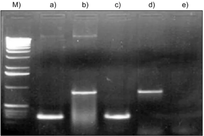
The pACCMVpLpA and pJM17 were co-transfected to hu- man 293 cells, which express E1A protein, by the calcium- phosphate method to propagate the recombinant viruses.30 High titer of adenovirus stock was prepared by two rounds of CsCl-density gradient centrifugation by the published protocol.31 The presence of the Ad-p53 was confirmed by the two PCR ampli- fications of p53 exon4 (S: 5'-ATCTACAGTCCCCCTTGCCG-3', AS: 5'-GCAACTGACCGTGCAAGTCA-3') and E2B sequence in adenovirus (S: 5'-TCGTTTCTCAGCAGCTGTTG-3', AS:
5'-CATCTGAACTCAAAGCGTGG-3') (Fig. 1). The titer of vi- ral concentration was determined by plaque forming assay as described before.32
3. β-galactosidase staining assay
To determine the transduction efficiency of adenovirus vec- tor, 5×10⁴ cells/well of ovarian cancer were plated and cul- tured for 2 days in 6-well plates and then transfected with 0, 5, 10, 20, and 50 MOIs (multiplicity of infection) of Ad-LacZ
Fig. 1. PCR amplification of p53 target sequences in recombinant Ad-p53 shows 240 bp product of p53 exon 4 (a), and 850 bp prod- uct of adenovirus E2B (b). M: size marker, c) positive control for p53, d) Ad-Luc, e) negative control.
Fig. 2. Transduction efficiency of adenovirus in ovarian cancer cell lines. MOIs of adenovirus with 70-80% transduction efficiency were 20 in SKOV3, 5 in OVCAR-3, and 50 in PA-1 cells.
Fig. 3. Sublethal concentration of cisplatin in ovarian cancer cells.
Concentration of cisplatin with growth inhibition below 10% of the control was 0.05μg/ml in SKOV3, OVCAR-3 and PA-1 cells.
Fig. 4. Western blotting for p53 and p21 protein expressions after Ad-p53 shows effective p53 and p21 protein expressions in SKOV3 cells. OVCAR-3 and PA-1 cells already expressed p53 and p21 proteins.
for an hour with serum free media and incubated for addi- tional 48 hours. After fixation with 0.5% glutaldehyde sol- ution, β-galactosidase activity was evaluated by incubation with X-gal staining solution [5 mM K3Fe(CN)6, 5 mM K4Fe(CN)6symbol 183 \f "Symbol" \s 83H2O, 2 ml 5×de- tergent, 1 mg/ml X-gal, 6.75 ml phosphate buffered saline (PBS)](Fig. 2).
4. Western blot analysis
p53 and p21 protein expressions before and after Ad-p53 transfer were determined by Western blot analysis. 1×10⁶ cells were plated in 60 mm dishes for 2 days, transduced with Ad-p53, and harvested 2 days after Ad-p53 transfer as de- scribed before. After solubilization with RIPA buffer (20 mM Tris-HCl, pH 7.5, 150 mM NaCl, 0.1% SDS, 1% Triton X-100, 1% sodium deoxycholate, 2 mM PMSF, 1μg/ml Aprotinin, 10 mM NaF, 1 mM sodium orthovanadate), proteins were separated on 12% acrylamide gel and transferred to nitro-
cellulose filter paper. Specific protein bands were probed with anti-p53 and anti-p21 antibodies (Oncoscience p53 Ab-1, SC-397, USA) and labelled with anti-mouse antibody [Goat anti-mouse IgG (H+L)-AP Conjugated, Bio-Rad]. ECL kit (Asherman) was used to visualize the specific bands.
5. Sublethal concentration of cisplatin
To determine the sublethal concentration of cisplatin to in- hibit cell growth below 10% of the control, 5×10⁴ ovarian cancer cells were cultured in 6-well plates for 2 days, and treated with serially diluted concentrations of cisplatin. After 2 days, the cells were harvested and stained with Trypan blue solution, and the cell number was counted by hemocytometer (Fig. 3).
6. Cell growth assay
5×10⁴ ovarian cancer cells were cultured in 6-well plates for 2 days, and treated with Ad-p53 for an hour in serum free media. In p53+P group, sublethal concentration of cisplatin was added after 24 hours of Ad-p53 transduction, and before 4 hours of Ad-p53 transduction in P+p53 group. Three con-
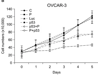
Fig. 5. The growth curves of ovarian cancer cells show growth in- hibition with Ad-p53 (p53 group) (A, B, C). The synergistic tumor growth suppression effect with combination of Ad-p53 and cispla- tin was observed only in p53+P group of SKOV3 (A) and P+p53 group of OVCAR-3 cells (B). Growth curve of PA-1 cells did not show a synergy with combination of Ad-p53 and cisplatin (C).
Points: mean; bars: standard error.
trol groups, C, P and Luc were treated with PBS, sublethal concentration of cisplatin, and Ad-Luc, respectively. Cells were counted daily for 5 days after Trypan blue staining by hemocytometer. Each plate was triplicated, and the cell growth was reconfirmed by Sulforhodamine B (SRB) assay.33
7. Flow cytometric analysis
1×10⁶ cells/well were cultured for 2 days and treated with Ad-p53. Twenty four hours later, the cells were treated with 5×10⁻⁸ gm/ml concentration of cisplatin, further cultured for 72 hours and analysed with flowcytometry for G2/M phase fraction after propidium iodide staining.34 In control and P groups, PBS and 5×10⁻⁸ gm/ml concentration of cisplatin were added to the media, respectively.
RESULTS 1. Transduction efficiencies
As shown in Fig. 2, the ovarian cancer cell lines showed 70-80% transduction efficiency with 20 multiplicity of in- fections (MOIs), 5 MOIs, and 50 MOIs in SKOV3, OVCAR-3,
and PA-1 cell lines, respectively.
2. Sublethal concentration of cisplatin
With 5×10⁻⁸ gm/ml concentration of cisplatin, all three ovarian cancer cells showed cytotoxic effect below 10% of the control (Fig. 3).
3. p53 and p21 expressions after Ad-p53
Western blotting analysis of p53 and p21 protein ex- pressions after Ad-p53 transfer indicated effective p53 and p21 protein expressions in SKOV3 cells. In OVCAR-3 and PA-1 cells, however, p53 and p21 proteins were already ex- pressed before Ad-p53 transfer (Fig. 4).
4. Cell growth assay
All three ovarian cancer cells manifested growth inhibition with Ad-p53 transfer alone, however, the synergy by combina- tion of Ad-p53 and cisplatin was detected in both SKOV3 and OVCAR-3 cells. In SKOV3 cells, Ad-p53 administration fol- lowed by cisplatin showed a synergistic cytotoxic effect, and synergistic cytotoxic effect with reverse treatment was ob-
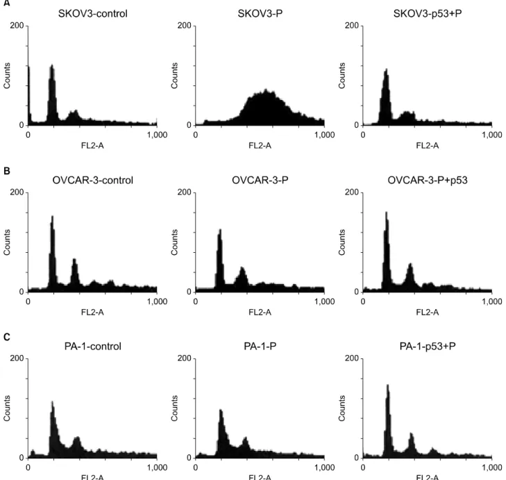
Fig. 6. Flow cytometric analysis show that cell fraction at G2/M phase increased after cisplatin treatment in p53 deleted SKOV3 cells, how- ever, G2/M phase fraction decreased dramatically after Ad-p53 transfer (A). The changes of G2/M phase fraction of OVCAR-3 and PA-1 cells were not definitive after cisplatin treatment, but G1/S phase fraction were slightly increased after Ad-p53 transfer (B, C). Legends are the same as described in Fig. 5.
served in OVCAR-3 cells (Fig. 5).
5. Change of cell fraction at G2/M phase with Ad-p53 transfer
Cell fraction at G2/M phase increased after cisplatin treat- ment in p53 deleted SKOV3 cells, however, G2/M phase frac- tion decreased dramatically after Ad-p53 transfer. G2/M phase cell fraction of OVCAR-3 and PA-1 cells did not change significantly with Ad-p53 transfer (Fig. 6).
DISCUSSION
The results described herein demonstrated that a combina- tion of Ad-p53 transfer and cisplatin had a synergistic in- hibitory effect on growth in ovarian cancer cell lines whose p53 gene was either deleted or mutated. In the cells with wild-type p53 gene, there was no synergistic cytotoxic effect, indicating that the synergy depended on p53 status of the
cells. The mechanism of synergistic growth inhibition has been known to be due to enhanced sensitivity of the cells to cisplatin with restored p53 gene function after wild-type p53 gene transfer.16,17 Indeed, in SKOV3 and OVCAR-3 cells with mutated or deleted p53 gene, restoration of p53 gene function with Ad-p53 most likely enhanced the sensitivity of the cells to cisplatin. On the other hand, in PA-1 cell line with wild type p53 gene, there was no synergistic growth inhibition by com- bination of Ad-p53 and cisplatin. The lack of synergy in PA-1 cell line might be due to the fact that there exist certain hither- to unknown impediments in the expression of the p53-p21 downstream genes or lack of induction of the chemo- sensi- tivity mediating factors. Since the whole genetic alteration in a specific cell line is not clearly defined, exact mechanisms in- volved in the response of PA-1 cells can not be offered at present.
Temporal sequence of Ad-p53 and cisplatin administration was found to be profoundly important to induce synergistic growth inhibition in ovarian cancer cells. In p53 deleted SKOV3 cells, the synergy was observed only in the sequential administration of Ad-p53 followed by cisplatin, whereas re- verse sequence was effective in OVCAR-3 cells. The mecha- nism of dependency of synergy on the sequence of admin- istration remains unclear, however, as explained below, it ap- peared to necessitate intact p53-p21 pathway. In p53 deleted SKOV3 cells, the synergy was observed after establishment of p53-p21 pathway with Ad-p53, which induced p21 protein expression. In OVCAR-3 cells, which were already highly ex- pressing p53 and p21 proteins, the synergy was observed with cisplatin administration before Ad-p53 transfer. The depend- ency of synergy on temporal sequence of Ad-p53 and cisplatin administration appeared to involve specific characteristics of cancer cells, nevertheless, establishment of p53 gene function before cisplatin administration seemed to be the most effec- tive means to evoke a synergistic growth inhibition in ovarian cancer cells.
Our data showed that the G2/M phase cell fraction of p53 gene deleted SKOV3 cells increased by cisplatin admin- istration, but this increase declined dramatically after Ad-p53 transfer. This result is in good agreement with recent studies that p53 protein is also involved in the G2/M cell cycle check point, inhibiting cells to progress to additional mitosis, re- sulting in aneuploidy or polyploidy cells which are sensitive to chemotherapeutic agents.25-27 The main mechanism of p53 gene transfer to cells with non-functioning p53 gene has been known to be due to enhanced induction of apoptosis by trans- ferred p53 protein.35,36 After DNA damage, cells are arrested at the G1/S phase of the cell cycle, thus preventing DNA repli- cations and mitosis in the presence of un-repaired chromoso- mal alterations.24,25 In the present study, we observed the change of DNA ploidy after Ad-p53 transfer in p53 gene de- leted ovarian cancer cell lines, however, we were not certain how much this contributed to the apparent synergistic growth inhibitory effect by the combination of Ad-p53 and cisplatin.
In OVCAR-3 cells with p53 gene mutated, there was no change of DNA ploidy with Ad-p53, however, still showed a synergy. This conflicting data left us at a loss to explain the role of DNA ploidy with regard to chemosensitivity. However, it is likely that the change of DNA ploidy might participate partly in the synergy of combination of Ad-p53 and cisplatin in p53 gene deleted ovarian cancer cell lines.
A limitation of our present study was that we selected three ovarian cancer cell lines with different p53 gene status and there was no prototype cell line of each p53 gene status. Even in a cell line with p53 gene deletion, there might be also other unknown genetic alterations necessary for chemosensitivity.
In addition to other unknown genetic alterations, the diver- sity of genetic alterations in cancer cell lines would prevent us from direct clinical application of knowledge on the combina- tion of Ad-p53 and cisplatin. Furthermore, we could hardly predict the outcome exactly when our study was applied to in vivo situations. Further studies with more cell lines of ovarian cancer with different p53 gene status are in need.
In conclusion, we demonstrated here that a combination of adenovirus mediated p53 gene transfer and cisplatin was syn- ergistic in ovarian cancer cell lines in vitro, and the synergy de- pended on the p53 gene status and the temporal sequence of cisplatin administration. Since p53 gene mutations are quite prevalent in ovarian cancer and cisplatin is a commonly used chemothrerapeutic agent in human ovarian cancer, the com- bination of cisplatin and Ad-p53 has been an attractive and clinically applicable gene therapy strategy in ovarian cancer.16 However, our results show that the combination of cisplatin and Ad-p53 should be applied judiciously, depending on p53 status of the cancer cells. Furthermore, the temporal se- quence of combination may also be selected carefully depend- ing on p53 gene status. Further study is in need to clarify the synergy in in vivo situations.
REFERENCES
1. Ozols RF, Schwartz PE, Eifel PJ, Ovarian cancer, fallopian tube carcinoma, and peritoneal carcinoma. In: DeVita VT, Hellman S, Rosenberg SA, editors. Cancer: Principle and practice of oncology. 5th ed. Philadelphia: Lippincott-Raven Publishers;
1997. p.1502-39.
2. Pecorelli S, Odicino F, Maisonneuve P, Creasman W, Shepard J, Sideri M, et al. Carcinoma of the ovary. J Epidemiol Biostat 1998; 3: 75-102.
3. Hamilton TC, Johnson SW, Godwin AK. Molecular biology of gynecologic malignancies. In: Ozols RF, editor. Gynecologic oncology. 1st ed. Boston: Kluwer Academic Publisher; 1998.
p.103-14.
4. Kohler MF, Marks JR, Wiseman RW, Jacobs IJ, Davidoff AM, Clarke-Pearson DL, et al. Spectrum of mutation and frequency of allelic deletion of the p53 gene in ovarian cancer. J Natl Cancer Inst 1993; 85: 1513-9.
5. Kupryjanczyk J, Thor AD, Beauchamp R, Merritt V, Edgerton SM, Bell DA, et al. p53 gene mutations and protein accumu- lation in human ovarian cancer. Proc Natl Acad Sci U S A 1993;
90: 4961-5.
6. Berchuck A, Kohler MF, Bast RC Jr. Molecular genetic features of ovarian cancer. Prog Clin Biol Res 1996; 394: 269-84.
7. Chen PL, Chen YM, Bookstein R, Lee WH. Genetic mecha- nisms of tumor suppression by the human p53 gene. Science 1990; 250: 1576-80.
8. Williams GT, Smith CA. Molecular regulation of apoptosis:
Genetic controls on cell death. Cell 1993; 74: 777-9.
9. Shaw P, Bovey R, Tardy S, Sahli R, Sordat B, Costa J. Induction of apoptosis by wild-type p53 in a human colon tumor-derived cell line. Proc Natl Acad Sci U S A 1992; 89: 4495-9.
10. Eastham JA, Hall SJ, Sehgal I, Wang J, Timme TL, Yang G, et al.
In vivo gene therapy with p53 or p21 adenovirus for prostate cancer. Cancer Res 1995; 55: 5151-5.
11. Roth JA, Nguyen D, Lawrence DD, Kemp BL, Carrasco CH, Ferson DZ, et al. Retrovirus-mediated wild-type p53 gene transfer to tumors of patients with lung cancer. Nat Med 1996;
2: 985-91.
12. Mujoo K, Maneval DC, Anderson SC, Gutterman JU. Adenoviral- mediated p53 tumor suppressor gene therapy of human ovarian carcinoma. Oncogene 1996; 12: 1617-23.
13. Polyak K, Waldman T, He TC, Kinzler KW, Vogelstein B. Genet- ic determinants of p53-induced apoptosis and growth arrest.
Genes Dev 1996; 10: 1945-52.
14. von Gruenigen VE, Santoso JT, Coleman RL, Muller CY, Miller DS, Mathis JM. In vivo studies of adenovirus-based p53 gene therapy for ovarian cancer. Gynecol Oncol 1998; 69: 197-204.
15. Yang B, Eshleman JR, Berger NA, Markowitz SD. Wild-type p53 protein potentiates cytotoxicity of therapeutic agents in human colon cancer cells. Clin Cancer Res 1996; 2: 1649-57.
16. Fujiwara T, Grimm EA, Mukhopadhyay T, Zhang WW, Owen- Schaub LB, Roth JA. Induction of chemosensitivity in human lung cancer cells in vivo by adenovirus-mediated transfer of the wild-type p53 gene. Cancer Res 1994; 54: 2287-91.
17. Kanamori Y, Kigawa J, Minagawa Y, Irie T, Oishi T, Shimada M, et al. A newly developed adenovirus-mediated transfer of a wild- type p53 gene increases sensitivity to cis-diamminedichloro- platinum (II) in p53-deleted ovarian cancer cells. Eur J Cancer 1998; 34: 1802-6.
18. McGuire WP, Hoskins WJ, Brady MF, Homesley HD, Creasman WT, Berman ML, et al. Assessment of dose-intensive therapy in suboptimally debulked ovarian cancer: a Gynecologic Oncology Group study. J Clin Oncol 1995; 13: 1589-99.
19. Randomised comparison of cisplatin with cyclophosphamide/
cisplatin and with cyclophosphamide/doxorubicin/cisplatin in advanced ovarian cancer. Gruppo Interegionale Cooperativo Oncologico Ginecologia. Lancet 1987; 2: 353-9.
20. Ogawa N, Fujiwara T, Kagawa S, Nishizaki M, Morimoto Y, Tanida T, et al. Novel combination therapy for human colon cancer with adenovirus-mediated wild-type p53 gene transfer and DNA-damaging chemotherapeutic agent. Int J Cancer 1997; 73: 367-70.
21. Nguyen DM, Spitz FR, Yen N, Cristiano RJ, Roth JA. Gene
therapy for lung cancer: enhancement of tumor suppression by a combination of sequential systemic cisplatin and adenovirus- mediated p53 gene transfer. J Thorac Cardiovasc Surg 1996;
112: 1372-6.
22. Hamada M, Fujiwara T, Hizuta A, Gochi A, Naomoto Y, Takakura N, et al. The p53 gene is a potent determinant of che- mosensitivity and radiosensitivity in gastric and colorectal cancers. J Cancer Res Clin Oncol 1996; 122: 360-5.
23. Cross SM, Sanchez CA, Morgan CA, Schimke MK, Ramel S, Idzerda RL, et al. A p53-dependent mouse spindle checkpoint.
Science 1995; 267: 1353-6.
24. Kastan MB, Canman CE, Leonard CJ. P53, cell cycle control and apoptosis: implications for cancer. Cancer Metastasis Rev 1995; 14: 3-15.
25. Waldman T, Lengauer C, Kinzler KW, Vogelstein B. Uncoupling of S phase and mitosis induced by anticancer agents in cells lacking p21. Nature 1996; 381: 713-6.
26. Bunz F, Dutriaux A, Lengauer C, Waldman T, Zhou S, Brown JP, et al. Requirement for p53 and p21 to sustain G2 arrest after DNA damage. Science 1998; 282: 1497-501.
27. Duesberg P, Rausch C, Rasnick D, Hehlmann R. Genetic in- stability of cancer cells is proportional to their degree of aneu- ploidy. Proc Natl Acad Sci U S A 1998; 95: 13692-7.
28. Yaginuma Y, Westphal H. Abnormal structure and expression of the p53 gene in human ovarian carcinoma cell lines. Cancer Res 1992; 52: 4196-9.
29. Graham FL, Smiley J, Russell WC, Nairn R. Characteristics of a human cell line transformed by DNA from human adenovirus type 5. J Gen Virol 1977; 36: 59-74.
30. Sambrook J, Fritsch EF, Maniatis T. Molecular cloning: a labo- ratory manual. 2nd ed. Cold Spring Harbor: Cold Spring Harbor Laboratory Press; 1989.
31. Kanegae Y, Makimura M, Saito I. A simple and efficient method for purification of infectious recombinant adenovirus. Jpn J Med Sci Biol 1994; 47: 157-66.
32. Tang DC, Johnston SA, Carbone DP. Butyrate-inducible and tu- mor-restricted gene expression by adenovirus vectors. Cancer Gene Ther 1994; 1: 15-20.
33. Skehan P. Cytotoxicity and cell growth assays. In: Julio EC, editor. Cell biology. 2nd ed. San Diego: Academic Press; 1998.
p.313-8.
34. Juan G, Darzynkiewicz Z. Cell cycle analysis by flow and laser scanning cytometry. In: Julio EC, editor. Cell biology. 2nd ed.
San Diego: Academic Press; 1998. p.261-74.
35. Zhang WW, Fang X, Mazur W, French BA, Georges RN, Roth JA. High-efficiency gene transfer and high-level expression of wild-type p53 in human lung cancer cells mediated by recombi- nant adenovirus. Cancer Gene Ther 1994; 1: 5-13.
36. Roemer K, Friedmann T. Mechanisms of action of the p53 tu- mor suppressor and prospects for cancer gene therapy by re- constitution of p53 function. Ann N Y Acad Sci 1994; 716:
265-80.