ABSTRACT
BACKGROUND: Sequential changes in left ventricular (LV) systolic function over time in patients with recurrent episodes of Kawasaki disease (KD) remain unclear.
METHODS: Twenty-five children with recurrent KD were retrospectively studied. Using conventional echocardiographic parameters and myocardial deformation analysis, systolic LV function in children in initial and recurrent KD episodes were compared with separate control groups, comprising 15 controls each. Recurrent KD was defined as occurring at an interval of ≥2 months between the initial and recurrent episodes.
RESULTS: The interval range between initial and recurrent episodes of KD was 3–103 months.
In children with KD, 8 (32%) were <1 year of age at the initial episode, 10 (40%) had a recurrence within 1 year of the initial episode, and 4 (16%) and 5 (20%) were intravenous immune globulin nonresponders in initial and recurrent episodes, respectively. In both the initial and recurrent episodes of KD, the mean LV longitudinal peak systolic ε was all within normal range. However, when compared to controls, mean LV longitudinal peak systolic ε was decreased in patients with KD in the acute phases of both the initial and recurrent episodes. When compared to controls, mean LV longitudinal peak systolic ε was decreased in patients with KD in the convalescent phase of the recurrent episodes.
CONCLUSIONS: Subclinical decreases in myocardial systolic deformation, as evidenced by decreased LV longitudinal peak systolic ε, may persist in children in the convalescent phase of recurrent KD; further studies involving larger numbers of patients may be needed for verification.
Keywords: Kawasaki disease; Recurrent; Left ventricle; Deformation
INTRODUCTION
Kawasaki disease (KD) is a systemic vasculitis of childhood, with a recurrence rate of approximately 2%–4%.1) Left ventricular (LV) dysfunction has been reported to occur in 50% to 70% of children in the acute stage of KD,2) and has been shown to rapidly improve with high-dose intravenous immune globulin (IVIG) treatment.3) However, studies
Original Article
Received: Dec 4, 2017 Revised: Jul 13, 2018 Accepted: Aug 30, 2018 Address for Correspondence:
Soo Jung Kang, MD, PhD
Department of Pediatrics, CHA Bundang Medical Center, CHA University School of Medicine, 59 Yatap-ro, Bundang-gu, Seongnam 13496, Korea.
E-mail: kittysooni@chamc.co.kr Copyright © 2018 Korean Society of Echocardiography
This is an Open Access article distributed under the terms of the Creative Commons Attribution Non-Commercial License (https://
creativecommons.org/licenses/by-nc/4.0/) which permits unrestricted non-commercial use, distribution, and reproduction in any medium, provided the original work is properly cited.
ORCID iDs Soo Jung Kang
https://orcid.org/0000-0003-1482-3086 Bo Kyeong Jin
https://orcid.org/0000-0002-9080-2001 Seo Jung Hwang
https://orcid.org/0000-0002-8874-1625 Hyo Jin Kim
https://orcid.org/0000-0002-3112-5805 Conflict of Interest
The authors have no financial conflicts of interest.
Soo Jung Kang , MD, PhD1, Bo Kyeong Jin , MD1, Seo Jung Hwang , MT2, and Hyo Jin Kim , MT2
1 Department of Pediatrics, CHA Bundang Medical Center, CHA University School of Medicine, Seongnam, Korea
2 Department of Diagnostic Laboratory Medicine, CHA Bundang Medical Center, CHA University School of Medicine, Seongnam, Korea
Sequential Changes in Left Ventricular Systolic Myocardial Deformation
Mechanics in Children with Recurrent
Kawasaki Disease
on the incidence of cardiac sequelae in children with recurrent episodes of KD have reported conflicting results. One study reported an increased incidence of coronary artery abnormalities in recurrent KD,4) while another reported similar risk of coronary artery abnormalities in children in initial and recurrent KD episodes.5)
By analyzing myocardial strain, a parameter that indicates percentage of change in the length of myocardial fibers,6) systolic and diastolic dysfunction in children in the acute phase of KD has been assessed.7-9) Myocardial strain can be assessed with velocity vector imaging, a technique using endocardial contour tracking to determine cardiac function.10) Previous studies on cardiac function in children with KD utilized conventional echocardiography to analyze changes in LV function, and demonstrated recovery of LV systolic function over time.2)3) However, more sensitive methods such as myocardial deformation imaging may be able to detect possible LV dysfunction over time in children with recurrent KD. To date, sequential changes in systolic LV function over time in patients who experience recurrent episodes of KD remain unclear. We aimed to analyze sequential changes in LV systolic function in children with recurrent episodes of KD.
METHODS
Study population
The clinical and available echocardiographic data of 25 children who experienced recurrent episodes of KD were retrospectively studied. Children with KD who were initially admitted to CHA Bundang Medical Center between March 2006 and January 2012 for treatment of KD and who subsequently had recurrent episodes of KD were recruited from the medical database. The diagnosis of KD was established according to the American Heart Association guidelines.11) Recurrent KD was defined as occurring at an interval of ≥ 2 months between the first and second episodes.12) For controls, the echocardiograms of those who visited the outpatient clinic for evaluation of murmurs or noncardiac chest pain were analyzed. Infants with structural congenital heart defects and children who presented with incomplete KD11) were excluded from this study. All children with KD underwent echocardiography at the time of admission before IVIG treatment for the episode.9) Separate control groups were used for comparison with children in the initial and recurrent episodes of KD, with each group comprising 15 children. For children with KD who had more than one recurrent episode, the clinical and echocardiographic data from the most recent recurrent episode were analyzed. This study was approved by the Institutional Review Board of CHA University Bundang Medical Center, and informed consent was waived due to the retrospective nature of the study.
Clinical data
In all children with KD, echocardiography was performed at the time of admission for initial and recurrent episodes of KD. Immediately after echocardiography was performed and the diagnosis was made, all children with KD were treated with IVIG and aspirin.9) Laboratory data including hemoglobin levels, white blood cell count, percentage of segmented neutrophils, platelet count, C-reactive protein levels, total bilirubin, transaminase levels, and albumin and serum sodium levels obtained before IVIG treatment, as well as duration of fever before IVIG treatment at the initial and recurrent episodes of KD, were studied. Nonresponders to IVIG were documented as children having fever ≥36 h after initial IVIG treatment.11)
Echocardiographic data
All echocardiograms were obtained using commercially available ultrasound equipment (Acuson SC 2000, Siemens Medical, Mountain View, CA, USA). Conventional echocardiographic parameters including LV ejection fraction (LVEF) and LV mass indexed to body surface area were measured according to previously published guidelines.13) Children with KD were defined as having coronary artery lesions (CAL) when the calculated z-scores of the coronary arteries were ≥ 2.5 in one or more of the internal diameters of the proximal right coronary artery, proximal left anterior descending coronary artery, and left main coronary artery.14) For myocardial deformation data, digital images acquired at 70 frames per second (fps) were analyzed offline using velocity vector imaging software (version 3.0, Siemens Medical).9) All offline analysis was performed by two investigators who were not aware of the clinical characteristics or the allocations of the study populations (KD group versus controls).
LV longitudinal peak systolic ε was obtained from an apical four-chamber view by manually tracing the endocardial border of the LV at the onset of the QRS wave, and through automatic tracking with the velocity vector imaging software.9)15) An average of LV longitudinal
peak systolic ε curves from six segments (three from the LV free wall and three from the interventricular septum) was derived, and LV longitudinal peak systolic ε was identified as the highest point of the average curve.9)15)
Statistical analysis
All values are expressed as mean ± standard deviation or minimum – maximum range, as appropriate. SPSS version 24 (IBM SPSS Statistics 24, Armonk, NY, USA) was used to analyze data. Continuous data of KD groups and controls were compared using Student's t-test or the Mann-Whitney U test, as appropriate. Comparisons between acute and convalescent phases of KD were performed with the paired t-test. Categorical variables were analyzed using chi-square tests. A p-value < 0.05 was considered significant. Pearson's or Spearman's correlation was used, as appropriate, to determine associations between laboratory
parameters and LV longitudinal peak systolic ε. To assess intraobserver variability, one investigator repeated an analysis of LV longitudinal peak systolic ε in 20 children after 4 weeks. To assess interobserver variability, two independent investigators blinded to the clinical characteristics and the allocations of the study populations at the time of analysis, separately analyzed LV longitudinal peak systolic ε in 20 children. Intraobserver and interobserver variabilities were expressed as the mean percentage error.9)16)
RESULTS
Clinical data
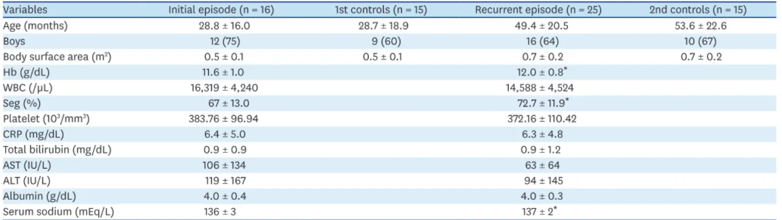
The clinical data of children with KD in the initial and recurrent episodes and those of controls are shown in Table 1. Age, body surface area at echocardiography, and heart rate were similar among the KD and control groups at both episodes. In children with KD, 8 (32%) were < 1 year of age at the initial episode, 10 (40%) had a recurrence within 1 year of the initial episode, 4 (16%) were IVIG nonresponders in the initial episode, and 5 (20%) were IVIG nonresponders in the recurrent episode. In the initial and recurrent episodes, 4 and 5 children with KD, respectively, were IVIG nonresponders. In initial episodes, children had total duration of fever for 6.3 ± 2.2 days, while children had total duration of fever for 6.0 ± 1.3 days in recurrent episodes. Five children (20%) had CAL in the acute phase of the initial
episode, with return to normal in the convalescent phase. Two children (8%) had CAL in the acute and convalescent phase of the recurrent episode. The interval range between the initial and recurrent episodes of KD was 3–103 months. Among children with recurrent KD, 15 (60%) were boys, 22 (88%) had 2 episodes, 2 (8%) had 3 episodes, and 1 (4%) had 4 episodes.
In recurrent episodes of KD, the mean values of hemoglobin, segmented neutrophils, and serum sodium levels were higher than those on the initial episode of KD.
Echocardiographic data
The echocardiographic data of children with KD in the initial and recurrent episodes and those of controls are shown in Table 2. Among patients with recurrent KD, the initial episode echocardiograms were available for only 16 patients. The duration of fever at the time of admission in both episodes of KD was the time of acquisition (fever days) of myocardial strain parameters (5.9 ± 1.9 days and 5.6 ± 1.0 days in the initial and recurrent episodes, respectively). All children with KD underwent follow-up echocardiograms 2-5 months after treatment for each episode (convalescent phase). No significant differences in the LVEF and LV mass index were found between the KD and control groups in both episodes. The mean LV longitudinal peak systolic ε in the initial and recurrent episodes of KD was within normal range in the acute and convalescent phases. However, in the initial episode in children with KD, LV longitudinal peak systolic ε was decreased in the acute phase, compared with that in controls, but was improved in the convalescent phase. In children with recurrent KD, mean heart rate was significantly higher than that of controls in the acute phase, and LV longitudinal peak systolic ε was decreased, in comparison with that in controls. However, apart from the initial episode, LV longitudinal peak systolic ε in children with recurrent KD was significantly decreased in the convalescent phase, compared with that in controls.
No significant differences were observed in the LV longitudinal peak systolic ε between acute and convalescent phases in initial and recurrent KD.
No significant correlations were found between laboratory parameters and LV longitudinal peak systolic ε in children with KD in initial and recurrent episodes.
The mean percentage error for intraobserver and interobserver variability in LV longitudinal peak systolic ε was 15% and 16%, respectively.
Table 1. Clinical and initial laboratory data of children in the initial and recurrent episodes of Kawasaki disease and respective controls
Variables Initial episode (n = 16) 1st controls (n = 15) Recurrent episode (n = 25) 2nd controls (n = 15)
Age (months) 28.8 ± 16.0 28.7 ± 18.9 49.4 ± 20.5 53.6 ± 22.6
Boys 12 (75) 9 (60) 16 (64) 10 (67)
Body surface area (m2) 0.5 ± 0.1 0.5 ± 0.1 0.7 ± 0.2 0.7 ± 0.2
Hb (g/dL) 11.6 ± 1.0 12.0 ± 0.8*
WBC (/µL) 16,319 ± 4,240 14,588 ± 4,524
Seg (%) 67 ± 13.0 72.7 ± 11.9*
Platelet (103/mm3) 383.76 ± 96.94 372.16 ± 110.42
CRP (mg/dL) 6.4 ± 5.0 6.3 ± 4.8
Total bilirubin (mg/dL) 0.9 ± 0.9 0.9 ± 1.2
AST (IU/L) 106 ± 134 63 ± 64
ALT (IU/L) 119 ± 167 94 ± 145
Albumin (g/dL) 4.0 ± 0.4 4.0 ± 0.3
Serum sodium (mEq/L) 136 ± 3 137 ± 2*
Data are expressed as mean ± standard deviation or number (%).
CRP: C-reactive protein, Hb: hemoglobin, Seg: percentage of segmented neutrophils.
*p < 0.05 when compared with initial episode.
DISCUSSION
In children in the initial episodes of KD, LV longitudinal peak systolic ε was decreased in the acute phase when compared with that in controls. In children in the recurrent episodes of KD, LV longitudinal peak systolic ε was decreased in both the acute and convalescent phase, compared with that in controls.
To our knowledge, this is the first study to compare the sequential changes in LV longitudinal peak systolic ε in initial and recurrent episodes of KD. Relatively few studies of recurrent KD have been reported.4)5)12)17) LV longitudinal peak systolic ε has been reported to be a more sensitive parameter than conventional echocardiographic parameters for the detection of early myocardial dysfunction in children.18-20) Our results support these findings, as LV longitudinal peak systolic ε in children with acute phase of KD was decreased in comparison with that in controls, in initial and recurrent episodes, while no significant differences in the LVEF or LV mass index were observed between children with KD and controls.
In the acute phase of KD, the degree of myocardial inflammation has been shown to be associated with the severity of myocardial dysfunction.21) Therefore, recurrent episodes of KD may result in repeated bouts of inflammation due to myocarditis, possibly resulting in more severe myocardial dysfunction. The results of our study seem to support this, since the mean LV longitudinal peak systolic ε was decreased in the convalescent phase of recurrent episodes of KD, compared to that in controls.
A previous study reported that the KD recurrence rate was higher in children who had cardiac sequelae after the initial episode.12) However, this study did not specify the method of assessing coronary artery abnormalities. In addition, when KD recurs in children with CALs in the initial episode, the risk of CALs in recurrent episodes may be higher.17) However, no such association of the severity of CALs between initial and recurrent episodes of KD was observed in our study. This might be explained by the difference in the interval range between initial and recurrent episodes of KD, which was longer in our study compared to that in a previous study.17) The relatively longer interval range between initial and recurrent episodes in our study may have allowed CALs in the initial episode to more fully recover. In addition, the small number of patients with KD with CALs might have affected the statistical results of our study.
In all 5 children with KD who had CALs in the acute phase of the initial episode, the values of LV longitudinal peak systolic ε increased from the acute to the convalescent phase (acute to convalescent, respectively: 17.2% → 23.0%, 16.0% → 24.1%, 21.0% → 22.0%, 16.8% → 27.0%, 17.0% → 24.6%). In addition, all 2 children who had CALs in the acute and convalescent phase of the recurrent episode showed values of LV longitudinal peak systolic ε in the normal range in the convalescent phase (acute to convalescent, respectively: 23.5% → 19.9%, 20.7% → 21.8%).
Table 2. Echocardiographic data of children in the initial and recurrent episodes of Kawasaki disease and respective controls
Variables Initial acute
(n = 16) Initial convalescent
(n = 16) 1st controls
(n = 15) Recurrent acute
(n = 25) Recurrent
convalescent (n = 25) 2nd controls (n = 15)
Heart rate (bpm) 112 ± 16 102 ± 15 109 ± 20 108 ± 17* 102 ± 21 93 ± 12
LV EF (%) 62.6 ± 5.0 67.2 ± 4.3 65.9 ± 7.3 63.4 ± 9.3 64.4 ± 7.4 66.0 ± 4.2
LV MI (g/m2) 59.2 ± 14.9 57.9 ± 13.6 56.5 ± 11.7 65.7 ± 10.9 56.9 ± 13.8 60.5 ± 8.0
LV peak longitudinal systolic ε (%) 20.5 ± 4.1* 23.8 ± 4.3 24.1 ± 3.0 21.0 ± 3.9* 22.1 ± 3.1* 23.8 ± 2.7 Data are expressed as mean ± standard deviation.
Strain values are presented as absolute values.
bpm: beats per minute, EF: ejection fraction, LV: left ventricular, MI: mass index, ε: strain.
*p < 0.05 when compared between respective controls.
Further studies involving larger numbers of children who experienced recurrent episodes of KD may be needed in order to elucidate the association of CALs and recurrence of KD.
Even though the mean values of LV longitudinal peak systolic ε were decreased in both the acute and convalescent phases in recurrent episodes of KD, when compared with those in controls, LV longitudinal peak systolic ε was within normal range in all episodes of KD. Since more patients with KD had LV longitudinal peak systolic ε within normal range in the acute phase of initial and recurrent episodes than those with decreased values, the overall mean values of LV longitudinal peak systolic ε in the acute phase of KD in initial and recurrent episodes may fall within the normal range.
Another possible explanation for why the mean values of LV longitudinal peak systolic ε were within normal range in all episodes of KD may be that during the long interval between initial and recurrent episodes (3-103 months), myocardial dysfunction in a subset of initial KD patients may have improved over time. The normalization of myocardial function over time has also been shown in a previous study on prognosis of left atrial reservoir function at long term follow-up in children with a history of KD.22) An additional explanation could be related to age at diagnosis of recurrent KD episodes. Since children were older when they experienced recurrent episodes of KD, we could speculate that they could tolerate myocardial inflammation more easily and that subsequent LV dysfunction could recover more rapidly compared to that in initial episodes of KD. Another additional explanation for the similar LV longitudinal peak systolic ε in recurrent KD and controls may be the small number of children with CALs or IVIG nonresponders, factors that have been associated with decreased LV function.23)24) Moreover, as treatment with IVIG has been associated with accelerated recovery of LV function3) and since all children with KD in our study received IVIG, we could speculate that the decreased LV function caused by myocarditis from repeated recurrences of KD recovered with IVIG treatment.
Limitations
We could not establish causality due to the retrospective nature of our study. The small numbers of children in our study group may have limited our statistical results, especially for subgroup analysis, such as patients with KD with CALs, age < 1 year at KD diagnosis, or the effect of recurrence within < 1 year after the initial KD episode. Even though recovery of LV function over time in KD has been reported,2)3) the number of patients with KD and decreased LV longitudinal peak systolic ε in the acute phase was small in our study; therefore, there were limitations in the ability to show recovery of LV systolic function between the acute and convalescent phase in these subgroups of patients.
Not all children with recurrent KD had available initial episode echocardiograms, thus affecting our statistical results. In our study, LV longitudinal peak systolic ε was obtained as an average of six segments from an apical four-chamber view only.9) Longer follow-up of children with recurrent episodes of KD beyond the convalescent phase may be needed in the future.
CONCLUSIONS
Subclinical decreases in myocardial systolic deformation, as evidenced by decreased LV longitudinal peak systolic ε, may persist in children in the convalescent phase of recurrent KD; further studies involving larger numbers of patients may be needed for verification.
REFERENCES
1. Sudo D, Nakamura Y. Nationwide surveys show that the incidence of recurrent Kawasaki disease in Japan has hardly changed over the last 30 years. Acta Paediatr 2017;106:796-800.
PUBMED | CROSSREF
2. Moran AM, Newburger JW, Sanders SP, et al. Abnormal myocardial mechanics in Kawasaki disease: rapid response to gamma-globulin. Am Heart J 2000;139:217-23.
PUBMED
3. Newburger JW, Sanders SP, Burns JC, Parness IA, Beiser AS, Colan SD. Left ventricular contractility and function in Kawasaki syndrome. Effect of intravenous gamma-globulin. Circulation 1989;79:1237-46.
PUBMED | CROSSREF
4. Maddox RA, Holman RC, Uehara R, et al. Recurrent Kawasaki disease: USA and Japan. Pediatr Int 2015;57:1116-20.
PUBMED | CROSSREF
5. Chahal N, Somji Z, Manlhiot C, et al. Rate, associated factors and outcomes of recurrence of Kawasaki disease in Ontario, Canada. Pediatr Int 2012;54:383-7.
PUBMED | CROSSREF
6. Haque U, Stiver C, Rivera BK, et al. Right ventricular performance using myocardial deformation imaging in infants with bronchopulmonary dysplasia. J Perinatol 2017;37:81-7.
PUBMED | CROSSREF
7. Yu JJ, Choi HS, Kim YB, et al. Analyses of left ventricular myocardial deformation by speckle-tracking imaging during the acute phase of Kawasaki disease. Pediatr Cardiol 2010;31:807-12.
PUBMED | CROSSREF
8. McCandless RT, Minich LL, Wilkinson SE, McFadden ML, Tani LY, Menon SC. Myocardial strain and strain rate in Kawasaki disease. Eur Heart J Cardiovasc Imaging 2013;14:1061-8.
PUBMED | CROSSREF
9. Kang SJ, Kwon YW, Hwang SJ, Kim HJ, Jin BK, Yon DK. Clinical utility of left atrial strain in children in the acute phase of Kawasaki disease. J Am Soc Echocardiogr 2018;31:323-32.
PUBMED | CROSSREF
10. Pirat B, Khoury DS, Hartley CJ, et al. A novel feature-tracking echocardiographic method for the quantitation of regional myocardial function: validation in an animal model of ischemia-reperfusion. J Am Coll Cardiol 2008;51:651-9.
PUBMED | CROSSREF
11. Newburger JW, Takahashi M, Gerber MA, et al. Diagnosis, treatment, and long-term management of Kawasaki disease: a statement for health professionals from the Committee on Rheumatic Fever, Endocarditis, and Kawasaki Disease, Council on Cardiovascular Disease in the Young, American Heart Association. Pediatrics 2004;114:1708-33.
PUBMED | CROSSREF
12. Hirata S, Nakamura Y, Yanagawa H. Incidence rate of recurrent Kawasaki disease and related risk factors:
from the results of nationwide surveys of Kawasaki disease in Japan. Acta Paediatr 2001;90:40-4.
PUBMED | CROSSREF
13. Lang RM, Bierig M, Devereux RB, et al. Recommendations for chamber quantification: a report from the American Society of Echocardiography's Guidelines and Standards Committee and the Chamber Quantification Writing Group, developed in conjunction with the European Association of Echocardiography, a branch of the European Society of Cardiology. J Am Soc Echocardiogr 2005;18:1440-63.
PUBMED | CROSSREF
14. McCrindle BW, Li JS, Minich LL, et al. Coronary artery involvement in children with Kawasaki disease:
risk factors from analysis of serial normalized measurements. Circulation 2007;116:174-9.
PUBMED | CROSSREF
15. Voigt JU, Pedrizzetti G, Lysyansky P, et al. Definitions for a common standard for 2D speckle tracking echocardiography: consensus document of the EACVI/ASE/Industry Task Force to standardize deformation imaging. J Am Soc Echocardiogr 2015;28:183-93.
PUBMED | CROSSREF
16. Kutty S, Padiyath A, Li L, et al. Functional maturation of left and right atrial systolic and diastolic performance in infants, children, and adolescents. J Am Soc Echocardiogr 2013;26:398-409.e2.
PUBMED | CROSSREF
17. Yang HM, Du ZD, Fu PP. Clinical features of recurrent Kawasaki disease and its risk factors. Eur J Pediatr 2013;172:1641-7.
PUBMED | CROSSREF
18. Dogan V, Öcal B, Orun UA, et al. Strain and strain rate echocardiography findings in children with asymptomatic congenital aortic stenosis. Pediatr Cardiol 2013;34:1152-8.
PUBMED | CROSSREF
19. Perk G, Tunick PA, Kronzon I. Non-Doppler two-dimensional strain imaging by echocardiography--from technical considerations to clinical applications. J Am Soc Echocardiogr 2007;20:234-43.
PUBMED | CROSSREF
20. Dandel M, Hetzer R. Echocardiographic strain and strain rate imaging--clinical applications. Int J Cardiol 2009;132:11-24.
PUBMED | CROSSREF
21. Kao CH, Hsieh KS, Wang YL, Wang SJ, Yeh SH. The detection of ventricular dysfunction and carditis in children with Kawasaki disease using equilibrium multigated blood pooling ventriculography and 99Tcm- HMPAO-labelled WBC heart scans. Nucl Med Commun 1993;14:539-43.
PUBMED | CROSSREF
22. Kang SJ, Ha J, Hwang SJ, Kim HJ. Long term outcomes of left atrial reservoir function in children with a history of Kawasaki disease. J Cardiovasc Ultrasound 2018;26:26-32.
PUBMED | CROSSREF
23. Printz BF, Sleeper LA, Newburger JW, et al. Noncoronary cardiac abnormalities are associated with coronary artery dilation and with laboratory inflammatory markers in acute Kawasaki disease. J Am Coll Cardiol 2011;57:86-92.
PUBMED | CROSSREF
24. Phadke D, Patel SS, Dominguez SR, et al. Tissue Doppler imaging as a predictor of immunoglobulin resistance in Kawasaki disease. Pediatr Cardiol 2015;36:1618-23.
PUBMED | CROSSREF