Copyrights © 2018 The Korean Society of Radiology
88
Case Report
pISSN 1738-2637 / eISSN 2288-2928 J Korean Soc Radiol 2018;79(2):88-91 https://doi.org/10.3348/jksr.2018.79.2.88
INTRODUCTION
Petersen’s hernia was first described in 1900 by German sur- geon, Dr. Walther Petersen. It is a specific type of internal her- nia in which the small bowel moves into a potential space bounded by the caudal surface of the transverse mesocolon, the retroperitoneum and the mesentery of the gastrojejunostomy’s limb (1). This type of internal hernia after laparoscopic gastrec- tomy with Roux-en-Y reconstruction for the treatment of obe- sity have been reported frequently (2, 3). However, reports about Petersen’s hernia after gastrectomy with Billroth II gas- trojejunostomy for gastric cancer are a few.
We report a case of Petersen’s hernia after radical subtotal gas- trectomy with Billroth II gastrojejunostomy through antecolic route for gastric cancer and demonstrate an unreported specific
computed tomography (CT) finding as a predictor of Petersen’s hernia.
Case RepORT
A 37-year-old woman presented with intermittent abdominal pain. She had undergone a robot-assisted radical subtotal gas- trectomy with Billroth II gastrojejunostomy for advanced gas- tric cancer 8 months prior to her presentation. After undergo- ing gastrectomy, she was followed up with outpatient visits and received XELOX for 6 months. She did not have any other note- worthy symptoms and her physical examination results were also unremarkable. Laboratory investigations revealed nonspe- cific findings (C-reactive protein < 0.3 mg/L, white blood cell 6240/μL). Abdominal contrast-enhanced CT images showed
Petersen’s Hernia after Subtotal Gastrectomy with Billroth II Gastrojejunostomy for Gastric Cancer: A Specific CT Finding
위암으로 Billroth II 위공장문합술을 통한 위아전절제술을 받고 난 후 발생한 Petersen 탈장: 특징적인 CT 소견
Hwan Kim
1, Yong Eun Chung, MD
2*, Woo Jin Hyung, MD
3, Joon Seok Lim, MD
21Yonsei University College of Medicine, Seoul, Korea
Departments of 2Radiology, 3Surgery, Severance Hospital, Yonsei University College of Medicine, Seoul, Korea
Petersen’s hernia is a rare internal hernia occurring after any type of gastrojejunal anastomosis. This type of internal hernia after laparoscopic gastrectomy with Roux- en-Y reconstruction for obesity treatment has been reported frequently. However, reports about Petersen’s hernia after gastrectomy with Billroth II gastrojejunostomy for gastric cancer are relatively rare. To diagnose internal hernia, whirl sign has been reported to be a sensitive predictor of internal hernia. However, it is hard to diagnose a specific type of internal hernia. We report a case of Petersen’s hernia after radical subtotal gastrectomy with Billroth II gastrojejunostomy for gastric cancer with an unreported specific computed tomography finding that suggests Petersen’s hernia.
Index terms Postperative Hernia
Computed Tomography, X-Ray Gastroenterostomy
Gastric Cancer
Received October 13, 2017 Revised January 28, 2018 Accepted March 16, 2018
*Corresponding author: Yong Eun Chung, MD Department of Radiology, Severance Hospital, Yonsei University College of Medicine, 50-1 Yonsei-ro, Seodaemun-gu, Seoul 03722, Korea.
Tel. 82-2-2228-7400 Fax. 82-2-393-3035 E-mail: yelv@yuhs.ac
This is an Open Access article distributed under the terms of the Creative Commons Attribution Non-Commercial License (https://creativecommons.org/licenses/by-nc/4.0) which permits unrestricted non-commercial use, distri- bution, and reproduction in any medium, provided the original work is properly cited.
Hwan Kim, et al
89
jksronline.org 대한영상의학회지 2018;79(2):88-91
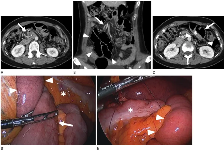
the whirl sign, which indicates mesenteric vessel rotation, with mesenteric fat haziness (Fig. 1A, arrow). On a coronal image, the vessels showed engorgement (Fig. 1B, arrow) and the small bowel showed diffuse dilatation without evidence of strangula- tion (Fig. 1B, arrowheads). Furthermore, the interposed small bowel appeared (Fig. 1C, asterisk) between the transverse colon (Fig. 1C, arrowheads) and the afferent limb (Fig. 1C, arrow).
A diagnostic laparoscopy was performed, and revealed inter- nal herniation of the efferent limb (Fig. 1D, arrow) through the Petersen defect, a space between the transverse mesocolon (Fig.
1D, E, asterisks) and the afferent limb (Fig. 1D, E, arrowheads), confirming the diagnosis of Petersen’s hernia. A dilated small
bowel loop was observed without any sign of strangulation. The herniated small bowel was successfully reduced and the Peters- en defect was closed by running absorbable sutures (Fig. 1E).
The patient was followed up and no recurrence of intestinal ob- struction was observed thereafter.
DIsCUssION
Petersen’s hernia is a rare specific type of internal hernia in which the small bowel moves into a potential space bounded by the caudal surface of the transverse mesocolon, the retroper- itoneum and the mesentery of the gastrojejunostomy’s limb (1).
Fig. 1. Petersen’s hernia in a 37-year-old woman.
A. Abdominal contrast enhanced CT showing whirl sign which indicates mesenteric vessel rotation and mesenteric fat haziness (arrow).
B. Abdominal contrast enhanced CT coronal image showing engorged vessels (arrow) and small bowel with diffuse dilatation without evidence of strangulation (arrowheads).
C. Abdominal contrast enhanced CT showing small bowel (asterisk) interposed between the transverse colon (arrowheads) and the afferent limb (arrow) like a sandwich.
D. A diagnostic laparoscopy showing internal herniation of the efferent limb (arrow) through the Petersen defect, a space between the transverse mesocolon (asterisk) and the afferent limb (arrowheads).
E. The herniated small bowel was successfully reduced and the Petersen defect between the transverse mesocolon (asterisk) and the afferent limb (arrowheads) was closed using absorbable suture.
CT = computed tomography
D E
A B C
petersen’s Hernia after subtotal Gastrectomy
90
대한영상의학회지 2018;79(2):88-91 jksronline.orgThis type of internal hernia usually occurs within 1 year after bariatric surgery or (subtotal) gastrectomy performed to treat cancer, although it can develop several years after surgery (2).
Internal hernias more frequently develop after laparoscopic surgery than laparotomy. This might be because more adhesion occurs after laparotomy which can reduce the mobility of the small bowel, and this in turn, prevents the internal herniation of the small bowel (2, 3). Loss of body weight and abdominal fat after gastrectomy which may widen potential spaces is another predisposing factor (3). Closure of all the mesenteric defects with running non-absorbable sutures can reduce the risk of an inter- nal hernia developing after gastrectomy (2). Petersen’s hernia can lead to the small intestinal obstruction and necrosis of the small bowel necessitating intestinal resection. When Petersen’s hernia is diagnosed, the herniated intestinal loop should be re- duced with the closure of all the mesenteric defect (2, 4-6).
The diagnosis of internal hernia including Petersen’s hernia is challenging due to nonspecific clinical symptoms, laboratory findings and radiological findings (2, 3). The most common clinical symptoms are abdominal pain, nausea and vomiting, and these might be transient and self-resolving (2, 3). On CT, rotation of the mesenteric vessels, mesenteric fat haziness, in- testinal distention in the upper abdomen, and herniated intesti- nal loop above the gastric level can be seen (7). The whirl sign has been reported to be the most sensitive single predictor of internal hernia (3, 7). However, even if these aforementioned signs are present on CT, it is hard to diagnose a specific type of internal hernia. In this case report, we demonstrate a specific CT finding, which is an interposed small bowel between the transverse colon and the gastrojejunostomy’s limb as a predictor
of Petersen’s hernia. It is a reasonable finding considering that Petersen’s hernia is an internal hernia through a space between the transverse mesocolon and the gastrojejunostomy’s limb.
RefeReNCes
1. Rogers AM, Ionescu AM, Pauli EM, Meier AH, Shope TR, Haluck RS. When is a Petersen's hernia not a Petersen's her- nia. J Am Coll Surg 2008;207:121-124
2. Faria G, Preto J, Oliveira M, Pimenta T, Baptista M, Costa- Maia J. Petersen’s space hernia: a rare but expanding diag- nosis. Int J Surg Case Rep 2011;2:141-143
3. Baba A, Yamazoe S, Dogru M, Okuyama Y, Mogami T, Kobashi Y, et al. Petersen hernia after open gastrectomy with Roux- en-Y reconstruction: a report of two cases and literature review. Springerplus 2015;4:753
4. Comeau E, Gagner M, Inabnet WB, Herron DM, Quinn TM, Pomp A. Symptomatic internal hernias after laparoscopic bariatric surgery. Surg Endosc 2005;19:34-39
5. Somville FJ, De Moitié E, Fransen L. Atypical clinical presen- tation of internal herniation after laparoscopic Roux-en-Y gastric bypass. Acta Chir Belg 2013;113:152-154
6. Iannelli A, Facchiano E, Gugenheim J. Internal hernia after laparoscopic Roux-en-Y gastric bypass for morbid obesity.
Obes Surg 2006;16:1265-1271
7. Ximenes MA, Baroni RH, Trindade RM, Racy MC, Tachibana A, Moron RA, et al. Petersen’s hernia as a complication of bariatric surgery: CT findings. Abdom Imaging 2011;36:126- 129
Hwan Kim, et al
91
jksronline.org 대한영상의학회지 2018;79(2):88-91
위암으로 Billroth II 위공장문합술을 통한 위아전절제술을 받고 난 후 발생한 Petersen 탈장: 특징적인 CT 소견
김 환
1· 정용은
2* · 형우진
3· 임준석
2Petersen 탈장은 위공장문합술 후에 드물게 발생하는 내탈장이다. 비만의 치료를 위해 Roux-en-Y 위우회술을 시행한 후에 발생한 Petersen 탈장은 상대적으로 빈번하게 보고되었으나, 위암으로 Billroth II 위공장문합술을 통한 위아전절제 술을 받고 난 후에 발생한 경우는 거의 보고되지 않았다. 컴퓨터단층촬영에서 whirl sign이 내탈장을 진단할 수 있는 가장 민감한 소견으로 보고되어 있다. 그러나 이러한 소견이 보이더라도, 구체적인 내탈장 타입을 진단하는 것은 어려운 경우가 많다. 저자들은 위암으로 Billroth II 위공장문합술을 통한 위아전절제술을 받고 난 후에 발생한 Petersen 탈장을 보고하 며, 내탈장 중에서도 Petersen 탈장을 나타내는 이전에 보고되지 않았던 특징적인 영상소견을 보고한다.
1연세대학교 의과대학, 연세대학교 의과대학 세브란스병원 2영상의학과, 3외과