www.jpis.org
pISSN 2093-2278 eISSN 2093-2286 Copyright © 2012 Korean Academy of PeriodontologyThis is an Open Access article distributed under the terms of the Creative Commons Attribution Non-Commercial License (http://creativecommons.org/licenses/by-nc/3.0/).
Mucosal dehiscence coverage for dental implant using sprit pouch technique: a two-stage approach
Toyohiko Hidaka1, Daisuke Ueno2,*
1Second Department of Comprehensive Care, Tsurumi University School of Dental Medicine, Yokohama, Japan
2Unit of Oral and Maxillofacial Implantology, Tsurumi University School of Dental Medicine, Yokohama, Japan
Purpose: Soft tissue recessions frequently cause esthetic disharmony and dissatisfaction. Compared with soft tissue coverage around a tooth, the coverage of an implant site is obviously unpredictable. Particularly in the cases of thin mucosa, a significant greater amount of recession takes place compared to thick mucosa. To overcome this problem, this case report demonstrates a two-step mucosal dehiscence coverage technique for an endosseous implant.
Methods: A 33-year-old female visited us with the chief complaint of dissatisfaction with the esthetics of an exposed implant in the maxillary left cental incisor region. A partial-thickness pouch was constructed around the dehiscence. A subepithelial connective tissue graft was positioned in the apical site of the implant and covered by a mucosal flap with normal tension. At 12 months after surgery, the recipient site was partially covered by keratinized mucosa. However, the buccal interdental papilla between implant on maxillary left central incisor region and adjacent lateral incisor was concave in shape. To resolve the mu- cosal recession after the first graft, a second graft was performed with the same technique.
Results: An esthetically satisfactory result was achieved and the marginal soft tissue level was stable 9 months after the second graft.
Conclusions: The second graft was able to resolve the mucosal recession after first graft. This two-step approach has the po- tential to improve the certainty of esthetic results.
Keywords: Case report, Dental Esthetics, Mouth mucosa, Oral surgical procedures.
INTRODUCTION
Soft tissue recessions around dental implants have frequent- ly been observed. A recession during the early phase after implant crown placement originates from the process of modeling of the peri-implant mucosa [1]. Cardaropoli et al. [2]
reported that bone resorption and soft tissue recession were manifested as 0.7 and 0.6 mm at the buccal aspect of the im- plants during the period between implant placement and abutment connection. Muller et al. [3] has suggested that thin mucosa is friable and recesses more readily following me-
chanical stress and surgical procedures than does thick mu- cosa. Furthermore, the quantity, quality, and position of the existing peri-implant bone also affects soft tissue recession [4].
Soft tissue recessions often cause esthetic disharmony and dissatisfaction. Burkhardt et al. [5] surgically covered soft tis- sue recessions using a coronally advanced flap (CAF) in com- bination with a free connective tissue graft. Although the im- plant sites revealed a substantial, clinically significant im- provement following coronal mucosal displacement, clinical- ly significant soft tissue shrinkage was observed after one month of healing. Thin mucosa underwent significantly more
Received: Mar. 22, 2012; Accepted: Apr. 16, 2012
*Correspondence: Daisuke Ueno
Unit of Oral and Maxillofacial Implantology, Tsurumi University School of Dental Medicine, 2-1-3 Tsurumi, Tsurumi-ku, Yokohama, Japan E-mail: [email protected], Tel: +81-45-581-1001, Fax: +81-45-581-0024
approach is useful in acquisition of esthetic results because the first graft can increase the thickness of the peri-implant mucosa. Furthermore, the second graft probably compen- sates for the mucosal recession after first graft. The present case demonstrates a two-step mucosal dehiscence coverage technique in an endosseous implant. Sufficient soft tissue regeneration was achieved and the marginal soft tissue level was stable with an esthetically satisfactory result.
CASE DESCRIPTION
The patient was a 33-year-old female who visited us on Oc- tober 2008 with a chief complaint of esthetic dissatisfaction
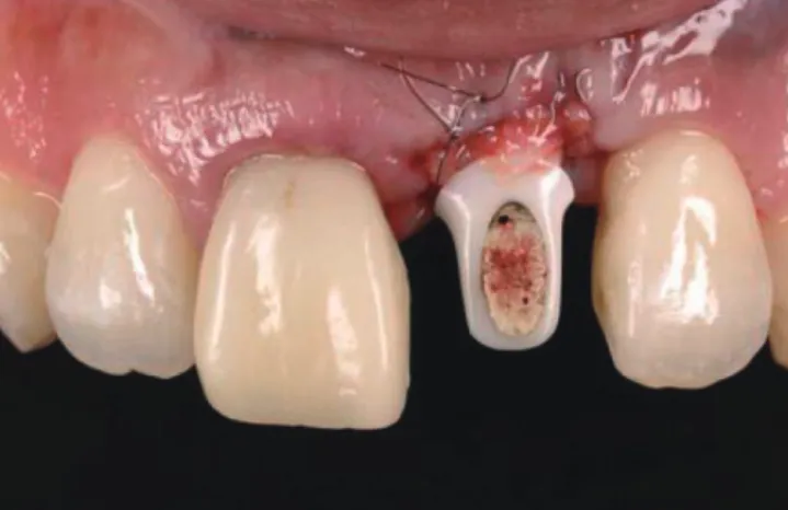
gion. A zirconia abutment and temporary crown had been already fitted to the implant. Periodontal examination re- vealed a healthy peri-implant condition with a probing depth ranging from 2 to 3 mm and satisfactory oral hygiene was observed. There was 3 mm abutment exposure in the buccal mucosa around the implant (Fig. 1). An intraoral periapical radiograph and cone beam computed tomography were tak- en to evaluate peri-implant bone resorption. Because only 1 mm of marginal peri-implant bone resorption was observed (Fig. 2) and the implant was a bit buccally angulated, no bone graft or removal of the implant was performed, but it was de- cided to carry out only soft tissue augmentation to restore esthetics.
Following local anesthesia, a circumferential partial-thick- ness incision was performed using a CK3 stainless steel blade (Blade Round Tip Angled 10 Deg, SWAN Analytical USA Inc., Wheeling, IL, USA) to achieve a 17 mm wide×12 mm height pouch around the dehiscence (Fig. 3). A 10×6 mm subepithe- lial connective tissue graft (SCTG) was harvested from the palate in the right second premolar to second molar region.
The graft tissue was trimmed to fit the formerly prepared re- cipient bed. A horizontal incision to the bone was made 5 mm from the palatal gingival margin and the blade (Razor Blade, FEATHER Safety Razor Co., Osaka, Japan) was subsequently placed parallel to the long axis of the roots. Another horizon- tal incision was made 2 mm coronal to the first incision and the periosteum was dissected before removing the wedge of soft tissue. The SCTG was trimmed precisely to adapt to the Figure 1. Preoperative intraoral view: 3 mm of vertical abutment ex-
posure was observed.
Figure 2. Preoperative radiographical examination of the left maxillary central incisor region: intraoral periapical radiograph (A) and come beam computed tomography (B, axial view; C, coronal view; D, sagittal view).
A
C B
D
ent pouch and sutured with 7.0-nylon (Nicho Kogyo Ltd., To- kyo, Japan) to stabilize the soft tissue graft and supplemental site. The graft was positioned and fixed by a 7.0-nylon suture in apical site of the implant. After covering the SCTG graft with a mucosal flap, the flap was coronally stretched with a 7.0-nylon suture (coronally positioned flap, CPF). The donor site was sutured with 5-0-nylon (LOOK Suture 774B, Angio- tech, Vancouver, BC, Canada) (Fig. 4).
At 12 months after surgery, the recipient site was partially covered by keratinized mucosa (Fig. 5A). However, the buccal interdental papilla between implant on maxillary left cental incisor region and adjacent lateral incisor was in a concave shape. To achieve an esthetic result, we planned a second graft.
Following local anesthesia, a circumferential partial-thick- ness incision was performed using CK3 around the dehis- cence. An SCTG was harvested from the palate in the left second premolar to second molar region. Then, the SCTG was inserted into the recipient pouch with the same tech- nique. The graft was stabilized with a 7.0-nylon suture (Fig.
5B). No complications had arisen within 2 weeks post-sur- gery (Fig. 5C).
Because proper vertical mucosal gain was achieved at 8
ed. After an impression of implant on maxillary left central incisor region and prepared adjacent lateral incisor was tak- en, a zirconia custom abutment was fabricated (Fig. 6). The crown-abutment margin was set up more apically than the previous abutment. The all-ceramic crowns (zirconia frame- work and glass-ceramic veneer material) were fixed with res- in cement.
The intra-oral picture shows that the peri-implant mucosa
Figure 4. Intraoral appearance immediately after the first subepithe- lial connective tissue graft.
Figure 3. Surgical steps of the subepithelial connective tissue graft (SCTG) with split pouch technique: (A) A circumferential partial-thickness incision was performed using a round tip blade. (B) A partial-thickness pouch was constructed around the dehiscence. (C) The SCTG was po- sitioned and sutured in the pouch with normal tension.
A B C
Figure 5. (A) Intraoral appearance at 12 months after the first subepithelial connective tissue graft (SCTG): The mucosal dehiscence was par- tially covered by epithelium. (B) Intraoperative view in the second stage surgery: The SCTG was positioned and sutured in the pouch with normal tension. (C) Intraoral appearance at 2 weeks after the second SCTG.
A B C
is harmonious with the right central incisor at 9 months after graft (Fig. 7). Although the interdental papilla between implant and adjacent tooth was more apical than the contralateral side, it was esthetically acceptable to the patient.
DISSCUSSION
A two-step split pouch technique using an SCTG demon- strated that it was possible to achieve a substantial area of soft tissue dehiscence coverage around the endosseous im- plant. Burkhardt et al. [5] evaluated soft tissue dehiscence coverage using a CPF in combination with a free connective tissue graft around the implants. The soft tissue recessions were covered with a coronal overcompensation at a mean of 0.5 mm after the graft. After 1 and 6 months, shrinkage of 75% and 66%, respectively, was observed. Compared with the percentages of soft tissue coverage around the tooth [6], the implant sites clearly could not be covered consistently. A folding process further hampers vascularization of the graft and could induce extensive shrinkage.
To overcome these problems, we considered the anatomi- cal and physiological properties of the buccal peri-implant
mucosa. Burkhardt et al. [5] reported that the preoperative mucosal thickness in the implant site was positively correlat- ed with the height of recession coverage. Nozawa et al. [7] also reported that marginal soft tissue dimensions indicated that there may be a relationship between the thickness and the height of about 1.5:1 at the platform level. The results may support the approach of acquiring thick mucosa to improve vertical mucosal regeneration. Thick soft tissue, which is ob- tained from the first graft, can increase the blood supply to the graft; thus, it may prevent necrosis and recession after the second graft. Furthermore, the second graft can compen- sate for mucosal recession after the first graft.
Coverage procedures can be categorized into pedicle grafts and free soft tissue grafts. Although pedicle grafts, such as the laterally positioned flap, double pedicle flap, oblique rotational flap and CPF have an advantage over free soft tissue grafts, there is only a limited increase in the tissue thickness and width of the keratinized gingiva. On the other hand, free soft tissue grafts, such as the free gingival graft (FGG) [8] and SCTG [9] can increase the width of the keratinized gingiva and tissue thickness. Oates et al. [10] and Roccuzzo et al. [6]
reported on the advantage of the SCTG in combination with a CPF. The SCTG was statistically superior in achieving root coverage when compared to the FGG, CPF alone, guided tis- sue regeneration, and allogeneic tissue grafts. The CAF with envelope technique has the advantages of increasing kera- tinized mucosa, a better postoperative course, and a more positive esthetic evaluation than CAF with vertical releasing incisions (VRIs) [11]. The longer surgical time to complete the CAF with VRIs may have been responsible for the greater in- cidence of swelling and pain [12]. Furthermore, VRIs often cause unesthetic visible scars after healing that patients can find unsatisfactory. From a biologic standpoint, VRIs might damage the lateral blood supply to the flap. The blood supply to soft tissue grafts is critical for the success of the surgery.
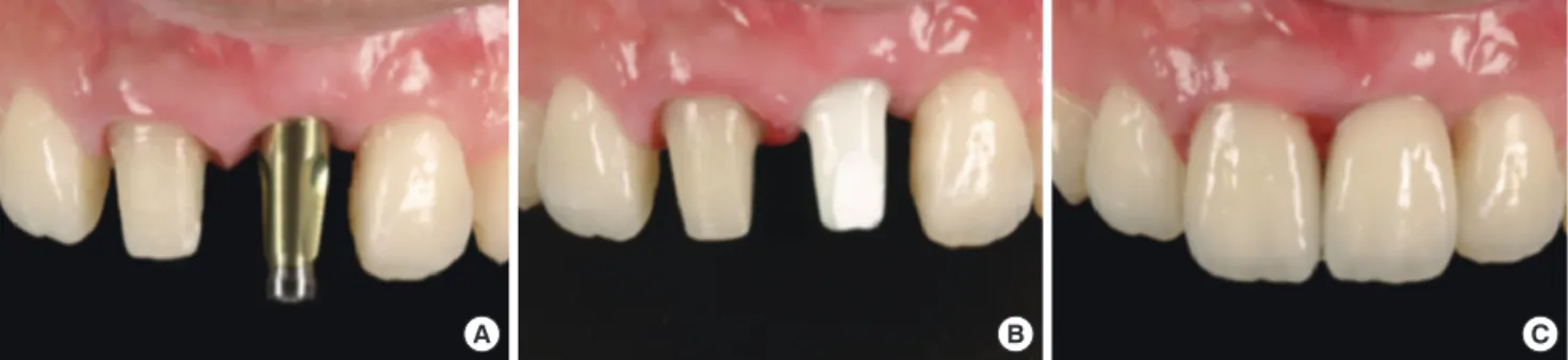
Site-related factors such as the dimensions of the recession defect, vestibulum depth, and the level of interdental or in- terimplant papilla should be considered. A systematic review Figure 6. Prosthetic procedures: (A) intraoral view after connection of impression coping, (B) intraoral view after connection of custom zir- conia abutment, and (C) intraoral view immediately after placement of all-ceramic crowns.
A B C
Figure 7. Nine months after the second SCTG. Sufficient soft tissue regeneration was achieved and the marginal soft tissue level was stable and esthetically satisfactory.
complete root coverage [13]. Miller [14] classified the oral mu- cosal recession by the marginal soft tissue level and inter- proximal bone loss. Based on the literature, complete root coverage can be expected in class 1 and 2. However, there are no reports on the classification of soft tissue recession in dental implants. Soft tissue dehiscence of plural implants is further complicated and technically sensitive [5]. Therefore, site related factors, surgical methods, and the patient’s expec- tations should be cautiously taken into account.
CONFLICT OF INTEREST
No potential conflict of interest relevant to this article was reported.
ACKNOWLEDGEMENTS
The authors thank Dr. Asiri Jayawardena, Department of General Education, Tsurumi University, School of Dental Medicine, Yokohama, Japan, for editorial assistance.
REFERENCES
1. Berglundh T, Lindhe J. Dimension of the periimplant mu- cosa. Biological width revisited. J Clin Periodontol 1996;23:
971-3.
2. Cardaropoli G, Lekholm U, Wennstrom JL. Tissue altera- tions at implant-supported single-tooth replacements: a 1-year prospective clinical study. Clin Oral Implants Res 2006;17:165-71.
3. Muller HP, Heinecke A, Schaller N, Eger T. Masticatory mucosa in subjects with different periodontal phenotypes.
J Clin Periodontol 2000;27:621-6.
4. Belser UC, Buser D, Hess D, Schmid B, Bernard JP, Lang NP. Aesthetic implant restorations in partially edentulous patients: a critical appraisal. Periodontol 2000 1998;17:132-
5. Burkhardt R, Joss A, Lang NP. Soft tissue dehiscence cov- erage around endosseous implants: a prospective cohort study. Clin Oral Implants Res 2008;19:451-7.
6. Roccuzzo M, Bunino M, Needleman I, Sanz M. Periodon- tal plastic surgery for treatment of localized gingival re- cessions: a systematic review. J Clin Periodontol 2002;29 Suppl 3:178-94.
7. Nozawa T, Enomoto H, Tsurumaki S, Ito K. Biologic height- width ratio of the buccal supra-implant mucosa. Eur J Es- thet Dent 2006;1:208-14.
8. Nabers JM. Free gingival grafts. Periodontics 1966;4:243-5.
9. Soileau KM, Brannon RB. A histologic evaluation of vari- ous stages of palatal healing following subepithelial con- nective tissue grafting procedures: a comparison of eight cases. J Periodontol 2006;77:1267-73.
10. Oates TW, Robinson M, Gunsolley JC. Surgical therapies for the treatment of gingival recession. A systematic re- view. Ann Periodontol 2003;8:303-20.
11. Zucchelli G, Mele M, Mazzotti C, Marzadori M, Monte- bugnoli L, De Sanctis M. Coronally advanced flap with and without vertical releasing incisions for the treatment of multiple gingival recessions: a comparative controlled randomized clinical trial. J Periodontol 2009;80:1083-94.
12. Cortellini P, Tonetti M, Baldi C, Francetti L, Rasperini G, Rotundo R, et al. Does placement of a connective tissue graft improve the outcomes of coronally advanced flap for coverage of single gingival recessions in upper anterior teeth? A multi-centre, randomized, double-blind, clinical trial. J Clin Periodontol 2009;36:68-79.
13. Clauser C, Nieri M, Franceschi D, Pagliaro U, Pini-Prato G.
Evidence-based mucogingival therapy. Part 2: Ordinary and individual patient data meta-analyses of surgical treat- ment of recession using complete root coverage as the outcome variable. J Periodontol 2003;74:741-56.
14. Miller PD Jr. A classification of marginal tissue recession.
Int J Periodontics Restorative Dent 1985;5:8-13.