ORIGINAL ARTICLE
위 이형성의 내시경 절제술 후 헬리코박터 제균요법이 이시성 재발 에 미치는 영향
전일영, 최치언, 신철민, 박영수, 김나영, 이동호
분당서울대학교병원 내과
Effect of Helicobacter pylori Eradication on Subsequent Dysplasia Development after Endoscopic Resection of Gastric Dysplasia
Ilyoung Chon, Chiun Choi, Cheol Min Shin, Young Su Park, Nayoung Kim and Dong Ho Lee Department of Internal Medicine, Seoul National University Bundang Hospital, Seongnam, Korea
Background/Aims: Eradication of Helicobacter pylori reduces the incidence of gastric cancer, and may inhibit gastric dysplasia progression into gastric cancer. The aim of this study was to investigate the effect of eradication of Helicobacter on the incidence of subsequent gastric dysplasia development after endoscopic resection.
Methods: Medical records of patients who underwent endoscopic resection for gastric dysplasia were retrospectively reviewed.
Presence of H. pylori was assessed by the Campylobacter-like organism test and histology. The rate of subsequent dysplasia development after endoscopic resection between the eradication group and non-eradication group was compared.
Results: Total of 129 patients positive for H. pylori infection were included for analysis. Of these, 85 patients received successful eradication therapy and 44 patients did not receive eradication therapy or failed to achieve successful eradication. Sex, mean age and pathologic grade of dysplasia did not differ between the two groups. In univariate analysis, the grade of intestinal metaplasia (p=0.013) significantly differed between metachronous dysplasia group and non-metachrounous dysplasia group.
In multivariate analysis, eradication of H. pylori (p=0.014) was related to reduced incidence of subsequent gastric dysplasia development after endoscopic resection.
Conclusions: Eradication of H. pylori likely has a beneficial effect in preventing the development of subsequent gastric dysplasia, a premalignant lesion of gastric cancer, after endoscopic resection. (Korean J Gastroenterol 2013;61:307-312)
Key Words: Helicobacter pylori; Gastric dysplasia; Disease eradication; Endoscopic resection
Received April 13, 2013. Revised May 15, 2013. Accepted May 16, 2013.
CC This is an open access article distributed under the terms of the Creative Commons Attribution Non-Commercial License (http://creativecommons.org/licenses/
by-nc/3.0) which permits unrestricted non-commercial use, distribution, and reproduction in any medium, provided the original work is properly cited.
교신저자: 이동호, 463-707, 성남시 분당구 구미로 166, 분당서울대학교병원 소화기내과
Correspondence to: Dong Ho Lee, Department of Internal Medicine, Seoul National University Bundang Hospital, 166 Gumi-ro, Bundang-gu, Seongnam 463-707, Korea. Tel: +82-31-787-7006, Fax: +82-31-787-4051, E-mail: [email protected]
Financial support: None. Conflict of interest: None.
INTRODUCTION
Helicobacter pylori infection is a well-established cause of gastric cancer worldwide.1 It is believed that H. pylori causes gastric cancer progressively starting from atrophic gastritis to intestinal metaplasia, dysplasia, and finally cancer.2 Gastric dysplasia is defined as noninvasive neoplastic pro- liferation of the gastric epithelium without evidence of tissue
invasion, characterized by histologic alteration.3,4 As it repre- sents the penultimate stage of gastric carcinogenesis, endo- scopic resection is recommended.3,5,6 A previous study from Japan suggested that eradication of H. pylori after endo- scopic resection of early gastric cancer has a prophylactic ef- fect on the development of metachronous gastric cancer.7 Although gastric dysplasia is considered to be a premalig- nant lesion of gastric cancer and its development is thought
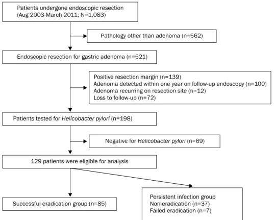
Fig. 1. Patient selection criteria.
to be closely linked to H. pylori infection, the effect of erad- ication of H. pylori on the natural course of gastric dysplasia remains largely unknown. Moreover, no study has assessed the effect of eradication of H.pylori after endoscopic re- section of gastric dysplasia.
The present study was undertaken to determine the effect of eradication of H. pylori on the development of gastric dys- plasia after endoscopic resection of the affected lesions.
SUBJECTS AND METHODS
1. Patients
This was a retrospective study that reviewed medical re- cords for patient selection. Patients who had undergone en- doscopic resection at the Seoul National University Bundang Hospital (Seongnam, Korea) between August 2003 and March 2011 (n=1,083) were assessed for enrollment.
Patients were eligible for enrollment if they were diagnosed with gastric dysplasia and had undergone endoscopic resection. Patient selection criteria is shown in Fig. 1.
Endoscopic resection included both endoscopic mucosal re- section (EMR) and endoscopic submucosal dissection. We defined metachronous dysplasia as dysplasia subsequently
detected in different location more than one year after endo- scopic resection of the first lesion. Exclusion criteria were pre- vious history of gastric cancer, previous gastric surgery, pos- itive resection margin, absence of active H. pylori infection, endoscopic resection performed for lesions other than dys- plasia such as benign gastric polyp, dysplasias detected with- in one year on follow-up endoscopy, and absence of follow-up endoscopy after endoscopic resection. All patients had given their written informed consent, and this study was approved by the institutional review board of Seoul National University Bundang Hospital.
2. Determination of H. pylori infection status and erad- ication of bacterium
To assess the H. pylori infection status, the rapid urease test (Campylobacter-like organism test; Delta West, Bently, WA, Australia) and histologic evaluation of biopsy samples taken during endoscopy were used. Patients were regarded as currently H. pylori-infected when both tests showed pos- itive results. Those who received an antimicrobial treatment were given the standard triple therapy of the standard dose proton pump inhibitor (PPI) and 500 mg of clarithromycin twice daily and 1 g of amoxicillin twice daily for 7 days as the
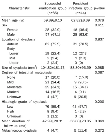
Table 1. Baseline Characteristics of Patients and Lesions accor- ding to Eradication Status
Characteristic
Successful eradication group
(n=85)
Persistent infection group
(n=44)
p-value
Mean age (yr) 59.89±9.10 62.82±8.39 0.078 Sex
Female Male
28 (32.9) 57 (67.1)
16 (36.4) 28 (63.6)
0.611
Location of dysplasia Antrum
Body Lower Mid Upper
62 (72.9)
19 (22.4) 2 (2.4) 2 (2.4)
31 (70.5)
12 (27.3) 1 (2.3) 0 (0)
0.837
Size of dysplasia (mm3) 104.82±181.73 88.80±93.59 0.585 Degree of intestinal metaplasia
7 (15.9)
0.087
None 17 (20.0)
Mild 21 (24.4) 9 (20.5)
Moderate 29 (34.1) 15 (34.1)
Marked 14 (16.5) 4 (9.1)
Unknown 4 (4.7) 9 (20.5)
Histologic grade of dysplasia 0.200
Low 76 (89.4) 43 (97.7)
High 8 (9.4) 1 (2.3)
Unknown 1 (1.2) 0 (0)
Mean duration of follow-up (mo) Metachronous dysplasia
42.99±20.31
4 (4.7)
36.00±20.85
5 (11.4)
0.069
0.272 Values are presented as mean±SD or n (%).
first-line therapy. Thereafter, patients were given ulcer medi- cations for 8 weeks to treat iatrogenic ulcer. After 4 weeks, eradication was evaluated using the urea breath test. If erad- ication was not achieved, a quadruple therapy (standard dose PPI twice daily, 500 mg tetracycline qid daily, 500 mg metronidazole tid daily, 300 mg tripotassium dicitrato bis- muth three times daily for 7 days) was given as the second line-therapy. After second line-therapy, patients were re- assessed with the urea breath test to confirm eradication.
Only those who had successfully achieved eradication were included for analysis. Patients who did not achieve erad- ication after second line-therapy were included as control in persistent infection group.
3. Histologic evaluation
Biopsy specimens were examined by two expert patholo- gists, and histologic criteria of intestinal metaplasia and dys- plasia were graded according to the updated Sydney system and the Vienna classification, respectively. Intestinal meta- plasia was graded by biopsy specimens taken from antrum and body of each subjects. Low-grade dysplasia (category 3) and high-grade dysplasia (category 4) were included in the study.8,9 The size of dysplasia was measured using endo- scopicaly resected tissue and applied in the analysis.
Patients were followed-up regularly with endoscopy every 3-12 months after endoscopic resection and biopsy was done at sites suspected of harboring metachronous dys- plastic lesions. New dysplasia occurring at the previously re- sected site was considered as recurrence and the associated data were censored.
4. Statistical analyses
Categorical variables were analyzed by the chi-square test.
Continuous variables were analyzed by the Student’s t-test.
Univariate analysis was done by the Student’s t-test and chi-square test and multivariate analysis was done using all the variables. Statistical significance was set at 0.05.
Cumulative survival was estimated by Cox’s proportional haz- ards model. All statistical analyses were performed by using PASW Statistics software version 18.0 (IBM Co., Armonk, NY, USA).
RESULTS
A total of 1,083 patients were assessed for eligibility, and 129 patients were included for analysis. Patients were div- ided into two groups based on whether they received suc- cessful eradication therapy for H. pylori (n=85) or not (n=44).
The baseline characteristics of these groups are summarized in Table 1. There were no significant differences between the successful eradication group and persistent infection group in terms of age, sex, location and size of dysplasia, degree of intestinal metaplasia, and duration of follow-up after endo- scopic resection. New gastric dysplasia was detected in four patients (4.7%) in the successful eradication group and in five patients (11.4%) in the persistent infection group after endoscopic resection.
The time taken to detect metachronous dysplasia was me- dian 26 months (range 16.5-36 months). We compared the characteristics of patients with metachronous dysplasia and those who did not develop dysplasia. The results are sum- marized in Table 2. There were statistically significant differ-
Table 2. Comparison of Baseline Characteristics of Patients and Lesions between Those Who Developed Metachronous Dysplasia and Who Did Not
Characteristic Metachronous dysplasia group (n=9)
Non-metachronous dysplasia group (n=120)
p-value
Univariate Multivariate
Mean age (yr) 64.44±10.35 60.63±8.82 0.218
Sex Female Male
1 (11.1) 8 (88.9)
43 (35.8) 77 (64.2)
0.165
Location of dysplasia Antrum
Body Lower Mid Upper
5 (55.6)
4 (44.4) 0 (0) 0 (0)
88 (73.3)
27 (22.5) 3 (2.5) 2 (1.7)
0.464
Size (mm3) 94.32±119.89 99.74±159.91 0.921
Degree of intestinal metaplasia None
Mild Moderate Marked Unknown
0 (0) 1 (11.1) 3 (33.3) 5 (55.6) 0 (0)
24 (20.0) 29 (24.2) 41 (34.2) 13 (10.8) 13 (10.8)
0.013 0.104
Histologic grade Low
High Unknown
9 (100.0) 0 (0) 0 (0)
110 (91.7) 9 (7.5) 1 (0.8)
>0.999
Successful eradication 5 (41.6) 81 (67.5) 0.110 0.014
Values are presented as mean±SD or number (%).
Fig. 2. Effect of Helicobacter pylori eradication on time to develop- ment of metachronous dysplasia.
ences the degree of intestinal metaplasia (p=0.013) be- tween the two groups in univariate anlysis. Multivariate anal- ysis showed that the eradication of H. pylori was correlated with a reduced incidence of subsequent gastric dysplasia af- ter endoscopic resection (p=0.048). Cox’s proportional haz- ard model showed that cumulative hazard ratio of sub- sequent gastric dyplasia differed between the successful
eradication group and the persistent infection group (hazard ratio=0.143, p=0.008) (Fig. 2).
DISCUSSION
Gastric premalignant lesions are commonly detected dur- ing routine endoscopy, but guidelines for follow-up or surveil- lance of such lesions are still lacking.4 Prevalence of gastric dysplasia varies between 9% to 20% in high-risk areas for gastric cancers and up to 9.3% even in asymptomatic sub- jects without indication for esophagogastroduodenosco- py.4,10 According to a recently published guideline in the man- agement of gastric precancerous lesions, both low-grade and high-grade dysplasia defined endoscopically, should be considered for endoscopic resection because of the possi- bility of upgraded histologic diagnosis after EMR.5,11,12 Considering the high incidence of gastric cancer in the coun- try and the possibility of dysplasia progression into cancer, endoscopic resection is widely performed for precancerous lesions.
The natural course of gastric premalignant lesions, espe- cially dysplasia, is not well established. Previous studies re-
ported that the local recurrence rate after EMR for early gas- tric cancer ranged between 2% and 35% and showed a higher curability and lower recurrence rate in en-bloc resection, rather than in piecemeal resection.4,13 However, there is no data regarding the recurrence rate of gastric dysplasia after endoscopic resection. To exclude the possibility of residual dysplasia being diagnosed as a new lesion, we only included patients with clear resection margin in the analysis. This in- clusion criterion would have also minimized the influence of individual physician’s endoscopic skill on therapeutic out- come. H. pylori causes chronic active gastritis, which pro- gresses through the carcinogenic cascade of atrophic gas- tritis, intestinal metaplasia and dysplasia into gastric adenocarcinoma.14 There was statistical significance be- tween the degree of intestinal metaplasia and metachronous dysplasia development in univariate analysis and this carci- nogenic cascade can be the explanation. Although multi- variate analysis showed no correlation between the degree of intestinal metaplasia and metachronous dysplasia (p=0.104), larger sample size could show different results.
Whether eradication of H. pylori can arrest or reverse carcino- genesis in premalignant gastric lesions remains controver- sial. Some studies suggest that it leads to an improvement of gastric histology or even inhibition of gastric dysplasia pro- gression into cancer.15-18 In our study, multivariate analysis showed that eradication of H. pylori can significantly inhibit the development of subsequent dysplasia after endoscopic resection of gastric dysplasia. When compared by Cox’ pro- portional hazard model, the hazard ratio of developing methachronous dysplasia was significantly greater in persis- tent infection group (Fig. 2).
There are some limitations to our study. First, this was a ret- rospective, single-institutional study with a small sample size, so the results cannot be generalized. But according to Trautmann et al.,19 the recruitment of control group is difficult in gastric cancer prevention studies because Helicobact- er-infected patients expect antimicrobial therapy rather than being in the control group. In Korea, gastric cancer is one of the most common primary cancer and therefore, patients di- agnosed of Helicobacter infection are very willing to receive antimicrobial therapy even in the absence of strongly recom- mended indications for eradication therapy due to concerns of developing gastric cancer.20 A prospective study in this set- ting would be extremely difficult to conduct. Second, since
there is no established guidelines concerning eradication of H. pylori in gastric dysplasia, no specific eradication protocol existed for this study concerning who to treat, and eradication was performed based on the physician’s preference or at pa- tient’s will. Considering this was a retrospective study, se- lection bias could have been occurred. Third, although the median follow-up period after eradication was relatively long (median of 36 months), taking into account that other stud- ies investigating the effect of eradication of H. pylori on gas- tric premalignant lesion had conflicting results may be due to the short follow-up periods.21,22 Thus, a more sufficient fol- low-up time may be needed to evaluate the long-term effect of eradication therapy on gastric dysplasia. But in our present study, we focused on the outcome itself rather than on changes over time.
Despite these limitations, this study demonstrates note- worthy findings as the first study to investigate the effect of eradication of H. pylori on the incidence of subsequent meta- chronous gastric dysplasia development after endoscopic resection. Our results demonstrate that eradication of H. py- lori is beneficial in reducing the incidence of subsequent gas- tric dysplasia after endoscopic resection of gastric dysplasia.
As gastric dysplasia is a premalignant lesion of gastric cancer and the preventive effect of eradication of H. pylori in such pa- tients are yet to be clarified, more well-designed, randomized controlled trials are warranted for the assessment of the long-term benefits of the treatment. If the present findings are confirmed, this could have great implications on gastric dysplasia treatment.
REFERENCES
1. Parkin DM, Bray F, Ferlay J, Pisani P. Global cancer statistics, 2002. CA Cancer J Clin 2005;55:74-108.
2. Correa P, Haenszel W, Cuello C, et al. Gastric precancerous proc- ess in a high risk population: cross-sectional studies. Cancer Res 1990;50:4731-4736.
3. Lauwers GY, Riddell RH. Gastric epithelial dysplasia. Gut 1999;45:784-790.
4. de Vries AC, Haringsma J, Kuipers EJ. The detection, surveillance and treatment of premalignant gastric lesions related to Helicobacter pylori infection. Helicobacter 2007;12:1-15.
5. Dinis-Ribeiro M, Areia M, de Vries AC, et al; European Society of Gastrointestinal Endoscopy; European Helicobacter Study Group; European Society of Pathology; Sociedade Portuguesa de Endoscopia Digestiva. Management of precancerous con- ditions and lesions in the stomach (MAPS): guideline from the
European Society of Gastrointestinal Endoscopy (ESGE), Euro- pean Helicobacter Study Group (EHSG), European Society of Pathology (ESP), and the Sociedade Portuguesa de Endoscopia Digestiva (SPED). Endoscopy 2012;44:74-94.
6. Dixon MF. Gastrointestinal epithelial neoplasia: Vienna revi- sited. Gut 2002;51:130-131.
7. Fukase K, Kato M, Kikuchi S, et al; Japan Gast Study Group.
Effect of eradication of Helicobacter pylori on incidence of meta- chronous gastric carcinoma after endoscopic resection of early gastric cancer: an open-label, randomised controlled trial.
Lancet 2008;372:392-397.
8. Dixon MF, Genta RM, Yardley JH, Correa P. Classification and grading of gastritis. The updated Sydney system. International Workshop on the Histopathology of Gastritis, Houston 1994. Am J Surg Pathol 1996;20:1161-1181.
9. Schlemper RJ, Riddell RH, Kato Y, et al. The Vienna classification of gastrointestinal epithelial neoplasia. Gut 2000;47:251-255.
10. den Hoed CM, van Eijck BC, Capelle LG, et al. The prevalence of premalignant gastric lesions in asymptomatic patients: predict- ing the future incidence of gastric cancer. Eur J Cancer 2011;47:1211-1218.
11. Kim YJ, Park JC, Kim JH, et al. Histologic diagnosis based on for- ceps biopsy is not adequate for determining endoscopic treat- ment of gastric adenomatous lesions. Endoscopy 2010;42:
620-626.
12. Cho SJ, Choi IJ, Kim CG, et al. Risk of high-grade dysplasia or car- cinoma in gastric biopsy-proven low-grade dysplasia: an analy- sis using the Vienna classification. Endoscopy 2011;43:
465-471.
13. Gotoda T, Yamamoto H, Soetikno RM. Endoscopic submucosal dissection of early gastric cancer. J Gastroenterol 2006;41:
929-942.
14. Correa P. Human gastric carcinogenesis: a multistep and multi- factorial process--First American Cancer Society Award Lecture on Cancer Epidemiology and Prevention. Cancer Res 1992;52:
6735-6740.
15. Saito K, Arai K, Mori M, Kobayashi R, Ohki I. Effect of Helicobacter pylori eradication on malignant transformation of gastric adenoma. Gastrointest Endosc 2000;52:27-32.
16. Correa P, Fontham ET, Bravo JC, et al. Chemoprevention of gas- tric dysplasia: randomized trial of antioxidant supplements and anti-Helicobacter pylori therapy. J Natl Cancer Inst 2000;92:
1881-1888.
17. You WC, Brown LM, Zhang L, et al. Randomized double-blind fac- torial trial of three treatments to reduce the prevalence of pre- cancerous gastric lesions. J Natl Cancer Inst 2006;98:974-983.
18. Mera R, Fontham ET, Bravo LE, et al. Long term follow up of pa- tients treated for Helicobacter pylori infection. Gut 2005;54:
1536-1540.
19. Trautmann K, Stolte M, Miehlke S. Eradication of H.pylori for the prevention of gastric cancer. World J Gastroenterol 2006;
12:5101-5107.
20. Shin HR, Jung KW, Won YJ, et al. National cancer incidence for the year 2002 in Korea. Cancer Res Treat 2007;39:139-149.
21. Ley C, Mohar A, Guarner J, et al. Helicobacter pylori eradication and gastric preneoplastic conditions: a randomized, dou- ble-blind, placebo-controlled trial. Cancer Epidemiol Biomar- kers Prev 2004;13:4-10.
22. Sung JJ, Lin SR, Ching JY, et al. Atrophy and intestinal metaplasia one year after cure of H. pylori infection: a prospective, random- ized study. Gastroenterology 2000;119:7-14.