J o u r n a l o f R h e u m a t i c D i s e a s e s V o l . 1 9 , N o . 6 , D e c e m b e r 2 0 1 2
http://dx.doi.org/10.4078/jrd.2012.19.6.341 □ Case Report □
341
<Received:December 6, 2011, Revised:December 22, 2011, Accepted:December 23, 2011>
Corresponding to:Seung Won Choi, Department of Internal Medicine, Ulsan University Hospital, College of Medicine, University of Ulsan, 877, Bangeojin Sunhwandoro, Dong-gu, Ulsan 682-714, Korea. E-mail:[email protected]
pISSN: 2093-940X, eISSN: 2233-4718
Copyright ⓒ 2012 by The Korean College of Rheumatology
This is a Free Access article, which permits unrestricted non-commerical use, distribution, and reproduction in any medium, provided the original work is properly cited.
Treatment with High Dose Dexamethasone of Severe Thrombocytopenia in Primary Sjögren’s Syndrome
Jae Wan Jun, Joong Ki Bae, Hyun Sung Lee, Jae Hee Kim, Hyung Wook Ju, Seung Won Choi
Department of Internal Medicine, Ulsan University Hospital, College of Medicine, University of Ulsan, Ulsan, Korea
Sjögren’s syndrome is a systemic autoimmune disease characterized by sicca symptoms and B lymphocyte hyperactivity. Hematological abnormalities are common in Sjögren’s syndrome, including anemia, leukopenia, throm- bocytopenia, and lymphoproliferative disorders. These hematologic involvements are mostly recognized as mild
laboratory abnormalities. Life-threatening, severe throm- bocytopenia is very rare. We describe a patient with pri- mary Sjögren’s syndrome, who developed very severe thrombocytopenia during the clinical course and success- fully treated with high dose dexamethasone therapy.
Key Words. Sjögren’s syndrome, Thrombocytopenia, Purpura
Introduction
Sjögren’s syndrome is a chronic autoimmune disease with broad spectrum manifestations, the most prevalent being de- creased lacrimal and salivary gland function. It can occur alone (primary Sjögren’s syndrome) or in association with other rheumatic diseases (secondary Sjögren’s syndrome).
Patients with primary Sjögren’s syndrome have a wide range of hematologic abnormalities, including hemocytopenia, hy- pergammaglobulinemia, and monoclonal gammopathy (1).
Hemocytopenia is detected in 1/3 of patients, may develop as isolated cytopenia or pancytopenia (2,3). Although these alter- ations are usually mild without need for treatment, some pa- tients may present severe symptomatic cytopenia (3-5).
Thrombocytopenia in Sjögren’s syndrome seems to be caused by platelet destruction due to either antiplatelet antibody or immune complex formation (6). Clinically significant severe thrombocytopenia is very rare, only a few cases have been reported in the literature (5,7-9).
We report a rare case of patient with primary Sjögren’s syn- drome who developed very severe thrombocytopenia.
Case Report
A 50-year-old woman was admitted to the hospital for evalu- ation of purpura and mucosal bleeding. She had been given a diagnosis of primary Sjögren’s syndrome 6 years ago and treated with pilocarpine and tear substitutes. Over this fol- low-up period, no other rheumatic disease has developed. Two weeks ago, she noticed epistaxis, gum bleeding, and multiple ecchymoses and purpura on her body.
On admission, blood pressure was 120/80 mmHg, temperature 36.8oC, and pulse 75/min. Physical examination revealed wide- spread purpura and petechiae over trunk and extremities. Heart and lung were normal. Neither organomegaly nor lymphaden- opathy was noted. Laboratory tests revealed hemoglobin 10.4 g/dL, white blood cell 7,260/mm3, and platelet 4,000/mm3. Blood chemistry showed protein 8.2 g/dL, albumin 4.1 g/dL, total bilirubin 0.8 mg/dL, AST 25 IU/L, ALT 27 IU/L, crea- tinine 0.78 mg/dL, LDH 278 IU/L, iron 24 μg/dL, (normal range 70∼180) TIBC 294 μg/dL (250∼437), and ferritin 22.6 ng/mL (10∼290). The prothrombin time and activated partial thromboplastin time were normal. Complement component of C3, C4 and CH50 hemolytic activity were normal range.
342 Jae Wan Jun et al.
Figure 2. Bone marrow biopsy specimen shows adequate numbers of megakaryocytes without myelodysplasia or myelofibrosis (H&E,
×400).
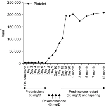
Figure 3. Clinical course of this case. Platelet counts were increased significantly after dexamethasone pulse therapy.
Figure 1. Peripheral blood smear is notable for severe throm- bocytopenia (H&E, ×400).
Antibodies against syphilis, hepatitis B, hepatitis C, or human immunodeficiency virus were not detected. Antinuclear anti- body was positive at a titer of 1:1,280 with a speckled pattern. Anti-SSA/Ro antibody and rheumatoid factor were pos- itive also. Other antibodies including anti-dsDNA, anti-Smith, anti-SSB/La, anti-Scl70, anti-Jo-1, ANCA (anti-neutrophil cyto- plasmic autoantibody), anti-phospholipid, anti-cardiolipin, anti- β2-GP1, lupus anticoagulant, and anti-platelet antibody were all negative. Both direct and indirect Coombs’ tests were negative. Urinalysis showed prominent microscopic hematuria.
Peripheral blood smear revealed scanty platelets without evi- dence of hemolysis (Figure 1). Bone marrow examination dem- onstrated adequate cellularity of megakaryocytes without mye- lodysplastic or myelofibrotic features (Figure 2). The chest ra- diography was unremarkable. Ultrasonography of the abdomen revealed no splenomegaly. Schirmer’s test was bilaterally pos- itive (1 mm/2 mm), associated with positive Rose Bengal cor- neal staining. Salivary glands scan disclosed decreased uptake
and secretory functions. Therapy with prednisolone (1.0 mg/kg/day) was started. Despite of therapy, the platelet count increase only transiently to 6,000/mm3 and dropped back to 3,000/mm3. Following the failure to respond, she received high dose dexamethasone pulse therapy (40 mg/day for 4 consec- utive days) followed by prednisolone 60 mg/day. After this therapy, the purpura and petechiae disappeared and platelet number gradually increased with count of 195,000/mm3. The dose of corticosteroid was slowly tapered with continued stable hematological status. At 12 months after therapy, the platelet count was still normal (208,000/mm3) with prednisolone 2.5 mg/every other day (Figure 3).
Discussion
Patients with primary Sjögren’s syndrome often have diverse hematologic manifestations. Ramos-Casals et al. reported the characteristics of the hematologic features in a cross-sectional study. The prevalence of cytopenia was up to 30%. The com- mon cytopenias were anemia (20%), leukopenia (16%), and thrombocytopenia (13%). The degree of thrombocytopenia was mild in most patients, although only 0.4% presented severe thrombocytopenia (platelet count less than 50,000/mm3) (3).
Several reports have described the characteristics of thrombo- cytopenia in patients with Sjögren’s syndrome (4,10,11). Most cases of thrombocytopenia were mild and fluctuating over many years. Few cases of symptomatic severe thrombocytope- nia have reported (2,5,7-9). One was caused by digitoxin in- toxication and treated with corticosteroid and discontinuation
Thrombocytopenia in Primary Sjögren’s Syndrome 343
of the culprit drug, and the other case was developed as a com- ponent of pancytopenia and managed with intravenous immunoglobulin.
Although the cause of thrombocytopenia was not completely defined, it was suggested that peripheral platelet destruction due to either platelet autoantibody or immune-complex reaction (2,6). In the multivariate analysis, cytopenias in Sjögren’s syn- drome were correlated with several autoantibodies, including antinuclear antibodies (anemia), rheumatoid factor (leukopen- ia), anti-SSA/Ro (leukopenia), and anti-SSB/La (thrombocyto- penia) (3). The causal relation between cytopenia and an- ti-SSA/Ro or anti-SSB/La antibodies has been suggested.
Abnormal cell membrane expression of SSA/Ro or SSB/La an- tigens might be induced by external agents such as viral infections. A certain viral agent with a specific bone marrow tropism may affect the bone marrow, inducing cell membrane expression of SSA/Ro or SSB/La leading to an autoantibody mediated blood cell destruction (12). Kinds of viruses, such as parvovirus B19 and hepatitis C virus, have been linked to Sjögren’s syndrome with cytopenias (6).
The goal of treatment is to raise the platelet count to high enough to prevent bleeding. Conventionally, medium to low dose corticosteroid therapy is the first-line treatment in patients without serious bleeding. In patients experiencing life-threat- ening bleeding or extremely low platelet counts, high dose cor- ticosteroid pulse therapy is recommended. Cheng et al. re- ported that dexamethasone pulse therapy was effective for a large number of patients with immune thrombocytopenia (13).
Mechanisms of action of corticosteroids on immune thrombo- cytopenia may be explained as follows: has a direct lympho- cytic effect, accelerates lymphocyte apoptosis, inhibits T-cell activation, suppresses B-cell differentiation, and regulates mac- rophage Fc receptors (13). In patients who are refractory to these treatments, newer therapies including rituximab or the thrombopoetin receptor agonists should be considered.
Summary
We describe a rare case of patient with primary Sjögren’s syndrome who developed very severe thrombocytopenia dur- ing the clinical course and successfully treated with high dose dexamethasone therapy.
References
1. Fox RI, Howell FV, Bone RC, Michelson P. Primary Sjogren syndrome: clinical and immunopathologic features. Semin Arthritis Rheum 1984;14:77-105.
2. Ramakrishna R, Chaudhuri K, Sturgess A, Manoharan A.
Haematological manifestations of primary Sjögren's syn- drome: a clinicopathological study. Q J Med 1992;83:
547-54.
3. Ramos-Casals M, Font J, Garcia-Carrasco M, Brito MP, Rosas J, Calvo-Alen J, et al. Primary Sjögren syndrome:
hematologic patterns of disease expression. Medicine (Baltimore) 2002;81:281-92.
4. Manganelli P, Fietta P, Quaini F. Hematologic manifes- tations of primary Sjögren's syndrome. Clin Exp Rheumatol 2006;24:438-48.
5. Schattner A, Friedman J, Klepfish A, Berrebi A. Immune cytopenias as the presenting finding in primary Sjögren's syndrome. QJM 2000;93:825-9.
6. Ramos-Casals M, Cervera R, García-Carrasco M, Vidal J, Trejo O, Jiménez S, et al. Cytopenia and past human par- vovirus B19 infection in patients with primary Sjögren's syndrome. Semin Arthritis Rheum 2000;29:373-8.
7. Haro T, Shimoike E, Horiuchi T, Maruyama T, Niho Y.
Severe thrombocytopenia caused by digitoxin intoxication in a patient with heart failure associated with Sjögren's syndrome. Jpn Circ J 2000;64:309-11.
8. Choung BS, Yoo WH. Successful treatment with intra- venous immunoglobulin of severe thrombocytopenia com- plicated in primary Sjögren's syndrome. Rheumatol Int 2012;32:1353-5.
9. Wada M, Kamimoto H, Park SY, Shintani S, Nakasho K.
Autoimmune hepatitis concomitant with hypergamma- globulinemic purpura, immune thrombocytopenia, and Sjögren's syndrome. Intern Med 2001;40:308-11.
10. Ramos-Casals M, Solans R, Rosas J, Camps MT, Gil A, Del Pino-Montes J, et al; GEMESS Study Group. Primary Sjögren syndrome in Spain: clinical and immunologic ex- pression in 1010 patients. Medicine (Baltimore) 2008;87:
210-9.
11. García-Carrasco M, Ramos-Casals M, Rosas J, Pallarés L, Calvo-Alen J, Cervera R, et al. Primary Sjögren syndrome:
clinical and immunologic disease patterns in a cohort of 400 patients. Medicine (Baltimore) 2002;81:270-80.
12. Baboonian C, Venables PJ, Booth J, Williams DG, Roffe LM, Maini RN. Virus infection induces redistribution and membrane localization of the nuclear antigen La (SS-B):
a possible mechanism for autoimmunity. Clin Exp Immunol 1989;78:454-9.
13. Cheng Y, Wong RS, Soo YO, Chui CH, Lau FY, Chan NP, et al. Initial treatment of immune thrombocytopenic purpura with high-dose dexamethasone. N Engl J Med 2003;349:831-6.