ISSN 2234-3806 • eISSN 2234-3814
http://dx.doi.org/10.3343/alm.2015.35.1.132
Clinical Relevance of High-Resolution Single Nucleotide Polymorphism Array in Patients with
Relapsed Acute Lymphoblastic Leukemia with Normal Karyotype: A Report of Three Cases
Sang Hyuk Park, M.D.1,2,*, Seung-Hee Lee, M.D.1,*, Shine Young Kim, M.D.1, Sun Min Lee, M.D.3, Jongyoun Yi, M.D.1, In-Suk Kim, M.D.3, Hyung Hoi Kim, M.D.1,2, Chulhun Ludgerus Chang, M.D.3, and Eun Yup Lee, M.D.1,2
Department of Laboratory Medicine1, Pusan National University School of Medicine, Pusan National University Hospital, Busan; Biomedical Research Institute2, Pusan National University Hospital, Busan; Department of Laboratory Medicine3, Pusan National University School of Medicine, Pusan National University Yangsan Hospital, Yangsan, Korea
We report three patients with normal karyotype (NK) ALL, who showed genetic aberrations as determined by high-resolution single nucleotide polymorphism array (SNP-A) analysis at both diagnosis and relapse. We evaluated the clinical relevance of the SNP-A assay for the detection of subtle changes in the size of affected genetic lesions at relapse as well as the prognostic value of the assay. In our patients, application of the SNP-A assay enabled sensitive detection of cryptic changes affecting clinically important genes in NK ALL.
Therefore, this assay seems to be more advantageous compared to other conventional methods such as FISH assay, HemaVision (DNA Technology, Denmark), and conventional karyotyping for the detection of an “unstable genotype” at relapse, which may be associ- ated with microscopic clonal evolution and poor prognosis. Further comprehensive stud- ies are required to confirm the issues presented by our case patients in this report.
Key Words: Acute lymphoblastic leukemia, Array, Clonal evolution, Normal karyotype, Prognosis, Single nucleotide polymorphism
Received: May 19, 2014 Revision received: July 15, 2014 Accepted: October 28, 2014 Corresponding author: Eun Yup Lee Department of Laboratory Medicine and Biomedical Research Institute, Pusan National University School of Medicine, Pusan National University Hospital, 179 Gudeok-ro, Seo-gu, Busan 602-739, Korea Tel: +82-51-240-7419
Fax: +82-51-247-6560 E-mail: [email protected]
*These two authors contributed equally to this work.
© The Korean Society for Laboratory Medicine This is an Open Access article distributed under the terms of the Creative Commons Attribution Non-Commercial License (http://creativecom- mons.org/licenses/by-nc/3.0) which permits unrestricted non-commercial use, distribution, and reproduction in any medium, provided the original work is properly cited.
INTRODUCTION
A genome-wide single nucleotide polymorphism array (SNP-A) assay can detect not only the copy number alterations but also the copy-neutral loss of heterozygosity (CN-LOH) on a chromo- somal segment with high sensitivity, as demonstrated in MDS/
MPN and NK (normal karyotype) AML [1-5]. However, no study has evaluated SNP-A assay in NK ALL, except for one that re- ported detection of copy number alterations and CN-LOH by
SNP-A in 62% of NK ALL patients as well as smaller deletions compared with those in other leukemia subtypes [6]. In addi- tion, the clinical relevance of unstable genotype (subtle changes in the size of detected alterations at relapse) has not been eval- uated. We report three patients with NK ALL who harbored ge- nomic alterations, as detected by a high-resolution SNP-A assay at both diagnosis and relapse. We also assessed the clinical rel- evance of slight changes in the size of affected genetic lesions at relapse with the maintenance of normal cytogenetics.
CASE REPORT
1. Case 1A 38-yr-old man was diagnosed as having NK ALL on the basis of the results of negative HemaVision (DNA Technology; Aarhus, Denmark) and metaphase cytogenetics (MC) analysis. He was diagnosed as having B-cell ALL with NK and achieved complete remission (CR) at 10 weeks of treatment. He experienced relapse at 8 weeks after CR and, at relapse, his karyotype was maintained as normal and HemaVision results were also negative.
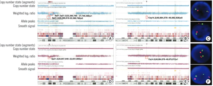
A genome-wide SNP-A assay was performed by using the Af- fymetrix Cytogenetics 2.7M Array (Affymetrix; Santa Clara, CA, USA) with bone marrow samples obtained both at diagnosis and relapse. At diagnosis, SNP-A analysis revealed homozygous de- letions of 9p21.3 (associated with CDKN2A) and 13q14.2 (as- sociated with RB1) and heterozygous deletions of 6p22.2, 8q22.1, 9p21.3p21.2, 14q11.2, 18q22.2, and 19p13.2 (Fig.
1A, B). FISH analysis for the detection of RB1 deletions showed nuc ish (RB1,13q14)×2[200] (Fig. 1C). An identical assay per- formed at relapse showed loss of heterozygous deletions in 6 af- fected lesions along with subtle size changes in deleted lesions of 9p21.3 (1,944-1,752 kb) and 13q14.2 (96-89 kb), indicating the persistence of CDKN2A and RB1 deletion at relapse (Fig.
1D, E). FISH analysis for the detection of RB1 deletions also
showed nuc ish (RB1,13q14)×2[200] (Fig. 1F).
2. Case 2
A 57-yr-old woman was diagnosed as having NK ALL on the ba- sis of the results of HemaVision (negative) and MC analysis. SNP- A analysis at diagnosis revealed heterozygous interstitial dele- tions of 3q13, 4q23q24, 5q15q21.3, 5q21.3q22.1, 5q22.3q23.1, 5q34, 6q16.3q21, 7q11.23, and 13q14.2q14.3, including RB1 (Fig. 2A). She achieved CR after 4 weeks of treatment but expe- rienced relapse at 6 months after CR. At relapse, her karyotype was normal and HemaVision results were also negative. Results of SNP-A analysis at relapse showed heterozygous deletions of 3q13, 4q23q24, 5q15q21.3, 5q21.3q22.1, 5q22.3q23.1, 5q34, 6q16.3q21, 7q11.23, and 13q14.2q14.3, including RB1 (Fig. 2B), which is similar to those at diagnosis. However, the deleted genomic lesions of 3q13, 4q23q24, 5q15q21.3, 5q21.3q22.1, 5q34, and 13q14.2q14.3 at relapse were slightly different from those at di- agnosis.
3. Case 3
A 58-yr-old man was diagnosed as having NK ALL as per the results of HemaVision (negative) and MC analysis. He achieved CR after 4 weeks of chemotherapy but experienced relapse at follow-up 32 months after remission. At relapse, the HemaVi-
Fig. 1. High-resolution single nucleotide polymorphism array (HR SNP-A) analysis results for chromosome 9 (A) and 13 (B) performed at diagnosis in Case 1. Deleted lesions are indicated with a red box, and detailed array results in each lesion are provided. The results showed a homozygous 1,944 kb deletion of 9p21.3, including CDKN2A; a 96 kb deletion of 13q14.2, including RB1; and a heterozygous 5,262 kb deletion of 9p21.3p21.2. The FISH analysis for the detection of RB1 deletion at diagnosis showed nuc ish (RB1,13q34)×2[200], which in- dicates no deletion of RB1(C). Identical HR SNP-A analysis results for chromosome 9 (D) and 13 (E) performed at relapse in Case 1 showed a heterozygous 1,752 kb deletion of 9p21.3, including CDKN2A and an 89 kb deletion of 13q14.2, including RB1. FISH analysis for the detection of RB1 deletion at relapse also showed nuc ish (RB1,13q34)×2[200], which indicates no deletion of RB1(F).
Copy number state (segments)
Copy number state (segments) Copy number state
Copy number state Weighted log2 ratio
Weighted log2 ratio Allele peaks
Allele peaks Smooth signal
Smooth signal
A
D
B
E
C
F
sion results were also negative. However, SNP-A analysis dem- onstrated an interstitial 451 kb deletion of 9p21.3, which in- cludes interferon α-1/13 (IFNA13) and micro-RNA 31 (MIR31) in a mosaic pattern, at both diagnosis and relapse (Fig. 2C). De- mographic findings of the deleted genetic lesions and the af- fected genes associated with ALL in the three cases are repre- sented in Table 1.
DISCUSSION
In Case 1, SNP-A analysis could detect the presence of intersti- tial microdeletion including both RB1 and CDKN2A, which is not otherwise detectable by MC or FISH analysis. Because the deletions of CDKN2A and RB1 are associated with poor progno- sis in ALL [7, 8], this may suggest that SNP-A assay can provide prognostic information in NK ALL. The SNP-A assay could detect Fig. 2. High-resolution single nucleotide polymorphism array (HR SNP-A) analysis results in whole chromosomes at diagnosis (idiograms of six chromosomes harboring aberrations are provided) in Case 2 (A). The array results showed heterozygous interstitial deletions of 3q13, 4q23q24, 5q15q21.3, 5q21.3q22.1, 5q22.3q23.1, 5q34, 6q16.3q21, 7q11.23, and 13q14.2q14.3, including RB1 gene. Deleted lesions are indicated separately with red arrows, and the detailed array results for each lesion are provided. Identical array results for whole chro- mosome at relapse in Case 2 (B) showed similar results, but slight size changes in deleted genomic lesions at 3q13, 4q23q24, 5q15q21.3, 5q21.3q22.1, 5q34, and 13q14.2q14.3 were identified at relapse compared to those at diagnosis. Deleted lesions are also indicated sepa- rately with red arrows, and detailed array results for each lesion are provided. In addition, HR SNP-A analysis results of chromosome 9 per- formed at both diagnosis and relapse in Case 3 (C) are provided. The deleted lesion is indicated with a red box, and detailed array result is provided. The array result showed an interstitial 451 kb deletion of 9p21.3, which includes IFNA13 and MIR31 in a mosaic pattern.
A
B
C
subtle size changes in the affected lesions (microscopic clonal evolution) and recovery of small-sized interstitial deletions at re- lapse, which are also not detectable by MC. Thus, the SNP-A assay is more advantageous in detecting microscopic clonal evolution than other methods. In addition, in Case 2, SNP-A as- say could detect subtle changes in the size of the affected ge- netic lesions at relapse (unstable genotype). We may speculate that unstable genotype may indicate microscopic clonal evolu-
tion at relapse. The early relapse in Case 2 patient supports this speculation.
The association between ALL and the loss of IFNA13 and MIR31 is unclear. However, as abnormal 9p is an adverse prog- nostic factor for B-ALL [9], the interstitial deletion of 9p21.3 de- tected by SNP-A analysis may contribute to poor prognosis, as demonstrated by early relapse in Case 3.
Our report has some limitations. First, the SNP-A assay at re- Table 1. Characteristics of abnormal lesions detected by high-resolution single nucleotide polymorphism array (SNP-A) assay in 3 cases
Case No. Stage Chromosome Affected point (Start)* Affected point (End)* Deletion size (kb) Affected genes associated with ALL†
1 Diagnosis 6p22.2 26,174,861 26,566,470 382 None
8q22.1 95,054,989 95,486,831 422 None
9p21.3 20,369,818 22,360,786 1,944 CDKN2A
9p21.3p21.2 22,360,786 27,749,366 5,262 None
13q14.2 48,984,578 49,082,926 96 RB1
14q11.2 22,447,554 23,017,964 557 TRA, TRD
18q22.2 66,977,443 67,361,947 375 None
19p13.2 12,519,413 12,738,738 214 None
Relapse 9p21.3 20,857,948 22,651,989 1,752 CDKN2A
13q14.2 48,984,578 49,075,572 89 RB1
2 Diagnosis 3q13 107,063,285 107,790,364 710 None
4q23q24 100,661,470 104,376,163 3,628 None
5q15q21.3 97,852,943 104,983,198 6,963 None
5q21.3q22.1 106,627,589 110,384,109 3,668 None
5q22.3q23.1 113,640,675 115,817,032 2,125 None
5q34 161,446,706 164,775,139 3,250 None
6q16.3q21 104,892,215 112,813,414 7,736 None
7q11.23 72,848,598 74,628,840 1,739 None
13q14.2q14.3 48,966,145 53,262,014 4,195 RB1
Relapse 3q13.12 107,081,719 107,787,107 689 None
4q23q24 100,661,470 104,385,396 3,637 None
5q15q21.3 97,840,121 105,039,827 7,031 None
5q21.3q22.1 106,655,459 110,384,109 3,641 None
5q22.3q23.1 113,640,675 115,817,032 2,125 None
5q34 161,418,668 164,775,139 3,278 None
6q16.3q21 104,892,215 112,813,414 7,736 None
7q11.23 72,848,598 74,628,840 1,739 None
13q14.2q14.3 48,984,803 53,243,124 4,159 RB1
3 Diagnosis 9p21.3 21,355,664 21,817,917 451 None
Relapse 9p21.3 21,355,664 21,817,917 451 None
*The locations of affected genetic lesions were aligned using the human genome browser–hg19 assembly (http:// genome.ucsc.edu/cgi-bin/hgGateway),
†The list of affected genes associated with ALL within each genetic lesion was determined from the review of a web-based database (http:// atlasgeneticson- cology.org/index.html) and browser (http:// genome.ucsc.edu/cgi-bin/hgGateway).
Abbreviations: CDKN2A, cyclin-dependent kinase 2a/p16; RB1, retinoblastoma; TRA, T cell receptor α; TRD, T cell receptor δ.
lapse in Case 2 did not show any additional abnormalities and slight size changes in the detected genetic lesions as evidence of clonal evolution have not been demonstrated. Therefore, the presence of unstable genotype should be regarded only as a phenomenon suggesting various events, including clonal evolu- tion. Second, we could neither perform SNP-A analysis using samples with CR or fibroblasts, which is important for the dis- crimination of acquired somatic events from germline aberra- tions, nor confirm whether all genetic abnormalities detected in our cases were somatic. Third, we could not perform FISH anal- ysis in RB1 in Case 2 as well as detailed evaluations of the dis- crepancies between the results of FISH and SNP-A analysis. The discrepancy in the results of FISH and SNP-A analysis regard- ing the RB1 gene in Case 1 may be explained from different de- tection sensitivity of both methods. However, more comprehen- sive analysis is required for comprehending this discrepancy.
In conclusion, our case report demonstrates that a SNP-A as- say can allow sensitive detection of cryptic changes affecting clinically important genes. A SNP-A assay may be more advan- tageous than other methods in detecting unstable genotype at relapse, which may be associated with microscopic clonal evo- lution and poor prognosis in ALL.
Authors’ Disclosures of Potential Conflicts of Interest
No potential conflicts of interest relevant to this article were re- ported.
Acknowledgments
This work was supported by the 2013 clinical research grant from Pusan National University Hospital.
REFERENCES
1. Bullinger L, Krönke J, Schön C, Radtke I, Urlbauer K, Botzenhardt U, et al. Identification of acquired copy number alterations and uniparental disomies in cytogenetically normal acute myeloid leukemia using high- resolution single-nucleotide polymorphism analysis. Leukemia 2010;
24:438-49.
2. Barresi V, Romano A, Musso N, Capizzi C, Consoli C, Martelli MP, et al.
Broad copy neutral-loss of heterozygosity regions and rare recurring copy number abnormalities in normal karyotype-acute myeloid leuke- mia genomes. Genes Chromosomes Cancer 2010;49:1014-23.
3. Tiu RV, Gondek LP, O’Keefe CL, Huh J, Sekeres MA, Elson P, et al. New lesions detected by single nucleotide polymorphism array-based chro- mosomal analysis have important clinical impact in acute myeloid leu- kemia. J Clin Oncol 2009;27:5219-26.
4. Yi JH, Huh J, Kim HJ, Kim SH, Kim HJ, Kim YK, et al. Adverse prog- nostic impact of abnormal lesions detected by genome-wide single nu- cleotide polymorphism array-based karyotyping analysis in acute my- eloid leukemia with normal karyotype. J Clin Oncol 2011;29:4702-8.
5. Hahm C, Huh HJ, Mun YC, Seong CM, Chung WS, Huh J. Genomic ab- errations of myeloproliferative and myelodysplastic/myeloproliferative neoplasms in chronic phase and during disease progression. Int J Lab Hematol 2014 May 21. doi: 10.1111/ijlh.12257. [Epub ahead of print]
6. Huh J, Jung CW, Kim HJ, Kim YK, Moon JH, Sohn SK, et al. Different characteristics identified by single nucleotide polymorphism array anal- ysis in leukemia suggest the need for different application strategies de- pending on disease category. Genes Chromosomes Cancer 2013;52:
44-55.
7. Betts D. del(13q) in ALL. Atlas Genet Cytogenet Oncol Haematol 2005;
9:24-5. (http://AtlasGeneticsOncology.org/Anomalies/del13qALLID1188.
html).
8. Kim M, Yim SH, Cho NS, Kang SH, Ko DH, Oh B, et al. Homozygous de- letion of CDKN2A (p16, p14) and CDKN2B (p15) genes is a poor prog- nostic factor in adult but not in childhood B-lineage acute lymphoblastic leukemia: a comparative deletion and hypermethylation study. Cancer Genet Cytogenet 2009;195:59-65.
9. Heerema NA. 9p rearrangements in ALL. Atlas Genet Cytogenet Oncol Haematol 1999;3:153-4. (http://atlasgeneticsoncology.org/Anomalies/
9prearrALLID1156.html).