Letter to the Editor
380 Ann Dermatol
Received July 23, 2012, Revised September 26, 2012, Accepted for publication October 14, 2012
Corresponding author: Young Min Park, Department of Dermatology, Seoul St. Mary’s Hospital, College of Medicine, The Catholic University of Korea, 222 Banpo-daero, Seocho-gu, Seoul 137-701, Korea. Tel: 82-2-2258-6223, Fax: 82-2-599-9950, E-mail: yymmpark6301@hotmail.com This is an Open Access article distributed under the terms of the Creative Commons Attribution Non-Commercial License (http://
creativecommons.org/licenses/by-nc/3.0) which permits unrestricted non-commercial use, distribution, and reproduction in any medium, provided the original work is properly cited.
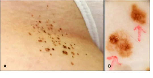
Fig. 1. (A) Asymmetric, multiple, grouped, irregular, flat, dark bro- wnish papules and macules on the inguinal area. (B) Dermoscopic fea- tures showing a patchy reticulated network.
http://dx.doi.org/10.5021/ad.2013.25.3.380
Agminated Acquired Melanocytic Nevi of the Common and Dysplastic Type
Yoon Jung Choi, Hei Sung Kim, Jun Young Lee, Hyung Ok Kim, Young Min Park
Department of Dermatology, Seoul St. Mary’s Hospital, College of Medicine, The Catholic University of Korea, Seoul, Korea
Dear Editor:
‘Agminated’ refers to circumscribed grouping of lesions confined to a localized area of the body. Pigmented lesions that have been described as agminated includes melanocytic nevi1, Spitz nevi2, nevi spilus3, blue nevi4, and multiple lentigines5. However, the presence of acqui- red common and dysplastic nevi (ACDN) arranged in an agminated pattern has not been well-established. Herein, we describe a patient with multiple agminated acquired melanocytic nevi, several of that were histologically cha- racterized as dysplastic nevi.
A 16-years-old female presented with multiple irregular moles on her right inguinal area (Fig. 1A). Her parents reported that this cluster of nevi developed at the age of 6
years, and continuously increased in numbers and sizes.
No family history of melanoma or multiple moles was present. Physical examinations revealed more than 40 melanocytic nevi clustered in a 5×5 cm skin area on her right inguinal area, where several of these nevi were irregular with variegation of color and a diameter greater than 5 mm (Fig. 1A). No background pigmentation within or surrounding the cluster was noted clinically or even with Wood’s light examination. Dermoscopy of the clustered nevi revealed a diffuse patchy reticulation (Fig.
1B). A biopsy from a clinically atypical nevus demonst- rated a lentiginous, compound-melanocytic nevus with architectural disorder in the epidermis. In the dermoepi- dermal junction, the nests of nevus cells were profused in
Letter to the Editor
Vol. 25 No. 3, 2013 381 Fig. 2. (A) There was elongation of rete ridges and increase in the number of junctional melanocytes, arranged in a nest. In the dermoepidermal junction, nests of nevus cells were profused on the tips and sides of elongated rete ridges. There were also scattered single cells in a lentiginous array without continuous proliferations. (B) Several melanocytes contained abnormally large nuclei, and the nuclei had irregular contours (arrows) (H&E; A: ×40, B: ×400).
the tips and sides of elongated rete ridges. In the center of the nevus, nests of melanocytes are present in the papi- llary dermis (Fig. 2A). There were scattered single cells in a lentiginous array without continuous proliferations (Fig.
2A). A few atypical melanocytes with large, irregularly shaped, hyperchromatic nuclei lay individually or within a small group (Fig. 2B). The diagnosis of dysplastic nevi was confirmed by the presence of an architectural disorders and cytologic atypia. Several nevi within the cluster had clinical, dermoscopic, and histologic features which are commonly attributed to dysplastic nevi.
Dysplastic nevi have been the subject of ongoing con- troversy regarding its definition and use of more than 20 years6. It is argued that dysplastic nevi are both acquired and common, and they should be regarded as nothing but common nevi. However, when considering a review article from Elder6, theses lesions have been only signi- ficant in relation to melanoma, as stimulants of melanoma, as markers of risks for melanoma, and as potential and occasional actual precursors of melanoma. Therefore, clinical and histological classifications of nevi have the most important purpose of categorizing these lesions so that dysplastic nevi can be distinguished, clinically and histologically, from melanoma. In our patient, the diag- nosis of dysplastic nevi was based on the clinical, dermoscopic and histologic features.
Agminated dysplastic lesions, similar to our case should be distinguished from other forms of dysplastic nevi, which show a segmental distribution without a definite clustering. Two cases of dysplastic nevi with segmental distributions have been previously reported in the lite- rature7,8. Both of the cases are not being described as agminated, because multiple dysplastic nevi were distributed throughout the patients’ upper left quadrant.
One thing that deserves the attention of clinicians is that malignant melanoma developed within the lesions of both cases.
There still exists much controversy about the presence of agminated ACDN. Marghoob et al.9 and Bragg et al.10 previously reported 5 cases of agminated ACDN. Unlike our case, however, agminated lesions were superimposed on an underlying dysplastic nevus syndrome phenotype in 4 out of 5 cases. In the 2 cases of the 5 reported as agmi- nated ACDN, malignant melanoma did not developed within the agminated lesion, but within the underlying dysplastic nevus syndrome phenotype. The authors beli- eved that agminated ACDN were a new, previously not described, clinical variant of dysplastic nevi. In conclu- sion, the uncertainty in the biological behaviors of agmi- nated ACDN suggests a strict follow-up for this unusual entity.
REFERENCES
1. Corradin MT, Alaibac M, Fortina AB. A case of malignant melanoma arising from an acquired agminated melanocytic naevus. Acta Derm Venereol 2007;87:432-433.
2. Glasgow MA, Lain EL, Kincannon JM. Agminated Spitz nevi:
report of a child with a unique dermatomal distribution.
Pediatr Dermatol 2005;22:546-549.
3. Marghoob AA, Blum R, Nossa R, Busam KJ, Sachs D, Hal- perin A. Agminated atypical (dysplastic) nevi: case reports and review of literature. Arch Dermatol 2001;137:917-920.
4. Bragg JW, Swindle L, Halpern AC, Marghoob AA. Agmi- nated acquired melanocytic nevi of the common and dysplastic type. J Am Acad Dermatol 2005;52:67-73.
5. Micali G, Nasca MR, Innocenzi D, Lembo D. Agminated lentiginosis: case report and review of the literature. Pediatr
Letter to the Editor
382 Ann Dermatol
Received October 9, 2012, Accepted for publication October 18, 2012
Corresponding author: Young Min Park, Department of Dermatology, Seoul St. Mary’s Hospital, The Catholic University of Korea College of Medicine, 222 Banpo-daero, Seocho-gu, Seoul 137-701, Korea. Tel: 82-2-2258-6223, Fax: 82-2-599-9950, E-mail: yymmpark6301@hotmail.com This is an Open Access article distributed under the terms of the Creative Commons Attribution Non-Commercial License (http://
creativecommons.org/licenses/by-nc/3.0) which permits unrestricted non-commercial use, distribution, and reproduction in any medium, provided the original work is properly cited.
Dermatol 1994;11:241-245.
6. Elder DE. Dysplastic naevi: an update. Histopathology 2010;
56:112-120.
7. Misago N, Takahashi M, Kohda H. Unilateral dysplastic nevi associated with malignant melanoma. J Dermatol 1991;18:
649-653.
8. Sterry W, Christophers E. Quadrant distribution of dysplastic
nevus syndrome. Arch Dermatol 1988;124:926-929.
9. Marghoob AA, Blum R, Nossa R, Busam KJ, Sachs D, Hal- pern A. Agminated atypical (dysplastic) nevi: case report and review of the literature. Arch Dermatol 2001;137:917-920.
10. Bragg JW, Swindle L, Halpern AC, Marghoob AA. Agmina- ted acquired melanocytic nevi of the common and dysplas- tic type. J Am Acad Dermatol 2005;52:67-73.
http://dx.doi.org/10.5021/ad.2013.25.3.382
Medallion-Like Dermal Dendrocyte Hamartoma:
Differential Diagnosis with Congenital Atrophic Dermatofibrosarcoma Protuberans
Minseok Cheon, Kyung Eun Jung, Hei Sung Kim, Jun Young Lee, Hyung-Ok Kim, Chan Kum Park
1, Young Min Park
Department of Dermatology, Seoul St. Mary’s Hospital, The Catholic University of Korea College of Medicine, 1Department of Pathology, Hanyang University College of Medicine, Seoul, Korea
Dear Editor:
Medallion-like dermal dendrocyte hamartoma (ML-DDH) is a recently described congenital benign dermal lesion, which was first reported by Rodríguez-Jurado et al.1 Clinically, ML-DDH presents as a solitary, several centi- meter-sized, round or oval, erythematous to yellow- brown, atrophic plaque on the neck or upper trunk. Histo- pathologically, ML-DDH is characterized by a prolifera- tion of CD34+ spindle-shaped cells or ovoid cells mainly in the reticular dermis and extending into the subcutis in some cases. Only a small number of ML-DDH has been reported in English literature1-3. Herein, we report a case of ML-DDH that was initially misdiagnosed as congenital atrophic dermatofibrosarcoma protuberans (DFSP).
A 6-year-old girl presented with symptoms of intermitt-
ently pruritic and painful, solitary, depressed, and erythe- matous to yellowish plaque along with fine wrinkles on her posterior neck (Fig. 1). The oval-shaped, 4.0×2.5 cm sized plaque had been present since birth. Other personal and family history was unremarkable. Routine laboratory tests were normal. The clinical impression was a scar or congenital atrophic DFSP. After obtaining an informed consent from the patient and her parents, a punch biopsy was performed on the depressed lesion. The skin biopsy specimen revealed dermal proliferation of spindle-shaped cells in a storiform-like pattern (Fig. 2A, B). The lesion was diffusely positive for CD34, but negative for S-100 protein on immunohistochemistry. Thus, the lesion was initially diagnosed as congenital atrophic DFSP. The patient was sent to a plastic surgeon for complete removal of the