Effect of Glutathione-Enriched Saccharomyces cerevisiae FF-8 on Tissues Lipid Peroxidation in Orotic Acid-Induced Fatty Liver Model Rats
Jae Young Cha1, Bo Kyung Park, Kyung Eun Eom, Hee Young Ahn and Young Su Cho*
Department of Biotechnology, Dong-A University, Busan 604-714, Korea
1Technical Research Institute, Daesun Distilling Co., Ltd. Busan 619-934, Korea Received December 16, 2008 /Accepted February 20, 2009
Glutathione is a well-known chemotherapeutic agent and a popular nutritional supplement for liver disease and oxidative stress. Our previous studies reported the suppressive effects of the gluta- thione-enriched Saccharomyces cerevisiae FF-8 (FF-8) strain on carbon tetrachloride- and alcohol-induced oxidative stress in rats. The purpose of the current study was to investigate the effect of the FF-8 strain on lipid peroxidation in tissues of rats with orotic acid (OA)-induced fatty liver. OA treatment showed a significant decrease in body weight gain compared to the normal diet, and simultaneous addition of FF-8 and OA had the same effect. OA treatment produced an increase in liver weight, however, this also increased with simultaneous addition of FF-8 and OA. Liver lipid peroxidation was significantly increased by OA, but was significantly decreased by FF-8 strain treatment. This same ten- dency was found in the kidney and heart. Concentration levels of hepatic glutathione and zinc are known to be closely associated with the antioxidant system, and OA treatment led to reductions in liver glutathione and zinc concentrations, whereas these were significantly increased by FF-8 strain treatment in OA feeding rats. These results suggest that the glutathione-enriched S. cerevisiae FF-8 strain may positively mediate orotic acid-induced oxidative stress by enhancing glutathione and zinc levels in rat livers.
Key words : Glutathione, zinc, yeast, orotic acid, lipid peroxidation
*Corresponding author
*Tel:+82-51-200-7586, Fax:+82-51-200-7505
*E-mail : [email protected]
Introduction
Glutathione is a ubiquitous tripeptide containing L-gluta- mate, L-cysteine, and glycine in living organisms and oxi- dized glutathione play a central role in the antioxidant de- fense process, while reduced glutathione is decreased by oxidative tissue damage [1,17,26,27]. It is involved in the maintenance of normal cell structure and functions [16,33].
S. cerevisiae have been found to produce high essential bio- active components for human health [5,6]. Many studies have found that glutathione is an antioxidant and it is con- tained high concentration in yeast strains [6,20,25]. Previous our study reported the suppressive effect of glutathione- enriched S. cerevisiae FF-8 strain treatment on carbon tetra- chloride-induced oxidative stress in rats [7]. A previous in vitro study with the intercellular glutathione-containing cell free extracts from S. cerevisiae FF-8 has also observed anti- oxidative effects [20]. As, glutathione is a well known che- motherapeutical agent and a popular nutritional supple- ment for liver disease and oxidative stress [14,19,34].
In addition, zinc is an essential trace element of all organ- isms and zinc plays important biological roles in hepatic in- jury and antioxidant properties against oxidative damage in organisms [2,15,23,26,27]. Recently have been reported that highly zinc containing yeast strain isolated from the tropical fruit rambutan and this strain was protected against alco- hol-induced hepatotoxicity and oxidative stress in rats [5].
However, the antioxidative effect of FF-8 strain in OA-in- duced fatty liver model rats has not been reported. Current study was to investigate the effect of glutathione-enriched S. cerevisiae FF-8 strain on the tissues lipid peroxidation of orotic acid (OA)-induced fatty liver model in rats.
Materials and Methods
S. cerevisiae FF-8 strain cultivation
S. cerevisiae FF-8 (KACC 93023) strain containing a high glutathione concentration used in this study was established in our laboratory [25]. S. cerevisiae FF-8 was aerobically cul- tured in a 100 l bioreactor containing the YM optimal me- dium (3.0% glucose, 3.0% yeast extract, 0.06% KH2PO4, and 0.06% L-cysteine) [6]. The harvested yeast cells were lyophi- lized to prepare experimental diet.
Animal experiments
Six-week-old male Sprague-Dawley strain rats (Hyochang Science Animals Co., Daegu, Korea) were randomly divided into three treatment dietary groups. The normal rats were fed a semisynthetic basal diet, the OA rats were fed a basal diet with 1% (w/w) OA, and the OA plus FF-8 rats were fed a basal diet with concomitant treatment OA and 5% FF-8.
The amount of FF-8 supplementation to the rats was esti- mated from the previous reports that S. cerevisiae had sig- nificant effects on the liver injury and oxidative stress in the rats [5,7,29,35]. Food and water were provided ad libitum for 10 days.
Assay of tissues lipid peroxidation
The tissues were quickly removed, weighed, and even- tually used for the estimation of lipid peroxidation, minerals, and glutathione. The tissues were homogenized in ice-cold 0.25 M sucrose solution containing 10 mM tris-HCl buffer (pH 7.4) and 1 mM ethylenediamine tetraacetate (EDTA) us- ing with IKA-ULTRA-TURRAX T25 basic homogenizer (IKA-WERKE GMBH & CO., KG, Staufen, Germany) as de- scribed previously [10]. Tissues lipid peroxidations were de- termined according to the spectroscopic technique by meas- uring thiobarbituric acid reactive substances (TBARS) [12].
The reaction mixture, containing hepatic homogenate sol- ution and thiobarbituric acid (TBA), was incubated under boiling water for 30 min. After the centrifugation at 1,000×
g for 10 min, the light absorbance of the upper layer was measured at 532 nm. The concentrations of TBARS were ex- pressed as nmole of malondialdehyde (MDA) per g tissue.
Assay of hepatic glutathione concentrations
The concentration of glutathione was determined by the method of Beutler et al. [3]. A 0.2 ml of liver homogenate was mixed well with 1.8 ml of EDTA solution then a 3.0 ml of the precipitating reagent (1.67 g of metaphosphoric acid, 0.2 g of EDTA disodium salt, 30 g sodium chloride in 1 l of distilled water) was added and mixed thoroughly then stood the mixture at 4oC for 5 min. The mixture was centrifuged at 3,000× g for 5 min and 2.0 ml of the super- natant was mixed with 4.0 ml of 0.3 M disodium hydrogen phosphate solution and 0.1 ml of 5,5'-dithiobis(2-nitro- benzoic acid) (DTNB) reagent then the concentration of glutathione was spectrophotometrically determined at 412 nm. Total glutathione concentrations were expressed as mg per g liver.
Assay of hepatic mineral concentrations
Mineral concentrations in hepatic were analyzed by a Perkin-Elmer (Uberlingen, Model 300) atomic absorption spectrophotometer [4]. The determination concentration was shown as ppm.
Statistical analysis
The data from animal experiments are presented as the mean±SE, and were analyzed using one way analysis of var- iance (ANOVA), with the differences analyzed using the Duncan's new multiple-range test [13]. A p value <0.05 was accepted as being a statistical significance of difference.
Results and Discussion
Glutathione is one of the most important protective fac- tors against oxidative damage. Reduction in cellular concen- trations of glutathione is used as the indication of oxidative stress in a organ system [26,27]. Many studies have found that glutathione is an antioxidant in organisms [2,15,19]. A highly contained glutathione from microorganisms have been found to inhibit those chemicals-induced oxidant stresses in vitro and in vivo such as ethanol, carbon tetra- chloride and acetaminophen [1,2,20,29]. Previous our ob- servation showed that glutathione-enriched FF-8 strain was effectively inhibited the alcohol-induced oxidative stress in rats [7]. A previous in vitro study with the intercellular gluta- thione-containing cell free extracts from FF-8 have also ob- served highly antioxidative effects [7]. In addition, admin- istration of FF-8 significantly inhibited lipid peroxidation of liver homogenate fractions in the carbon tetrachloride treat- ment rats [29]. In this respect, Mannaa et al. also reported that the powerful antioxidative components in S. cerevisiae effectively participated in attenuation of the oxidative stress caused by flutamide metabolites [21].
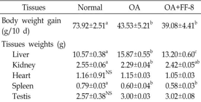
OA treatment showed a significant decrease in body weight gain compared to the normal diet, and simultaneous addition of FF-8 and OA was also decreased (Table 1). OA treatment produced an increase in the liver weight, however, this increase decreased by simultaneous addition of FF-8 and OA (Table 1).
The current study observed significant elevations of the lipid peroxidation in the liver homogenate of the OA treated rats, compared with the normal rats (Table 2). But, the ad- ministration of FF-8 strain was significantly inhibited the lip- id peroxidation of liver homogenate in the OA treatment
Table 1. Effect of glutathione-enriched FF-8 strain on body weight gain and tissues weights in OA feeding rats for 10 days
Tissues Normal OA OA+FF-8
Body weight gain
(g/10 d) 73.92±2.51a 43.53±5.21b 39.08±4.41b Tissues weights (g)
Liver Kidney Heart Spleen Testis
10.57±0.38a 2.55±0.06a 1.16±0.91NS 0.79±0.03a 2.57±0.38NS
15.87±0.55b 2.29±0.04b 1.15±0.03 0.60±0.04b 3.00±0.03
13.20±0.60c 2.42±0.05ab 1.05±0.03 0.58±0.03b 3.02±0.08 Orotic acid (1.0%, w/w) and FF-8 (5.0%, w/w) were supple- mented to the normal diet.
Each value is the mean±SE of six rats per experimental group.
Values with different letters are significantly different at p<0.05.
Table 2. Effect of glutathione-enriched FF-8 strain on the tissues lipid peroxidation in OA feeding rats for 10 days
(nmol/g)
Tissues Normal OA OA+FF-8
Liver Kidney Heart Spleen Testis
165.02±2.94a 126.66±4.18a 106.18±6.30a 107.32±11.78NS 159.90±4.28NS
178.98±4.76b 162.86±8.46b 127.50±7.99b 118.50±12.55 169.00±5.76
161.82±8.75b 78.04±10.59c 99.42±5.43a 110.44±2.28 167.18±5.15 Orotic acid (1.0%, w/w) and FF-8 (5.0%, w/w) were supple- mented to the normal diet.
Each value is the mean±SE of six rats per experimental group.
Values with different letters are significantly different at p<0.05.
rats. An increase in lipid peroxidation has been found in liver after poisoning with hepatotoxic substances and follow- ing dietary changes, i.e. choline-devoid diet and orotic acid-rich diet [9,11,18]. Treatment with 1% OA in rats did not elevate the lipid peroxidation content of fresh liver ho- mogenate when butylated hydroxytoluene (BHT) was pres- ent in the test system, however, when the antioxidant was omitted, the increased levels of TBARS were found which correlated with the triglyceride content [28]. This has been taken as indication for a prooxidative action of OA. A minor changes in lipid peroxidation occurred in the liver micro- somes of rats fed 5% OA-diet for 2 weeks, whereas after 6 weeks of treatment showed a more pronounced elevation [30]. It is also evidence that oral administration with S. cer- evisiae fermented substance, which produced significant quantities of glutathione and its related thiol-compounds suppressed dose-dependently acetaminophen-induced hep- atic damage [19,32].
Fig. 1. Effect of glutathione-enriched FF-8 strain on the liver glutathione concentration in OA feeding rats for 10 days.
Orotic acid (1.0%, w/w) and FF-8 (5.0%, w/w) were supplemented to the normal diet. Each value is the mean±SE of six rats per experimental group. Values with different letters are significantly different at p<0.05.
The OA treatment was slightly reduced the hepatic glu- tathione level in the current study without statistically sig- nificant, compared to the normal rats (Fig. 1). But, the hep- atic concentration of glutathione in the FF-8 strain fed rats was significantly increased compared to the OA treated rats (Fig. 1). The result suggest that high-glutathione S. cer- evisiae FF-8 would be useful for the treatment of hepatotox- icity and oxidative stress of which induced by OA-treat- ment in rat. Lipid peroxidation levels in kidney and heart were observed the same tendency as the liver. The concen- trations of lipid peroxidation in the homogenates of spleen and testis from orotic administered rats were similar to those of normals and this is in agreement with the result in the previous study [8].
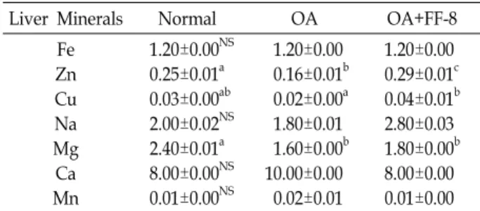
In addition, zinc play antioxidant properties against oxi- dative damage in organisms [2,15,23,26,27]. It was previous reported that the highest concentrations of zinc was found in the cell wall and membrane debris in yeast Yarrowia lip- olytica by electrochemical methods [31]. Present study also used the yeast whole cell containing a highly concentration of zinc, and the liver zinc concentrations were slightly low- ered in rats fed OA compared to the normal rats, but this reduction was significantly increased by FF-8 strain supple- mentation in OA feeding rats (Table 3). Recent rat studies reported an increase in serum and liver zinc due to an in- crease in the zinc absorption values [22,24]. Recently our study also demonstrated that the reduction of serum zinc concentration in rats fed ethanol was significantly increased by zinc-enriched yeast strain supplementation [4].
Table 3. Effect of glutathione-enriched FF-8 strain on the liver mineral concentrations in OA feeding rats for 10 days (ppm)
Liver Minerals Normal OA OA+FF-8
Fe Zn Cu Na Mg Ca Mn
1.20±0.00NS 0.25±0.01a 0.03±0.00ab 2.00±0.02NS 2.40±0.01a 8.00±0.00NS 0.01±0.00NS
1.20±0.00 0.16±0.01b 0.02±0.00a 1.80±0.01 1.60±0.00b 10.00±0.00 0.02±0.01
1.20±0.00 0.29±0.01c 0.04±0.01b 2.80±0.03 1.80±0.00b 8.00±0.00 0.01±0.00 Orotic acid (1.0%, w/w) and FF-8 (5.0%, w/w) were supple- mented to the normal diet.
Each value is the mean±SE of six rats per experimental group.
Values with different letters are significantly different at p<0.05.
The concentrations of Fe, Na, Ca, Mn in the liver were not significantly different among the experimental groups (Table 3). Zinc concentration was significantly decreased in the OA feeding rats compared with the normal rats, but this reduction was significantly elevated by concomitant with OA and FF-8 strain. This result was found to be a positive correlation between the zinc level and antioxidative action against lipid peroxidation in the liver.
In conclusion, OA treatment caused liver injury as oxida- tive stress by increased the liver lipid peroxidation level and administration of yeast strain containing highly glutathione and zinc concentrations provided antioxidative activity by reduced hepatic oxidative stress.
Acknowledgment
This study was supported by research funds from Dong-A university.
References
1. Aleynik, S. I. and C. S. Lieber. 2003. Polyenylphosphati- dylcholine corrects the alcohol-induced hepatic oxidative stress by restoring S-adenosylmethionine. Alcohol Alcoholism 38, 208-212.
2. Bao, B., A. S. Prasad, F. W. Beck, D. Snell, A. Suneja, F.
H. Sarkar, N. Doshi, J. T. Fitzgerald, and P. Swerdlow. 2008.
Zinc supplementation decreases oxidative stress, incidence of infection, and generation of inflammatory cytokines in sickle cell disease patients. Transl. Res. 152, 67-80.
3. Beutler, E., O. Duron, and B. M. Kelly. 1963. Improved method for the determination of blood glutathione. J. Lab.
Clin. Med. 61, 882-888.
4. Cha, J. Y., J. S Heo, B. K. Park, and Y. S. Cho. 2008. Effect of zinc-enriched yeast supplementation on serum zinc and testosterone concentrations in ethanol feeding rats. J. Life Sci. 18, 947-951.
5. Cha, J. Y., J. S. Heo, and Y. S. Cho. 2008. Effect of zinc-en- riched yeast FF-10 strain on the alcoholic hepatotoxicity in alcohol feeding rats. Food Sci. Biotechnol. 17, 1207-1213.
6. Cha, J. Y., S. H. Park, J. S. Heo, B. K. Park, J. W. Lee, and Y. S. Cho. 2008. Culture conditions for glutathione max- imum production by Saccharomyces cerevisiae FF-8 in bioreactor. J. Life Sci. 18, 620-624.
7. Cha, J. Y., S. H. Park, J. S. Heo, and Y. S. Cho. 2008.
Suppressive effect of administrated glutathione-enriched Saccharomyces cerevisiae FF-8 on the oxidative stress in alco- holic fatty liver. J. Life Sci. 18, 1053-1058.
8. Cha, J. Y., B. S. Jun, Y. B. Yi, J. C. Park, and Y. S. Cho.
2004. Effect of capsaicin on the lipid peroxidation in tissues of rats fed with orotic acid. J. Life Sci. 14, 541-546.
9. Cha, J. Y., B. S. Jun, and Y. S. Cho. 2004. Prevention of orotic acid-induced fatty liver in rats by capsaicin. Food Sci.
Biotechnol. 13, 597-602.
10. Catwright, I. J., A. M. Hebachi, and J. A. Higgins. 1993.
Transit and sorting of apolipoprotein B within the endoplas- mic reticulum and Golgi compartments of isolated hep- atocyte from normal and orotic acid-fed rats. J. Biol. Chem.
268, 20937-20949.
11. Dianzani, M. U., G. Muzio, M. E. Biocca, and R. A. Canuto.
1991. Lipid peroxidation in fatty liver induced by caffeine in rats. Int. J. Tissue React. 13, 79-85.
12. Draper, H. H. and M. Hadley. 1990. Malondialdehyde de- termination as index of lipid peroxidation. Methods in Enzymology 186, 421-431.
13. Duncan, D. B. 1957. Multiple range test for correlated and heteroscedastic means. Biometrics 13, 164-176.
14. George, A. Z., S. R. Efthymia, G. T. Demetrius, and A. S.
John. 2002. Determination of mineral content of active dry yeast used in pharmaceutical formulations. J. Pharm.
Biomedical. Anaylsis 28, 463-473.
15. Goel, A., V. Dani, and D. K. Dhawan. 2005. Protective ef- fects of zinc on lipid peroxidation, antioxidant enzymes and hepatic histoarchitecture in chlorpyrifos-induced toxicity.
Chem. Biol. Interact 156, 131-140.
16. Guerri, C. and S. Grisolia. 1980. Influence of prolonged etha- nol intake on the levels and turnover of alcohol and alde- hyde dehydrogenase and glutathione. Adv. Exper. Medi. Biol.
126, 365-384.
17. Jamieson, D. J. 1998. Oxidative stress responses in the yeast Saccharomyces cerevisiae. Yeast 14, 1511-1527.
18. Kadiska, M. B., B. C. Gladen, D. D. Baird, A. E. Dikaalova, R. S. Sohal, G. E. Hatch, D. P. Jones, R. P. Mason, and J.
C. Barrett. 2000. Biomarkers of oxidative stress study: Are plasma antioxidants markers of CCl4 poisoning? Free Radical Biol. Med. 28, 838-845.
19. Lai, J. T., H. L. Fang, W. H. Hsieh, and W. C. Lin. 2008.
Protective effect of a fermented substance from Saccharomyces cerevisiae on liver injury in mice caused by acetaminophen.
초록:지방간의 과산화지질에 미치는 글루타티온 고함유 효모 Saccharomyces cerevisiae FF-8 균주 급여의 영향
차재영1․박보경․엄경은․안희영․조영수*
(동아대학교 생명공학과, 1대선주조(주) 기술연구소)
간 질환 개선용 영양보충제로 시판되고 있는 글루타티온 고함유 Saccharomyces cerevisiae FF-8 효모 균체(FF-8) 급여가 사염화탄소 및 알코올-유발 흰쥐 조직 스트레스에 대하여 억제효과가 있다는 것을 보고한 바 있다. 본 실험에서는 글루타티온 고함유 효모 FF-8 급여에 의한 오르트산-유발 지방간에서 각 조직 중 과산화지질 농도와 간 조직 내 미네랄 성분과의 관계를 검토하였다. 체중 증가량은 정상군은 두 실험군에 비해 증가하였으나, 오르트산 급여 실험군들에서는 체중 증가량이 5% 수준에서 유의적으로 감소하였다. 각 조직 중량은 오르트산 급여군에서 간 조직에서 5% 수준에서 유의적으로 증가하였고 FF-8 투여에 의해 다소 감소하는 것으로 나타났다. 한편, 신장 및 비장에서도 오르트산 급여에 의해 5% 수준에서 유의적으로 감소하였다. 간 조직 중의 과산화지질 농도는 오르트 산 급여군에서 5% 수준에서 유의적으로 증가하였고, FF-8 균체 급여군에서 감소하였다. 이때 간 조직 중의 글루타티 온 농도도 유사한 경향을 보였다. 천연 항산화 미네랄로 알려진 간 조직 중의 아연 농도는 정상군에 비해 오르트산 유발 지방간에서 감소하였고, FF-8 균체 급여에 의해 다소 증가되는 것으로 나타났다. 이상의 실험 결과에서 오르트 산-유발 지방간에서 지질과산화 농도의 증가는 간 조직내 천연 항산화 물질인 글로타티온과 아연 농도의 현저한 감소에 기인하였고, 항산화 물질인 글루타티온을 고함유한 S. cerevisiae FF-8 효모 균체 동시 급여에 의해서는 글로타 티온과 아연 농도의 증가에 의해 과산화지질 농도가 경감됨으로서 오르트산 유발 산화스트레스를 경감시키는 효과가 있는 것으로 확인되었다.
Biosci. Biotechnol. Biochem. 72, 2514-2520.
20. Lee, C. H., J. Y. Cha, B. S. Jun, H. J. Lee, Y. C. Lee, Y.
L. Choi, and Y. S. Cho. 2005. Antioxidative activity of gluta- thione-enriched extract from Saccharomyces cerevisiae FF-8 in vitro model system. J. Life Sci. 15, 819-825.
21. Mannaa, F., H. H. Ahmed, S. F. Estefan, H. A. Sharaf, and E. F. Eskander. 2005. Saccharomyces cerevisiae intervention for reliving flutamide-induced hepatotoxicity in male rats.
Pharmazie 60, 689-695.
22. Mertz, W. (ed.) 1986. Zinc in Trace Elements in Human and Animal Nutrition. New York. Academic.
23. Murakami, M. and T. Hirano. 2008. Intracellular zinc ho- meostasis and zinc signaling. Cancer Sci. 99, 1515-1522.
24. Murillo-fuentes, M. L., R. Artillo, M. L. Ojeda, M. J.
Delgado, M. L. Murillo, and D. O. Carreras. 2007. Effects of prenatal or postnatal ethanol consumption on zinc in- testinal absorption and excretion in rats. Alcohol Alcoholism 42, 3-10.
25. Park, J. C., M. Ok, J. Y. Cha, and Y. S. Cho. 2003. Isolation and identification of the glutathione producing Saccharomyces cerevisiae FF-8 from Korean traditional rice wine and opti- mal producing conditions. J. Korean Soc. Agric. Chem.
Biochnol. 46, 348-352.
26. Powell, S. 2000. The antioxidant properties of zinc. J. Nutr.
130, 1447S-1454S.
27. Prasad, A. S., B. Bao, F. W. Beck, O. Kucuk, and F. H. Sarkar.
2004. Antioxidant effect of zinc in humans. Free Radic. Biol.
Med. 37, 1182-1190.
28. Scholz, W., A. Wolf, W. Kunz, R. Willenbrock, and C.
Steffen. 1991. Effect of orotic acid on the generation of
reactive oxygen and on lipid peroxidation in rat liver.
Toxicology 66, 197-212.
29. Shon, M. Y, J. Y. Cha, C. H. Lee, S. H. Park, and Y. S. Cho.
2007. Protective effect of administrated glutathione-enriched Saccharomyces cerevisiae FF-8 against carbon tetrachloride (CCl4)-induced hepatotoxicity and oxidative stress in rats.
Food Sci. Biotechnol. 16, 967-974.
30. Starkel, P., C. Sempoux, I. Leclercq, M. Herin, C. Deby, J.
P. Desager, and Y. Horsmana. 2003. Oxidative stress, KLF6 and transforming growth factor-beta up-regulation differ- entiate non-alcoholic steatohepatitis progressing to fibrosis from uncomplicated steatosis in rats. J. Hepatol. 39, 538-546.
31. Strouhal, M., R. Kizek, J. Vacek, L. Trnkova, and M. Nemec.
2003. Electrochemical study of heavy metals and metal- lothionein in yeast Yarrowia lipolytica. Bioelectrochemistry 60, 29-36.
32. Sugiyama, Y. and K. Yamamoto. 1998. The protective effect of glutathione-enriched yeast extract on acetaminophen-in- duced liver damage in rats. J. Jpan. Soc. Nutr. Food 51, 189-193.
33. Valko, M., D. Leibfritz, J. Moncol, M. T. Cronin, M. Mazur, and J. Telser. 2007. Free radicals and antioxidants in normal physiological functions and human disease. Int. J. Biochem.
Cell Biol. 39, 44-84.
34. Wei, G., Y. Li, and J. Chen. 2003. Application of a two-stage temperature control strategy for enhanced glutathione pro- duction in the batch fermentation by Candida utilis.
Biotechnol. Letter 25, 887-890.
35. Yasue, M. 2003. Brewer's yeast may prevent obesity.
Bioindustry 20, 38-43.