사람 동맥 평활근 세포에 대한 Rosmarinic Acid의 항동맥경화 활성
정재하2․윤현정1․허준영2․김재은1․박선동1,2*
1 : 심혈관계질환 천연물 연구개발센터, 2 : 동국대학교 한의과대학 방제학교실
Anti-Atherosclerosis Activity of Rosmarinic Acid in Human Aortic Smooth Muscle Cells
Jung Jae Ha2, Hyun Jeong Yun1, Joon Young Huh2, Jai Eun Kim1, Sun Dong Park1,2*
1 : Cardiovascular Medical Research Center, 2 : Department of Prescriptionology, Collage of Oriental Medicine, Dongguk University
Rosmarinic acid frequently found as a secondary metabolite in herbs and medicinal plants, has exhibited antimicrobial, antiviral, antioxidative, and anti-inflammatory activities. The proliferation and migration of human aortic smooth muscle cells (HASMC) in response to activation by various stimuli plays a critical role in the initiation and development of atherosclerosis. This study was conducted to examine the effects of Rosmarinic acid on the proliferation and migration of HASMC. Rosmarinic acid suppressed the proliferation of HASMC via induction of the expression of apoptotic proteins including cleaved poly ADP-ribose polymerase (PARP), and caspase-3. Rosmarinic acid decreased anti-apoptotic Bcl-2 and increased pro-apoptotic Bax. Moreover, treatment of rosmarinic acid decreased the G1/S cycle regulation proteins (cyclin D1, cyclin E, CDK2, CDK4 and CDK6) and increased p21, p27 and p53.
Rosmarinic acid also blocked HASMC migration via suppression of MMP-9 and MMP-2. Taken together, these results indicate that rosmarinic acid has the potential for use as an anti-atherosclerosis agent.
Key words : rosmarinic acid, HASMC, atherosclerosis, TNF-α, apoptosis, cell cycle, migration
* 교신저자 : 박선동, 경북 경주시 석장동 707, 동국대학교 한의과대학
․E-mail : [email protected], ․Tel : 054-770-2654
․접수 : 2009/10/26 ․수정 : 2009/11/15 ․채택 : 2009/12/01
서 론
Rosemarinic acid는 꿀풀과 (Lamiaceae herbe) 계통의 Salvia officinalis (sage), 로즈마리 (Rosmarinus officinalis), 하고 초 (Prunella vulgaris), 소엽 (Perilla frutescens), 바질 (Ocimum basilicum), 곽향 (Teucrium veronicoides), 형개 (Schizonepeta tenuifolia var. japonica), 향유 (Elsholtzia ciliata), 박하 (Mentha arvensis var. piperascens) 등에 함유되어 있다. Rosemarinic acid는 360.32 Da의 분자량을 갖는 polyphenolic compound로서 (α-[[3-(3,4-dihydroxyphenyl)-1-oxo-2-propenyl]-oxy]-3,4-dihydroxy -[R-(E)]-benzene-propanoic acid의 구조를 가지고 있다4). Rosmarinic acid에 대한 항산화 활성1,2), 항염증 활성3,4) 관한 많은 보고들이 있었고, 항염증 활성에 기인한 아토피 피부염에 대한 치료 효과5)가 보고되어 있었다. Rosmarinic acid의 항염증
활성은 비타민 E나 비타민 C의 10배 이상이라고 알려져 있다. 항 바이러스6), 항미생물 작용7)에 관한 보고도 몇몇 있었으며, alzheimer amyloid peptide로 유도된 cell neurotoxicity에 대한 방어효과가 있다고 보고되어 있었다8). 또한 여러 암세포주에 대 한 항암 활성에 대한 연구도 활발히 진행되고 있다9).
본 연구실에서 rosmarinic acid에 대한 선행 연구로서 사람 대장암 세포주인 HCT116에 대한 항암활성을 규명한 바 있다.
Rosmarinic acid가 다양한 생물학적 활성이 보고되고 있었기 때 문에 사람 동맥 평활근 세포인 HASMC에 대한 항동맥경화 활성 을 조사하게 되었다.
동맥경화 (atherosclerosis)는 뇌졸중과 관상동맥질환 같은 혈관질환의 원인이 된다. 따라서 동맥경화의 병인 기전 및 치료 에 관한 연구의 중요성이 점차 증대되고 있다. 동맥경화의 초기 병변은 혈관벽에 유입된 단구세포들이 대식세포로 분화하고, 나 아가서 지질을 탐식하여 foam cell이 되어 이들이 축적되어 혈관 벽에 fatty streak를 형성하게 된다. 활성화된 T-세포 및 거품세포 는 cytokines 및 성장인자를 비롯한 여러 염증성 매개물질들을
발현시켜 염증반응을 지속시키며 평활근세포 (smooth muscle cell ; SMC)의 증식을 유도하게 되어 혈관내벽이 두꺼워짐으로써 혈관내벽이 좁아져 협심증과 같은 심장질환의 원인이 된다.
MMP-2와 MMP-9의 단백질 분해 활성에 의해 평활근 세포 주변 의 세포외 기질 (extracellular matrix, ECM)이 분해되어 평활근 세포의 유주를 매개하게 된다. 따라서 평활근세포가 혈관의 내막 (intima)으로 유주하여 증식 (proliferation)하는데 MMP-2와 MMP-9이 중요한 역할을 하고, 이는 많은 혈관질환과 동맥경화 를 더욱 심화시키게 된다10).
본 연구에서는 rosmarinic acid의 활성을 조사하기 위하여 HASMC 세포의 증식과 apoptosis, cell cycle arrest에 관여하는 지 조사하였다. 또한 세포의 migration을 억제할 수 있는지 알아 보았고 그에 관여하는 효소인 MMPs의 활성을 측정하였다. 그 결과, rosmarinic acid가 항동맥경화 활성제로의 가능성이 있는 것으로 보여 보고하는 바이다.
재료 및 방법
1. 재료
세포 배양액인 Smooth Muscle Cell Medium (SMCM), SMC growth supplement는 ScienCell사 (Sandiego, USA)에서 구입하였다. 실험에 사용된 시약 중 sodium dodesyl sulfate (SDS), acrylamide, bis는 Bio-Rad사 (CA, USA)에서 구입하였고, traswell은 Corning사 (CA, USA)에서 구입하였다. Recombinant human TNF-α는 R&D Systems (MN, USA)에서 구입하였고, rosmarinic acid는 Sigma (CA, USA)에서 구입하였다. 5-bromo, 2'deoxyuridine (BrdU) kit는 Promega사 (WI, USA)에서 구입하 였다. 1차 항체 중 β-actin, cleaved Poly ADP-ribose polymerase (PARP), cleaved caspase-3, -8, Bcl-xL, Bax는 Cell Signaling Technology (MA, USA)에서 구입하였고 cytochrome c와 Bcl-2는 BD PharMingen (CA, USA)에서 구입하였다. 2차 항체인 Anti-rabbit IgG horseradish peroxidase (HRP)와 anti-mouse IgG HRP는 Santa Cruz Biotechnology (CA, USA)에서 구입하였 다. MMP-9의 발현량 측정을 위한 enzyme-linked immunosorbent assay (ELISA) kit는 R&D Systems (MN, USA) 에서 구입하였고, Protein assay reagent는 Bio-Rad사 (CA, USA) 에서 구입하였다. 실험에 사용된 모든 시약은 분석용 등급이상으 로 사용하였다.
2. 방법 1) 세포배양
사람의 동맥 평활근 세포주인 Human Aortic Smooth Muscle Cells (HASMC)은 ScienCell사 (CA, USA)에서 분양받아 SMCM에서 배양하였다. 각각의 세포는 37℃, 5% CO2 조건에서 배양하였다.
2) MTS assay
약재 추출물의 세포에 대한 독성 측정은 5-(3-caroboxymeth-oxyphenyl)-2H-tetra-zolium inner salt (MTS)
assay 방법11)으로 분석하였다. 이는 mitochondrial dehydrogenases에 의하여 MTS가 formazan으로 전환되는 것을 측정하는 것이다. 96 well plate에 1×10⁴/well의 HASMC를 분주 하고 약재를 농도별로 24 시간 동안 처리하였다. Well당 20 ㎕의 MTS solution을 첨가하여 37℃, 5% CO2 배양기에서 4시간 동안 반응시킨 후, microplate reader (DYNEX, Opsys MR, USA)를 이용하여 450 nm에서 흡광도의 변화를 측정하여 대조군에 대한 세포생존율을 백분율로 표시하였다. 각 농도별 약재가 갖는 흡광 도를 보정하기 위하여 세포를 뺀 배지를 같이 배양하여 대조군 과 실험군의 흡광도를 비교 보정하여 세포 생존율을 백분율로 표시하였다.
3) BrdU-(5'Bromo-2-deoxyuridine) ELISA assay
세포의 증식에 미치는 영향을 알아보기 위하여 BrdU-(5'Bromo-2-deoxyuridine) ELISA assay12)를 수행하였다.
96 well plate에 1×10⁴/well의 농도로 HASMC를 분주하고 하엽 추출물을 농도별로 1시간 전처리하고 100 ng/ml의 TNF-α를 처 리하여 24시간 동안 배양하였다. Well당 10 ㎕의 BrdU labeling solution을 첨가 (final concentration : 10 μM)하여 37℃, 5% CO2
배양기에서 2시간 동안 반응시킨 후, 상층액을 제거하고 anti-BrdU-POD working solution 100 ㎕ 넣어 상온에서 1시간 30분 반응시킨다. 3회 washing하고 substrate solution 100 ㎕ 넣 어 15분 반응시키고 1 M H2SO4으로 반응을 정지시킨 뒤 microplate reader (DYNEX, Opsys MR, USA)를 이용하여 450 nm에서 흡광도의 변화를 측정하여 세포의 증식율을 백분율로 표시하였다.
4) Western blot analysis
전기영동을 위한 단백질 시료의 추출은 세포를 ice-cold tris buffered saline (TBS ; 20 mM Tris-HCl, pH 8.0, 137 mM NaCl) 으로 3회 세척한 후, lysis buffer (TBS, 1% NP-40, 1 mM sodium orthovanadata, 10 ㎍/㎖ aprotinin, 10 ㎍/㎖ leupeptin 및 1 mM PMSF)를 넣어 4℃에서 30분간 반응시키고 12,000×g에서 10 분간 원심 분리하여 상층액을 모았다. 동일한 양의 단백질을 sodium dodecyl sulfate-polyacrylamide gel electrophoresis (SDS-PAGE)로 분리시킨 후, 단백질을 nitrocellulose membrane 에 transfer하였다. 이 membrane을 항체의 비특이적 결합을 차 단하기 위하여 blocking buffer (5% non-fat milk와 0.1% Tween 20을 함유한 TBS 용액)에서 1시간 동안 반응시킨 후, 각 검증 단 백질에 대한 항체를 가하여 1~2시간 동안 반응시켰다. 이어서 0.1% Tween 20을 함유한 TBST 용액으로 40분간 세척한 다음, secondary antibody로 반응시켰다. 이어서 ECL system으로 반 응 시킨 후 X-ray film상에서 단백질을 확인하였다. 각 시료의 단 백질 정량은 Bradford protein assay kit를 사용하여 595 nm에서 흡광도를 측정하여 실시하였다.
5) Migration assay
HASMC의 유주능은 transwell chamber를 사용하여 측정하 였다. 먼저 HASMC에 100 ng/ml의 TNF-α를 처리하여 24시간 배양한 후 배양액을 얻어 세포의 유주를 유도하는 conditioned medium을 준비한다. Upper chamber의 polycarbonate filter에
gelatin을 coating한 후 upper chamber 안에 세포를 1×10⁶ cells/ml로 seeding한다. Low chamber에는 conditioned medium을 1ml씩 넣은 후 37℃, 5% CO2 배양기에서 24시간 동 안 배양한다. 배양 후, filter를 4% paraformaldehyde 용액으로 고정시키고 PBS로 두 차례 세척한 다음 hematoxylin-eosin (H-E) 염색을 한다. 다시 PBS로 세척한 후 95% ethanol에서 탈수 시킨 다음 filter를 slide glass로 옮겨 permount solution으로 sealing하고 cover glass를 덮는다. 광학현미경을 통해 Filter로 유 주한 세포를 관찰하고 counting한다.
6) Gelatin zymography assay
MMP-2와 MMP-9의 activity 측정을 위하여 24 well plate에 1×10⁵/ml 로 세포를 분주하여 배양한 후 serum free medium으 로 교체하여 16시간 동안 starvation하였다. 여기에 각각의 약재 를 1시간동안 전처리한 후 100 ng/ml의 TNF-α를 24시간 처리하 였다. 각각의 세포 배양액을 수거하여 gelatin zymography를 수 행하였다. 20 ㎕의 배양액을 reducing agent가 없는 SDS-sample buffer에 섞은 후 전기영동을 한 후, gel을 2.5% Triton X-100에서 30분간 2회 soaking시킴으로써 SDS를 제거하고 3차 증류수로 20 분간 2회 세척하였다. 세척된 gel은 incubation buffer (50 mM Tris-HCl ; pH 7.6, 10 mM CaCl2, 150 mM NaCl, 0.01% NaN3) 에 넣어 37℃ incubator에서 24시간동안 반응시킨 후 0.1%
coomassie brilliant blue 용액으로 30분간 염색하여 gelatin이 분 해되는 정도를 관찰하였다.
7) Flow cytometry analysis
Cell cycle 분석을 하기 위해 PI/RNase staining을 하여 FACS analysis를 수행하였다. 1✕106 cells/ml의 세포에 rosmarinic acid와 TNF-α를 24시간 처리한 후 trypsin-EDTA를 처리하여 세포를 수거하여 1% paraformaldehyde로 15분간 상온 에서 고정시킨다. 0.2% BSA/PBS로 2회 washing한 다음, 0.1%
Triton X-100을 처리하여 permeability를 갖게 한 후 500 ㎕ PI/RNase를 첨가하여 상온에서 30분 동안 incubation하였다.
Becton Dickinson FACS calibur flow cytometer 와 CELL-Quest Pro (BD Biosciences)를 사용하여 cell cycle을 분석하였다.
3. 통계처리
실험결과는 means±SEM으로 표시하고 유의성 검증은 Prism 4.00을 이용하여 one-way ANOVA를 실시하였다.
Western blot과 gelatin zymography를 수행하여 얻은 band의 density 분석은 GelDoc-It BioImaging System (UVP, USA)을 사 용하여 측정하였다.
결 과
1. Rosmarinic acid의 HASMC에 대한 세포 독성
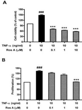
Rosmarinic acid가 HASMC 세포의 생존 및 증식에 미치는 영향을 알아보았다. 세포에 0.1, 1, 10 μM의 rosmarinic acid를 1 시간 전처리한 후 10 ng/ml TNF-α를 24시간 처리한 후 MTS assay를 수행하여 세포생존율을 측정한 결과 Fig. 1A에서 보듯이
TNF-α에 의해서 약 1.5배 세포생존율이 증가하였고 rosmarinic acid에 의해 control 수준이하까지 감소되는 것으로 나타났다. 또 한 BrdU assay를 통해 세포의 증식에 미치는 영향을 알아본 결 과에서도 TNF-α에 의해 증가되었던 증식율이 rosmarinic acid의 농도에 의존적으로 감소되는 것을 볼 수 있었다(Fig. 1B).
Fig. 1. Effect of rosmarinic acid on the cell viability and proliferation of HASMC. HASMC were treated with rosmarinic acid for 1 hr before treatment with or without 10 ng/ml TNF-α for 24 hr. HASMC proliferation was assayed by BrdU ELISA assay. Cell viability was measured by MTS assay as described in Materials and Methods. Data were chosen from three independent triplicate experiments.
2. Rosmarinic acid에 의해 유도되는 HASMC의 apoptosis Rosmarinic acid에 의해 apoptosis가 유도되는지 조사하였 다. HASMC에 rosmarinic acid를 농도별로 처리하고 1시간 후 10 ng/ml의 TNF-α를 처리하여 24시간 후 세포를 수거하였다.
Apoptosis의 지표가 되는 poly(ADP-ribose) polymerase (PARP) 의 cleavage되는 정도를 확인하기 위하여 western blot을 수행하 였다. 그 결과, rosmarinic acid의 농도가 증가할수록 cleaved PARP의 발현이 증가하는 것을 확인할 수 있었다. 또한 PARP의 upstream signal인 caspase-3가 rosmarinic acid에 의해 활성화되 는 것으로 나타났다(Fig. 2). Rosmarinic acid에 의해 유도되는 apoptosis가 mitochondrial pathway에 관여하는 단백질의 발현 에 미치는 영향을 조사하였다. 그 결과, pro-apoptotic Bax는 rosmarinic acid 처리 시 점차 증가하였고 anti-apoptotic Bcl-2는 감소하는 것을 확인하였다.
3. Rosmarinic acid에 의한 HASMC의 cell cycle arrest Rosmarinic acid가 HASMC의 cell cycle에 미치는 영향을 알 아보기 위하여 PI/RNase staining을 하여 FACS analysis를 수행 하였다. 그 결과, TNF-α에 의해 G0/G1 phase의 세포의 비율이
감소하였고 S phase의 세포비율이 약 455.3% 증가하였다.
Rosmarinic acid를 처리한 경우 감소되었던 G0/G1 phase의 비 율은 다시 회복되었고, 증가되었던 S phase의 비율은 거의 control 수준까지 감소되었다(Fig. 3).
Western blot을 수행하여 cell cycle에 관여하는 단백질들의 발현양상을 조사하였다. p53은 tumor suppressor gene으로서 자 극에 의해 DNA가 손상되었을 때 활성화되어 cell cycle arrest와 apoptosis의 두 가지 pathway에 관여하게 된다. p21과 p27은 S-phase를 유도하는 cyclin-CDK complex에 결합하여 CDK 2/4/6의 kinase 활성을 저해함으로써 세포는 G1 phase에 arrest 되면서 손상된 DNA를 repair한다. 만일 DNA 손상이 세포가 수 용하지 못할 정도로 심한 경우에는 p53이 apoptosis-inducing gene인 Bax 단백질의 gene을 발현 시켜 apoptosis를 유도한다.
Fig. 4에서 보듯이, TNF-α에 의해 감소되었던 p53, p21, p27 의 발현이 rosmarinic acid에 의해 다시 회복되었다. 또한 TNF-α 에 의해 증가되었던 cyclin D, cyclin E, CDK2, CDK4, CDK6의 발현은 rosmarinic acid에 의해 감소되는 것을 확인할 수 있었다.
A
B
Fig. 2. Rosmarinic acid induces apoptosis in TNF-α-treated HASMC.
HASMC was treated with indicated concentrations of rosmarinic acid for 1 hr and then treated with TNF-α (10 ng/ml) for 24 hr. The expression levels of cleaved PARP, cleaved caspase-3, Bax and Bcl-2 were measured by western blot assay. β -actin levels were used as a loading control.
Fig. 3. Rosmarinic acid induces cell cycle arrest in G0/G1 phase.
Cells were treated with indicated concentrations of rosmarinic acid for 1 hr and then treated with TNF-α (10 ng/ml) for 24 hr. DNA content was measured by propidium iodide staining on flow cytometry.
Fig. 4. Effect of rosmarinic acid on expression of cell cycle-regulatory proteins. Cells were treated with indicated concentrations of rosmarinic acid for 1 hr and then treated with TNF-α (10 ng/ml) for 24 hr. Cell extracts were prepared and cell cycle-regulatory proteins were detected by immunoblotting. b-actin levels were used as internal markers for loading variations.
The density of band was calculated by GelDoc-It BioImaging System.
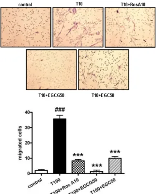
4. Rosmarinic acid가 HASMC의 유주능에 미치는 영향 Rosmarinic acid가 세포의 유주능에 미치는 영향을 알아보 기 위하여 transwell을 이용한 migtation assay를 수행하였다.
HASMC에 100 ng/ml의 TNF-α를 24시간 처리하여 얻은 conditioned medium을 transwell의 lower chamber에 넣고,
rosmarinic acid를 1시간 동안 전처리한 후 10 ng/ml의 TNF-α를 24시간 처리한 HASMC를 upper chamber에 loading하였다. 24 시간동안 37℃, 5% CO2 incubator에서 배양한 후 upper chamber에서 lower chamber로 유주한 세포를 관찰하기 위하여 upper chamber의 matrigel filter를 H-E 염색을 하여 광학 현미 경으로 관찰하였다. 그 결과, TNF-α 처리에 의해 세포의 유주가 약 17.3배 증가하였고, TNF-α에 의해 증가된 세포의 유주가 rosmarinic acid 전처리에 의해 약 76.6%, 96.3% 감소되었다(Fig.
5). 녹차의 성분으로서 MMP의 억제제로 알려져 있는 catechin의 유도체인 EGCG나 EGC와 비교해 보았을 경우 EGC보다 탁월한 유주능 저해 효과를 나타내었다.
Fig. 5. Inhibitory effect of rosmarinic acid on HASMC migration. Cells were stimulated with rosmarinic acid for 1 hr prior to treatment with 100 ng/ml of TNF-α for 24 hr. After 24 hr, the cells were collected and added to upper chamber.
TNF-α-treated conditioned medium was collected and added to lower chamber.
After 24 hr incubation, underside of matrigel filter was stained with hematoxylin and eosin. And total number of cells that migrated to the underside of the filter was counted. Data are represented as means±SEM. Significantly different from control (#) or TNF-α alone (*) ; ###, *** : P < 0.001.
5. Rosmarinic acid가 MMP-9, MMP-2의 활성에 미치는 영향 MMPs는 혈관을 형성하는 내막, 중막, 외막의 구성성분인 세포외 기질이나 기저막을 분해시킴으로써 평활근세포의 유주에 영향을 주는데, 이러한 MMP-9과 MMP-2의 활성을 조사하기 위 해 gelatin zymography assay를 수행하였고, 생성량에 대한 영향 을 조사하기 위해 MMP-9, MMP-2 ELISA를 수행하였다.
HASMC에서 MMP-9과 MMP-2가 TNF-α에 의해 증가한다
13,14)
. 따라서 본 실험에서는 TNF-α에 의해 유도되는 MMP-9, MMP-2가 rosmarinic acid에 의해 효과적으로 감소시키는 지 알 아보았다. Rosmarinic acid를 1시간 전처리하고 10 ng/ml의
TNF-α를 처리하여 24시간 후 세포 배양액을 얻어 gelatin zymography assay를 수행하였다. 그 결과, TNF-α에 의해 증가 되는 MMP-9과 MMP-2의 활성이 rosmarinic acid에 의해 효과적 으로 저해되었다. MMP-9은 TNF-α에 의해 약 1.7배 증가하였고, 이는 rosmarinic acid 1, 10 μM 처리에 의해 각각 23.5%, 76.5%
활성이 감소되었다(Fig 6A). MMP-2는 TNF-α에 의해 약 1.2배 증가하였고, 이는 rosmarinic acid에 의해 거의 control 수준까지 활성이 감소되었다(Fig 6B).
Fig. 6. Effect of rosmarinic acid on MMP-9 and MMP-2 activity in HASMC. Cells were stimulated with rosmarinic acid for 1 hr prior to treatment with 100 ng/ml of TNF-α for 24 hr. Gelatin zymography was performed with the medium. Inhibitory effect of rosmarinic acid on MMP-9 and MMP-2 was estimated in comparison with established MMP inhibitor, EGCG and EGC. The densitometric intensity of zymography bands was eatimated as described in materials and methods.
고 찰
동맥경화로 혈관이 좁아지거나 막히게 되면 그 혈관이 담당 하는 말초로의 혈액순환 장애가 생기므로, 좁아진 혈관에 따라 증상이 다르게 나타나게 된다. 죽상동맥경화는 주로 심장에 혈액
을 공급하는 관상동맥, 뇌에 혈액을 공급하는 뇌동맥과 경동맥, 신장의 신동맥 및 말초혈관을 침범하고, 이로 인해 협심증, 심근 경색 등의 허혈성 심장질환, 뇌경색과 뇌출혈 등의 뇌졸중, 신장 의 기능이 저하되는 신부전 및 허혈성 사지질환이 나타나게 된 다. 동맥경화로 좁아진 혈관은 수술을 통해서 확장하게 되는데 수술 후 혈관내의 상처로 인해 염증반응이나 재협착 같은 부작 용이 발생하게 된다. 이때 TNF-α 같은 염증성 사이토카인들이 생산되어 혈관내 염증반응을 유발하여 동맥경화를 더욱 심화시 키게 된다. 앞서 보고된 바에 의하면 TNF-α는 동맥평활근 세포 의 증식을 유도하여 혈관 내벽에 축적되어 혈관이 더욱 좁아지 게 만든다고 하였다15,16). 동맥평활근 세포의 증식은 고혈압, 동맥 경화, 혈관 내 재협착 등의 원인이 된다17). 따라서 혈관 내막에서 의 동맥평활근 세포의 증식을 억제하는 것이 혈관 질환 치료 및 예방에 중요하다.
Rosmarinic acid는 항산화, 항염증, 항바이러스, 항균, 항암 작용 등 많은 생물학적 활성이 보고되고 있다. 또한 최근에는 사 람 상피세포에서 NF-κB 활성을 저해함으로써 TNF-α에 의해 유 도되는 CCL1과 CCR3를 억제한다는 보고도 있다18). Rosmarinic acid가 동맥경화 질환의 예방 및 치료에도 활성을 가지는 지 알 아보기 위하여 사람동맥평활근세포인 HASMC에 처리하여 분자, 세포생물학적 실험을 수행하였다.
먼저, HASMC에 대한 rosmarinic acid의 독성 및 증식에 미 치는 영향을 조사하였다. TNF-α에 의해서 약 1.5배 세포생존율 이 증가하였고 rosmarinic acid에 의해 control 수준 이하까지 감 소되는 것으로 나타났다. 또한 TNF-α에 의해 증가되었던 증식율 이 rosmarinic acid의 농도에 의존적으로 감소되는 것을 볼 수 있 었다. Rosmarinic acid에 의해 apoptosis가 유도되는지 조사하였 다. Rosmarinic acid의 농도가 증가할수록 cleaved PARP의 발현 이 증가하였고 caspase-3가 활성화되는 것으로 나타났다. 또한 pro-apoptotic Bax는 rosmarinic acid 처리 시 점차 증가하였고 anti-apoptotic Bcl-2는 감소하는 것을 확인하였다. 이로써 rosmarinic acid에 의해 mitochondrial pathway을 통한 apoptosis가 유도되는 것을 확인할 수 있었다.
Cell cycle은 G1→S→G2→M으로 계속 순환19)되는데, S기는 DNA복제가 일어나고 M기에는 염색체의 분열과 세포 분열이 일어나고 이 사이를 각각 G1, G2라고 하며 이들 G1, G2 check point를 통해서 세포주기의 조절이 이루어지게 된다20). 이들 중 G1기에서는 CDK4, 6과 complex를 형성하는 cyclin D의 합성이 주로 G1 중기에 최고조에 달하며 이는 주로 mitogen같은 세포성 장인자 등에 의해 유도된다21). Cyclin D는 세 종류의 subtype (D1, 2, 3)이 있는데 세포의 종류에 따라서 발현되는 정도가 다르 다. Cyclin D의 합성을 저해하면 G1기에서 세포의 arrest가 일어 나게 된다. CDK2와 complex를 형성하는 cyclin E는 대부분 G1 후기에 발현되고 S기가 시작되면서 분해되기 시작한다22). 이렇게 세포주기의 각 단계에서 특이적인 CDK-cyclin complex가 활성 이 조절됨으로써 세포주기가 적절하게 조절된다. CDK-cyclin complex의 활성을 조절하는 cyclin-dependent kinase inhibitor (CKI)는 세포의 불필요한 증식을 억제하는 단백질로서 p21, p27,
p57 등이 있고 이들을 조절하는 단백질로 p53이 존재한다23). Rosmarinic acid가 HASMC의 cell cycle에 미치는 영향을 알아보 았다. PI/RNase staining을 통한 FACS analysis를 수행하한 결 과, TNF-α에 의해 G0/G1 phase의 세포의 비율이 감소하였고 S phase의 세포비율이 약 455.3% 증가하였다. Rosmarinic acid 처 리에 의해 감소되었던 G0/G1 phase의 비율은 다시 회복되었고, 증가되었던 S phase의 비율은 거의 control 수준까지 감소되었 다. Cell cycle 조절에 관여하는 단백질인 p53과 p21, p27의 발현 이 rosmarinic acid에 의해 증가하였고, TNF-α에 의해 증가되는 cyclin D, cyclin E, CDK2, CDK4, CDK6의 발현이 효과적으로 감소하였다. 이들의 결과로 보아, rosmarinic acid에 의해 G1 phase에서 arrest됨으로써 세포의 증식에 영향을 줄 수 있을 것 으로 보인다.
다음으로, rosmarinic acid가 HASMC의 유주능에 미치는 영 향을 알아보았다. 평활근세포의 유주는 세포간질 (interstitial tissue)이나 혈관 형성층 (내막, 중막, 외막)의 구성성분인 세포외 기질이나 기저막이 MMPs에 의해 분해됨으로써 중막에 있던 평 활근세포가 내막으로 이동하게 된다. 생체 내에는 MMPs를 조절 할 수 있는 단백질인 tissue inhibitor metalloproteinase (TIMP) 가 존재하는데, MMPs와 TIMP의 불균형이 생기면 혈관 평활근 세포의 유주 활성이 증가하게 되어 동맥경화 및 혈관질환의 원 인이 된다24). 따라서 이러한 MMPs의 억제제를 찾기 위한 연구 에 관심이 집중되고 있다. MMPs는 동맥경화에 관련된 사이토카 인인 tumor necrosis factor (TNF)-α, interleukin (IL)-1α, IL-1β과 키모카인인 IL-8, CD40L 발현 세포인 T 임파구 및 혈소판, 트롬 빈, 산화저밀도단백질 (ox-LDL)등에 의해 유도되며 주로 대식세 포, 혈관내피세포, 평활근세포에서 발현된다. MMPs 중 MMP-2와 MMP-9의 단백질 분해 활성에 의해 평활근 세포 주변의 세포외 기질 (extracellular matrix, ECM)이 분해되어 평활근 세포의 유주 를 매개하게 된다. 따라서 평활근세포가 혈관의 내막 (intima)으 로 유주하여 증식 (proliferation)하는데 있어서 MMP가 중요한 역할을 하고, 이는 많은 혈관질환의 원인이 되며 동맥경화를 더 욱 심화시킨다. MMP-2와 MMP-9은 유사한 기질을 분해하는 type Ⅳ collageanse (gelatinase)로서 pro-MMP-2는 HASMC에서 항상 발현되어 있고 MMP-2, pro-MMP-9, MMP-9은 거의 발현되 어 있지 않다가 TNF-α나 nerve growth factor (NGF)에 의해 증 가된다25). 혈관이 손상되면 HASMC의 migration의 증가와 함께 MMP-2, MMP-9의 발현이 증가한다는 보고26)와 MMP-2가 MMP-9의 활성을 증가시킨다는 보고도 있다27).
Transwell을 이용한 migration assay를 수행하였다. TNF-α 처리에 의해 세포의 유주가 증가하였고, 이는 rosmarinic acid 전 처리에 의해 크게 감소되었다. MMP의 억제제로 알려져 있는 catechin의 유도체인 EGCG나 EGC와 비교해 보았을 경우 EGC 보다 탁월한 유주능 저해 효과를 나타내었다. Rosmarinic acid는 MMP-9과 MMP-2의 활성 및 생성량 저해에도 효과적인 것으로 나타났다.
이러한 결과로 보아 rosmarinic acid는 HASMC의 세포의 생존 및 증식을 억제하며, mitochondrial pathway을 통한
apoptosis를 유도하고 cell cycle arrest에 관여하는 것으로 보인 다. 또한 MMP-9과 MMP-2의 활성 및 생성을 조절함으로써 세포 의 유주를 억제할 수 있는 것으로 보인다. 따라서 rosmarinic acid는 동맥경화 치료제 및 예방물질로서 가능성을 제시할 수 있 고, 이에 관여하는 메카니즘 연구 및 in vivo 실험 등 rosmarinic acid에 관한 심도 있는 연구가 수행되어야 할 것이다.
감사의 글
본 연구는 과학기술부/한국과학재단 기초의과학연구센터 육성사업(R13-2005-013-01000-0)의 지원으로 수행되었음.
참고문헌
1. Pirker, K.F., Kay, C.W., Stolze, K., Tunega, D., Reichenauer, T.G., Goodman, B.A. Free radical generation in rosmarinic acid investigated by electron paramagnetic resonance spectroscopy. Free Radic Res. 43(1):47-57, 2009.
2. Fujimoto, A., Sakanashi, Y., Matsui, H., Oyama, T., Nishimura, Y., Masuda, T., Oyama, Y. Cytometric analysis of cytotoxicity of polyphenols and related phenolics to rat thymocytes: potent cytotoxicity of resveratrol to normal cells. Basic Clin Pharmacol Toxicol. 104(6):455-462, 2009.
3. Inoue, K., Takano, H., Shiga, A., Fujita, Y., Makino, H., Yanagisawa, R., Kato, Y., Yoshikawa, T. Effects of volatile constituents of rosemary extract on lung inflammation induced by diesel exhaust particles. Basic Clin Pharmacol Toxicol. 99(1):52-57, 2006.
4. Sanbong,i C., Takano, H., Osakabe, N., Sasa, N., Natsume, M., Yanagisawa, R., Inoue, K.I., Sadakane, K., Ichinose, T., Yoshikawa, T. Rosmarinic acid in perilla extract inhibits allergic inflammation induced by mite allergen, in a mouse model. Clin Exp Allergy. 34(6):971-977, 2004.
5. Lee, J., Jung, E., Koh, J., Kim, Y.S., Park, D. Effect of rosmarinic acid on atopic dermatitis. J Dermatol.
35(12):768-771, 2008.
6. Swarup, V., Ghosh, J., Ghosh, S., Saxena, A., Basu, A.
Antiviral and anti-inflammatory effects of rosmarinic acid in an experimental murine model of Japanese encephalitis.
Antimicrob Agents Chemother. 51(9):3367-3370, 2007.
7. Petersen, M., Simmonds, M.S. Rosmarinic acid.
Phytochemistry. 62(2):121-125, 2003.
8. Iuvone, T., De Filippis, D., Esposito, G., D'Amico, A., Izzo, A.A. The spice sage and its active ingredient rosmarinic acid protect PC12 cells from amyloid-beta peptide-induced neurotoxicity. J Pharmacol Exp Ther. 317(3):1143-1149, 2006.
9. Aquilano, K., Filomeni, G., Di Renzo, L., Vito, M., Stefano,
C., Salimei, P.S., Ciriolo, M.R., Marfè, G. Reactive oxygen and nitrogen species are involved in sorbitol-induced apoptosis of human erithroleukaemia cells K562. Free Radic Res. 41(4):452-460, 2007.
10. Lin, S.J., Lee, I.T., Chen, Y.H., Lin, F.Y., Sheu, L.M., Ku, H.H., Shiao, M.S., Chen, J.W., Chen, Y.L. Salvianolic acid B attenuates MMP-2 and MMP-9 expression in vivo in apolipoprotein-E-deficient mouse aorta and in vitro in LPS-treated human aortic smooth muscle cells. J Cell Biochem. 100(2):372-384, 2007.
11. Desai, A., Vyas, T., Amiji, M. Cytotoxicity and apopotosis enhancement in brain tumor cells upon coadministration of paclitaxel and ceramide in nanoemulsion formulations.
97(7):2745-2756, 2008.
12. Magaud, J.P., Sargent, I., Mason, D.Y. Detection of human white cell proliferative responses by immunoenzymatic measurement of bromodeoxyuridine uptake. J Immunol Methods. 106(1):95-100, 1988.
13. Moon, S.K., Cha, B.Y., Kim, C.H. ERK1/2 mediates TNF-alpha-induced matrix metalloproteinase-9 expression in human vascular smooth muscle cells via the regulation of NF-kappaB and AP-1: Involvement of the ras dependent pathway. J Cell Physiol. 198(3):417-427, 2004.
14. Moon, S.K., Cha, B.Y., Kim, C.H. In vitro cellular aging is associated with enhanced proliferative capacity, G1 cell cycle modulation, and matrix metalloproteinase-9 regulation in mouse aortic smooth muscle cells. Arch Biochem Biophys. 418(1):39-48, 2003.
15. Moon, S.K., Cho, G.O., Jung, S.Y., Gal, S.W., Kwon, T.K., Lee, Y.C., Madamanchi, N.R., Kim, C.H. Quercetin exerts multiple inhibitory effects on vascular smooth muscle cells:
role of ERK1/2,·/cell-cycle regulation, and matrix metalloproteinase-9. Biochem Biophys Res Commun.
301(4):1069-1078, 2003.
16. Zhang, H.S., Wang, S.Q. Ginsenoside Rg1 inhibits tumor necrosis factor-alpha (TNF-alpha)-induced human arterial smooth muscle cells (HASMCs) proliferation. J Cell Biochem. 98(6):1471-1481, 2006.
17. Ross, R. Cell biology of atherosclerosis. Annu Rev Physiol.
57:791-804, 1995.
18. Lee, J., Jung, E., Kim, Y., Lee, J., Park, J., Hong, S., Hyun, C.G., Park, D., Kim, Y.S. Br J Pharmacol. Rosmarinic acid as a downstream inhibitor of IKK-beta in TNF-alpha-induced upregulation of CCL11 and CCR3.
148(3):366-375, 2006.
19. de Carcer, G., de Castro, I.P., Malumbres, M. Targeting cell cycle kinases for cancer therapy. Curr Med Chem.
14(9):969-985, 2007.
20. Singh, R.P., Agarwal, R. Natural flavonoids targeting deregulated cell cycle progression in cancer cells. Curr Drug Targets. 7(3):345-354, 2006.
21. Shukla, S., Gupta, S. Apigenin-induced cell cycle arrest is mediated by modulation of MAPK, PI3K-Akt, and loss of cyclin D1 associated retinoblastoma dephosphorylation in human prostate cancer cells. Cell Cycle. 6(9):1102-1114, 2007.
22. Gupte, R.S., Traganos, F., Darzynkiewicz, Z., Lee, M.Y.
Phosphorylation of RIalpha by cyclin-dependent kinase CDK 2/cyclin E modulates the dissociation of the RIalpha-RFC40 complex. Cell Cycle. 5(6):653-660, 2006.
23. Sanz-Gonzalez, S.M., Melero-Fernandez, de Mera R., Malek, N.P., Andres, V. Atheroma development in apolipoprotein E-null mice is not regulated by phosphorylation of p27 (Kip1) on threonine 187. J Cell Biochem. 97(4):735-43, 2006.
24. Raffetto, J.D., Khalil, R.A. Matrix metalloproteinases and
their inhibitors in vascular remodeling and vascular disease. Biochem Pharmacol. 75(2):346-359, 2008.
25. Kim, H.J., Yoo, E.K., Kim, J.Y., Choi, Y.K., Lee, H.J., Kim, J.K., Jeoung, N.H., Lee, K.U., Park, I.S., Min, B.H., Park, K.G., Lee, C.H., Aronow, B.J., Sata, M., Lee, I.K. Protective role of clusterin/apolipoprotein J against neointimal hyperplasia via antiproliferative effect on vascular smooth muscle cells and cytoprotective effect on endothelial cells.
Arterioscler Thromb Vasc Biol. 29(10):1558-1564, 2009.
26. Bendeck, M.P., Irvin, C., Reidy, M.A. Inhibition of matrix metalloproteinase activity inhibits smooth muscle cell migration but not neointimal thickening after arterial injury. Circ Res. 78(1):38-43, 1996.
27. Yong, V.W., Krekoski, C.A., Forsyth, P.A., Bell, R., Edwards, D.R. Matrix metalloproteinases and diseases of the CNS. Trends Neurosci. 21(2):75-80, 1998.