1
Background—The index of microcirculatory resistance (IMR) is a quantitative and specific index for coronary microcirculation. However, the distribution and determinants of IMR have not been fully investigated in patients with ischemic heart disease (IHD).
Methods and Results—Consecutive patients who underwent elective measurement of both fractional flow reserve (FFR) and IMR were enrolled from 8 centers in 5 countries. Patients with acute myocardial infarction were excluded. To adjust for the influence of collateral flow, IMR values were corrected with Yong’s formula (IMRcorr). High IMR was defined as greater than the 75th percentile in each of the major coronary arteries. FFR≤0.80 was defined as an ischemic value. 1096 patients with 1452 coronary arteries were analyzed (mean age 61.1, male 71.2%). Mean FFR was 0.84 and median IMRcorr was 16.6 U (Q1, Q3 12.4 U, 23.0 U). There was no correlation between IMRcorr and FFR values (r=0.01, P=0.62), and the categorical agreement of FFR and IMRcorr was low (kappa value=−0.04, P=0.10). There was no correlation between IMRcorr and angiographic % diameter stenosis (r=−0.03, P=0.25). Determinants of high IMR were previous myocardial infarction (odds ratio [OR] 2.16, 95% confidence interval [CI] 1.24–3.74, P=0.01), right coronary artery (OR 2.09, 95% CI 1.54–2.84, P<0.01), female (OR 1.67, 95% CI 1.18–2.38, P<0.01), and obesity (OR 1.80, 95%
CI 1.31–2.49, P<0.01). Determinants of FFR ≤0.80 were left anterior descending coronary artery (OR 4.31, 95% CI 2.92–6.36, P<0.01), angiographic diameter stenosis ≥50% (OR 5.16, 95% CI 3.66–7.28, P<0.01), male (OR 2.15, 95%
CI 1.38–3.35, P<0.01), and age (per 10 years, OR 1.21, 95% CI 1.01–1.46, P=0.04).
Conclusions—IMR showed no correlation with FFR and angiographic lesion severity, and the predictors of high IMR value were different from those for ischemic FFR value. Therefore, integration of IMR into FFR measurement may provide additional insights regarding the relative contribution of macro- and microvascular disease in patients with ischemic heart disease.
Clinical Trial Registration—URL: http://www.clinicaltrials.gov. Unique identifier: NCT02186093.
(Circ Cardiovasc Interv. 2015;8:e002857. DOI: 10.1161/CIRCINTERVENTIONS.115.002857.)
Key Words: coronary artery disease ◼ fractional flow reserve ◼ ischemic heart disease
◼ microcirculation ◼ physiology
© 2015 American Heart Association, Inc.
Circ Cardiovasc Interv is available at http://circinterventions.ahajournals.org DOI: 10.1161/CIRCINTERVENTIONS.115.002857 Received March 3, 2015; accepted September 30, 2015.
From the Department of Medicine, Seoul National University Hospital, Seoul, South Korea (J.M.L., J.-H.J., H.-J.L., B.-K.K.); Department of Cardiology, West of Scotland Heart and Lung Centre, Golden Jubilee National Hospital, Clydebank, United Kingdom (J.L., S.W., K.G.O.); BHF Glasgow Cardiovascular Research Centre, Institute of Cardiovascular and Medical Sciences, University of Glasgow, Glasgow, United Kingdom (J.L., S.W., K.G.O.); Servicio de Cardiología, Hospital Clinico San Carlos, Faculty of Medicine Complutense University of Madrid, Madrid, Spain (M.E.-P., J.E.);
Centro Nacional de Investigaciones Cardiovasculares Carlos III (CNIC), Madrid, Spain (M.E.-P., J.E.); Department of Cardiovascular Medicine, Stanford University Medical Center, Stanford, CA (A.S.Y., W.F.F.); Department of Medicine, Inje University Ilsan Paik Hospital, Goyang, South Korea (J.-H.D.);
Department of Medicine, Keimyung University Dongsan Medical Center, Daegu, South Korea (C.-W.N.); Department of Cardiology, Ulsan University Hospital, University of Ulsan College of Medicine, Ulsan, South Korea (E.-S.S.); Institute on Aging, Seoul National University, Seoul, South Korea (B.- K.K.); and Departments of Cardiology, Royal Prince Alfred and Concord Hospitals and University of Sydney, Sydney, Australia (M.K.N.).
The Data Supplement is available at http://circinterventions.ahajournals.org/lookup/suppl/doi:10.1161/CIRCINTERVENTIONS.115.002857/-/DC1.
Correspondence to Bon-Kwon Koo, MD, PhD, Department of Internal Medicine and Cardiovascular Center, Seoul National University Hospital, 101 Daehang-ro, Chongno-gu, Seoul, 110–744, Korea. E-mail [email protected]
Integrated Physiologic Assessment of Ischemic Heart Disease in Real-World Practice Using Index of Microcirculatory
Resistance and Fractional Flow Reserve
Insights From the International Index of Microcirculatory Resistance Registry
Joo Myung Lee, MD, MPH; Jamie Layland, MBCHB; Ji-Hyun Jung, MD;
Hyun-Jung Lee, MD; Mauro Echavarria-Pinto, MD; Stuart Watkins, MBChB, MD;
Andy S. Yong, MBBS, PhD; Joon-Hyung Doh, MD, PhD; Chang-Wook Nam, MD, PhD;
Eun-Seok Shin, MD, PhD; Bon-Kwon Koo, MD, PhD; Martin K. Ng, MBBS, PhD;
Javier Escaned, MD, PhD; William F. Fearon, MD; Keith G. Oldroyd, MBChB
by guest on April 19, 2017http://circinterventions.ahajournals.org/Downloaded from by guest on April 19, 2017http://circinterventions.ahajournals.org/Downloaded from by guest on April 19, 2017http://circinterventions.ahajournals.org/Downloaded from by guest on April 19, 2017http://circinterventions.ahajournals.org/Downloaded from by guest on April 19, 2017http://circinterventions.ahajournals.org/Downloaded from by guest on April 19, 2017http://circinterventions.ahajournals.org/Downloaded from
T
he coronary microcirculation is one of the major com- ponents of the coronary vascular system and an impor- tant contributor to the development of ischemic heart disease (IHD). However, limitations in the methods available to visualize or evaluate the microcirculatory system have been an obstacle to the diagnosis and treatment of microvascular disease. The index of microcirculatory resistance (IMR) is a pressure–temperature–derived parameter for quantifying microcirculatory resistance.1 Because distal coronary pressure is used in the calculation of IMR, this index can be used to interrogate selectively the microcirculation of vessels with a coronary stenosis, in contrast to coronary flow reserve, which is a combined assessment of the macro- and microcircula- tion. Previous studies have shown that IMR is independent of the severity of epicardial coronary stenosis as determined by angiographic or functional (fractional flow reserve [FFR]) criteria so long as the influence of collateral flow is taken into account.2,3 IMR is also relatively independent of hemody- namic conditions, such as blood pressure and heart rate.4Recent clinical studies have shown that IMR is useful for the risk stratification in patients with acute myocardial infarc- tion (MI).5–7 However, in the setting of IHD other than MI, the distribution and determinants of abnormal IMR remain to be clearly identified. Therefore, we sought to investigate the clinical relevance of microvascular assessment using IMR and the relative contribution of macro- and microvascular disease in non-MI patients enrolled from an international multicenter IMR registry.
Methods Patient Population
From April 2009 to September 2013, patients underwent FFR and IMR measurements and, with available clinical and angiographic in- formation, were registered from 8 hospitals from 5 countries (South Korea, United Kingdom, Spain, USA, and Australia). The investiga- tors were asked to provide the data of eligible patients, and those enrolled in other study protocols or those who underwent FFR and IMR measurements were both included in this study. The opera- tors involved in this study used FFR and IMR in their daily clinical practice. Patients with acute MI with cardiac enzyme elevation were excluded. All patients underwent preinterventional measurement of FFR and IMR, and the postinterventional data were excluded from the analysis. Institutional Review Board approval was obtained with regard to current regulations, and the study protocol was in accor- dance with the Declaration of Helsinki (clinicaltrials.gov identifier, NCT02186093).
Coronary Angiography and Coronary Physiological Measurements
Coronary angiography was performed by standard techniques.
Angiographic views were obtained after the administration of intra- coronary nitrate (100 or 200 μg). Quantitative coronary angiography was performed at each participating center using a contour-detection quantitative coronary angiography system and the guiding catheter tip as a scaling device. Percent diameter stenosis, minimum lumen diameter, reference vessel size, and lesion length were measured.
All coronary physiological measurements were obtained as pre- viously described.7–9 In brief, a 5–7F guiding catheter without side holes was used to engage the coronary artery, and a pressure–tem- perature sensor guidewire (St Jude Medical, St Paul, MN) was used for FFR and IMR measurement. The pressure sensor was positioned at the distal segment of the target vessel, and intracoronary nitrate (100 or 200 μg) was administered before each physiological mea- surement. To derive resting mean transit time (Tmn), a thermodilution curve was obtained by 3 injections of 3–4 mL of room temperature saline. Hyperemia was induced by intravenous infusion of adenos- ine (140 μg/kg/min) through a central or peripheral vein. Hyperemic proximal aortic pressure (Pa), distal arterial pressure (Pd), and hyper- emic Tmn were obtained during sustained hyperemia. FFR was cal- culated as mean Pd/Pa during hyperemia and apparent IMR (IMRapp) as Pd×Tmn during hyperemia. All IMR values were also corrected by Yong’s formula (IMRcorr=Pa×Tmn×([1.35×Pd/Pa]−0.32)) to adjust for the influence of collateral flow.9 At the end of the study, the guidewire was pulled back to the guiding catheter, and the presence of pressure drift was assessed.
Cut-Off Values for Physiological Indices
Because the distribution of IMRcorr was significantly different among the 3 epicardial coronary arteries, 75th percentile values in each of epicardial coronary arteries were used to define an elevated IMRcorr. Patients with at least 1 elevated IMRcorr were categorized as having high microvascular resistance. Ischemic FFR value was defined as FFR ≤0.80. All patients were classified according to the cut-off val- ues of FFR and IMRcorr to explore the relative contributions of macro- vascular and microvascular disease.
Statistical Analysis
Categorical variables are presented as numbers and relative frequen- cies (percentages), and continuous variables as means and standard deviations or median with interquartile range (first, third quartiles) according to their distribution and homogeneity in their variances, which was checked by the Kolmogorov–Smirov test. Data were ana- lyzed on a per-patient basis for clinical characteristics and classifica- tion according to FFR and IMRcorr and on a per-vessel basis for the rest of analysis.
For per-vessel analysis, including comparison of IMRapp or IMRcorr values according to the target vessels or comparison of FFR,
WHAT IS KNOWN
•
Fractional flow reserve–guided decision-making for patients with epicardial coronary stenosis is a well- validated approach.•
The index of microcirculatory resistance (IMR) is a quantitative and specific index for microcirculation.However, the distribution and determinants of IMR have not been fully investigated in patients with sta- ble coronary artery disease.
WHAT THE STUDY ADDS
•
In 1096 patients with 1452 vessels who underwent invasive coronary physiological assessment, frac- tional flow reserve and IMR showed no categorical agreement, and the presence of microvascular dis- ease was suspected to be a contributor for the myo- cardial ischemia in a significant portion of patients (17% of the population studied).•
The predictors of high IMR (previous myocardial infarction, right coronary artery, female, and obe- sity) and low fractional flow reserve (angiographic percent diameter stenosis, left anterior descending artery, male, and age) were substantially different.•
The integration of IMR into fractional flow reserve measurement can provide additional insights regard- ing the relative contribution of macro- and microvas- cular disease in patients with ischemic heart disease.by guest on April 19, 2017http://circinterventions.ahajournals.org/Downloaded from
IMRapp, or IMRcorr values according to the clinical diagnosis, the gen- eralized estimating equation with exchangeable correlation structure was used to adjust for intrasubject variability among vessels from the same patients. Estimated mean and 95% confidence interval (CI) were presented as summary statistics. No post hoc adjustment was performed. A model to determine the predictors of abnormal FFR or IMRcorr was constructed. The included covariates were variables having clinical importance or those with a single variable P value
<0.1, as follows: sex, hypertension, diabetes mellitus, hypercholes- terolemia, family history of coronary artery disease (CAD), obesity defined as body mass index ≥25 kg/m2 for the Asian population or
≥30 kg/m2 for the Western population, current smoking, acute coro- nary syndrome, history of MI or percutaneous coronary intervention, target vessel, diameter stenosis ≥50%, lesion length, reference vessel diameter, and age. The discriminant functions of each model were presented with c-index and 95% CI. The statistical package SPSS, version 18.0 (SPSS Inc., Chicago, IL), and R programming language, version 3.1.3 (R Foundation for Statistical Computing), were used for statistical analyses.
Results Baseline Characteristics
The enrolled population was 1096 patients with 1452 ves- sels. Table 1 and Table 2 show their baseline clinical char- acteristics and the details of the coronary stenoses studied.
Six hundred and thirty-five patients (58%) and 364 patients (42%) were from Asian and Western populations, respec- tively. 51% of patients presented as stable angina and 10% as asymptomatic CAD with evidence of myocardial ischemia in noninvasive tests. More than half of the vessels studied were left anterior descending artery (LAD), and most stenoses had an intermediate degree of severity (mean diameter stenosis,
40.5±17.3%). The reference diameter of right coronary artery (RCA) was larger than LAD or left circumflex artery (LCX;
3.32±0.59 mm in RCA, 2.90±0.55 mm in LAD, or 2.89±0.61 mm in LCX, P<0.001).
Distribution of Physiological Indices
Figure 1 shows the distribution of measured FFR and IMR values. The mean FFR was 0.84±0.14, and median IMRapp and IMRcorr were 17.0 U (13.0 U, 23.5 U) and 16.6 U (12.4 U, 23.0 U), respectively (Table 2). Compared with patients with stable CAD, patients with unstable angina had lower FFR val- ues (0.81, 95% CI 0.78–0.83 versus 0.85, 95% CI 0.84–0.86, P<0.01) and higher IMRcorr values (20.7 U [19.4 U, 22.0 U]
versus 18.9 U [18.3 U, 19.6 U], P=0.02).
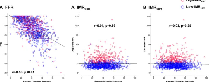
There was no correlation between IMRapp and FFR val- ues (r=0.01, P=0.62). Angiographic percent diameter steno- sis correlated modestly with FFR (r=−0.56, P<0.01), but not with IMRapp (r=0.01, P=0.86) or IMRcorr (r=−0.03, P=0.25) (Figure 2).
Regardless of IMRapp or IMRcorr, the RCA showed sig- nificantly higher IMR values than either the LAD or LCX.
There was no difference in IMR between LAD and LCX. The 75th percentile values of IMRcorr were 21.3 U, 23.0 U, 27.1 U for LAD, LCX, and RCA, respectively (Figure I in the Data Supplement). Therefore, the cut-off values for an abnormal IMRcorr were defined as greater than 22 U, 24 U, and 28 U for LAD, LCX, and RCA, respectively.
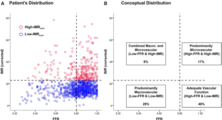
Populations Distribution According to the FFR and IMR Values
The categorical agreement of FFR and IMRcorr was low (kappa value=−0.04, P=0.10; Figure 3A). 55% of patients showed discordant results. Figure 3B shows the categorization of patients according to the cut-off values of FFR and IMRcorr. Overall, 17% of patients had stenoses with nonischemic FFR values but abnormally high microvascular resistance, whereas 8% of patients had both stenoses with ischemic FFR values and abnormally high microvascular resistance.
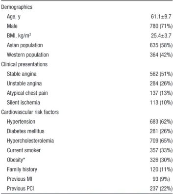
Table 1. General Characteristics of Study Population (n=1096) Demographics
Age, y 61.1±9.7
Male 780 (71%)
BMI, kg/m2 25.4±3.7
Asian population 635 (58%)
Western population 364 (42%)
Clinical presentations
Stable angina 562 (51%)
Unstable angina 284 (26%)
Atypical chest pain 137 (13%)
Silent ischemia 113 (10%)
Cardiovascular risk factors
Hypertension 683 (62%)
Diabetes mellitus 281 (26%)
Hypercholesterolemia 709 (65%)
Current smoker 357 (33%)
Obesity* 326 (30%)
Family history 120 (11%)
Previous MI 93 (9%)
Previous PCI 237 (22%)
Values are mean±SD, median (interquartile ranges, 25th–75th), or n (%).
BMI indicates body mass index; LVEF, left ventricular ejection fraction; MI, myocardial infarction; and PCI, percutaneous coronary intervention.
*Obesity was defined as body mass index ≥25 kg/m2 in Korean patients and
≥30 kg/m2 in Western patients.
Table 2. General Characteristics of Target Vessels and Physiological Parameters (n=1452)
Measured vessel location
Left anterior descending artery 784 (54%)
Left circumflex artery 295 (20%)
Right coronary artery 373 (26%)
Quantitative coronary angiography 1202
Reference diameter, mm 3.01±0.60
Minimum lumen diameter, mm 1.81±0.69
Diameter stenosis, % 40.5±17.3
Lesion length, mm 11.59±7.67
Coronary physiological indices
FFR 0.84±0.14
IMR, U 17.0 (13.0, 23.5)
IMRcorr, U 16.6 (12.4, 23.0)
Values are mean±SD, n (%), or median (Q1, Q3). CFR indicates coronary flow reserve; FFR, fractional flow reserve; IMR, index of microcirculatory resistance;
IMRcorr, calculated IMR with Yong’s formula (IMRcorr=Pa×Tmn×([1.35×Pd/Pa]−0.32).
by guest on April 19, 2017http://circinterventions.ahajournals.org/Downloaded from
Predictors of IMR and FFR
The multivariate generalized estimating equation model identified different predictors of IMR and FFR. Previous MI (odds ratio [OR] 2.16, 95% CI 1.24–3.74, P=0.01), RCA (OR 2.09, 95% CI 1.54–2.84, P<0.01), female sex (OR 1.67, 95%
CI 1.18–2.38, P<0.01), and obesity (OR 1.80, 95% CI 1.31–
2.49, P<0.01) were predictors of high IMRcorr (Table 3). The dichotomous data of IMRcorr in these cohorts are presented in Table I in the Data Supplement. LAD (OR 4.31, 95% CI
2.92–6.36, P<0.01), angiographic diameter stenosis ≥50%
(OR 5.16, 95% CI 3.66–7.28, P<0.01), male sex (OR 2.15, 95% CI 1.38–3.35, P<0.01), and age (per 10 years, OR 1.21, 95% CI 1.01–1.46, P=0.04) were predictors of a low FFR (Table 3).
Discussion
This study provides new insights into the overall characteristics of IMR in patients with coronary atherosclerosis without MI.
Figure 1. Distribution of fractional flow reserve (FFR) and index of microcirculatory resistance (IMR). The per-vessel distribution of preper- cutaneous coronary intervention values of FFR (A) and IMR (B) were presented. The apparent IMR and corrected IMR by Yong’s formula (IMRcorr=Pa×Tmn×([1.35×Pd/Pa]−0.32) were presented in B. IMRapp indicates apparent IMR; IMRcorr, corrected IMR; and IQR, interquartile range.
Figure 2. The association between angiographic % diameter stenosis and physiological indices. The association between percent diam- eter stenosis and FFR/IMRapp/IMRcorr was plotted. Angiographic diameter stenosis correlated modestly with FFR (r=−0.56, P<0.01), but not with IMRapp (r=0.01, P=0.86) or IMRcorr (r=−0.03, P=0.25). The correlations and P values in the figure did not account for intrapatient repeated measurements. FFR indicates fractional flow reserve; IMR, index of microcirculatory resistance; IMRapp, apparent IMR; and IMRcorr, corrected IMR.
by guest on April 19, 2017http://circinterventions.ahajournals.org/Downloaded from
The distinct value of these observations is that they are derived from a large, multiethnic patient population. The main findings of the study are (1) FFR and IMRcorr showed no categorical agreement, and the presence of microvascular disease was sus- pected to be a contributor for the myocardial ischemia in a sig- nificant portion of patients (17% of the population studied); (2) more than one quarter (26%) of vessels with functionally non- significant stenoses (FFR >0.80) had abnormally high IMRcorr
values; and (3) the predictors of high IMRcorr or ischemic FFR were substantially different. These findings suggest the need to integrate IMR with FFR when evaluating patients with CAD.
Relative Contribution of Macro- and Microvascular Disease to IHD
Currently, FFR-guided decision-making for epicardial coro- nary stenosis has been a well-validated approach to avoid unnecessary stent implantation and to enhance patient clinical outcomes.10–12 However, epicardial coronary stenosis is one of the components in the coronary circulatory system; there- fore, the presence of epicardial stenosis is not a sole cause of IHD. Furthermore, lesions with the same degree of stenosis may have different FFR values, according to the microvas- cular status. In our study, among patients with nonischemic FFR values (>0.80), 26% had high IMRcorr. These patients could potentially experience myocardial ischemia secondary to microvascular disease.13 A similar percentage of microvas- cular dysfunction based on IMR measurement was seen in a recent study evaluating patients with chest pain and nonob- structive coronary disease.14 This could at least partly explain why some patients with nonischemic FFR values continue to report symptoms consistent with angina. In the FAME II reg- istry population, (FFR >0.80 and deferral of percutaneous cor- onary intervention), ≈15% of patients had persistent Canadian Cardiovascular Society (CCS) class II-IV angina at 2-years follow-up. Additional evidence supporting the potential prog- nostic relevance of documenting coexisting coronary circula- tory abnormalities in patients with nonsignificant FFR values comes from recently published research on the long-term follow-up of patients interrogated with FFR and concomitant Figure 3. Classification of patients according to the FFR and IMR values. A, The patients’ distribution according to the cut-off values of FFR (≤0.80) and IMRcorr (cut-off: 75th percentile value according to the target vessels). B, The conceptual distribution of the patients. The proportions among the total population were presented in each section of the classification. FFR indicates fractional flow reserve; IMR, index of microcirculatory resistance; and IMRcorr, corrected IMR.
Table 3. Predictors for High IMR or Low FFR in Target Vessels*
Variables OR 95% CI P Value
High IMR (≥75th percentile)
Previous MI 2.16 1.24–3.74 0.01
RCA 2.09 1.54–2.84 <0.01
Female 1.67 1.18–2.38 <0.01
Obesity† 1.80 1.31–2.49 <0.01
Low-FFR (≤0.80)
LAD 4.31 2.92–6.36 <0.01
%DS≥50% 5.16 3.66–7.28 <0.01
Male 2.15 1.38–3.35 <0.01
Age (10 y) 1.21 1.01–1.46 0.04
CI indicates confidence interval; FFR, fractional flow reserve; IMR, index of microvascular resistance; LAD, left anterior descending artery; %DS, percent diameter stenosis; and RCA, right coronary artery.
*Generalized estimating equation model or maximum likelihood χ² tests were used to evaluate the predictors. C-indexes were 0.67 (95% CI 0.63–0.70) and 0.81 (95% CI 0.78–0.85) in predicted model for high IMR and low FFR, respectively.
†Obesity was defined as body mass index ≥25 kg/m2 in Korean patients and
≥30 kg/m2 in Western patients.
by guest on April 19, 2017http://circinterventions.ahajournals.org/Downloaded from
Doppler-derived flow measurements.15 In this report, van de Hoef et al compared 10-year MACE rates according to the FFR and coronary flow velocity reserve (CFVR) levels of target vessel from 157 patients with intermediate stenosis.
Compared with concordant normal FFR (>0.80) and CFVR (≥2.0) group, a normal FFR and abnormal CFVR (<2.0) group showed excess risk of 10-year MACE rates, regardless of FFR cut-off values (0.75 or 0.80). Although difference in MACE rates was mainly driven by excess risk of target vessel revas- cularization in a normal FFR and abnormal CFVR group, this report emphasized the importance of microvascular disease on future adverse events.15
It has been reported that ischemic FFR values are less likely to occur in a vessel with high IMR values.16 In our population, 7.5% of cases had both ischemic FFR values and high IMRcorr values. However, the mean FFR value in our study population was higher (0.84±0.14) and IMRcorr were lower (median 16.6 U, [12.4 U, 23.0 U]) than in the study from Echavarria-Pinto et al (mean FFR 0.81±0.12 and median IMR 18.1 U [12.1 U, 29.1 U]). IMR cut-off values in these studies are based on fre- quency distributions, and the FFR values will also influence IMRcorr values (through Yong’s correction). Accordingly, the observed differences between these 2 studies may have been influenced in part by their different study populations.
Determinants of High IMR
In the current study, multivariate generalized estimating equation model showed that previous MI, RCA, female sex, and obesity were significantly associated with high IMRcorr. The pathophysiologic mechanisms of primary microvascu- lar disease have not been well defined. Nonetheless, recent studies report sex differences in the presentation, diagnosis, management, and pathophysiological mechanisms of CAD.17 So-called cardiac Syndrome X, which is the presence of myo- cardial ischemia without significant epicardial coronary ste- nosis, is more common in females,18 and recent reports suggest that microvascular dysfunction might be the main pathophys- iological mechanism in this condition.18–20 Luo et al presented that the patients with cardiac syndrome X (female 72% of the patients) had significantly higher IMR compared with age- and sex-matched control patients (33.1±7.9 versus 18.8±5.6, P<0.01).19 Further reports suggest that estrogen deficiency, which is commonly observed in menopausal females, might be related to coronary microvascular disease.18,21 Our finding that female sex is a predictor of high IMRcorr is consistent with the previous studies which reported a higher prevalence of cardiac syndrome X in female patients, especially with postmenopausal status, and suggest microvascular disease as a potential mechanism of IHD in these patients.
Interestingly, obesity was also a determinant for high IMRcorr. There is evidence that obesity is a contributor to coro- nary microvascular disease.22 Expansion of adipose tissue leads to tissue hypoxia and results in an increased production of various cytokines (leptin, resistin, tumor necrosis factor, and interleukin-6). These secreted adipokines and proinflam- matory cytokines can induce oxidative stress, which leads to reduced availability of nitric oxide and limits vasodilatory capacity. However, Echavarria-Pinto et al reported a relation- ship between obesity (defined as body mass index ≥30 kg/
m2), lower IMRcorr values, and more frequent hypotensive responses to intravenous adenosine infusion in 79 patients with intermediate stenosis.23 Likewise, there have been con- troversies whether common cardiovascular risk factors which have been known to cause macrovascular disease are also risk factors for primary microvascular disease.24 In our results, conventional risk factors of macrovascular disease, as well as stenosis severity of epicardial coronary stenosis, were not associated with high IMR.
These results, along with the lack of correlation and categorical agreement between FFR and IMR, suggest the hypothesis that macro- and microvascular disease are differ- ent disease processes with different predisposing factors.1,24,25 In addition, these results reinforce the clinical relevance of IMR measurement in current FFR-guided strategy to enhance patient stratification and to clarify the relative contribution of macro- and microvascular disease as a cause of IHD.
Assessment for Microvascular Disease With IMR The major obstacle to integrate the concept of IMR into the clinical fields has been a lack of well-validated normal ranges, especially for IHD patients without MI. Most of the previous studies evaluated IMR in patients with acute MI.5–7 Recently, Fearon et al reported that in patients who underwent primary percutaneous coronary intervention for acute MI and had immediate post–percutaneous coronary intervention measure- ment of IMR and those with IMR >40 U had significantly higher 1-year rates of death or hospitalization with heart fail- ure than patients with an IMR <40 U.5
Previous studies suggested that the upper normal value of IMR is between 25 and 29 U. Melikian et al found that IMR values in a small normal control group without evidence of atherosclerosis were lower than 25 U.26 In patients with inter- mediate coronary stenoses, Echavarria-Pinto et al derived an IMRcorr cut-off value of 29 U based on the 75th percentile of observed IMRcorr values.16 In their study, 91 arteries in 78 patients were interrogated and 31% of patients were presented as post-MI. However, there have been no large studies explor- ing the real-world distribution of IMR among IHD patients without MI. In the current study, we evaluated 1096 patients with 1452 vessels who underwent invasive coronary physi- ological assessment. From the results, the IMRcorr showed median of 16.6 U (12.4 U, 23.0 U), and >75th percentile cut-off in all lesions was 23 U. It is reassuring that the upper limit of the normal value for IMR is within the same range as previous studies of patients with stable IHD. Nonetheless, it should be noticed that most patients enrolled to this registry had undergone FFR and IMR measurement with clinical indi- cations of invasive coronary angiography and FFR measure- ment. Therefore, the distribution of IMR values in a healthy population, without any symptoms and clinical indications of invasive coronary angiography, might be different.
In addition, the distribution of IMRcorr was different among the 3 major coronary arteries, even though the observed differ- ences were relatively small. As the IMR is the product of myo- cardial flow (1/Tmn) and distal arterial pressure (Pd), it can be influenced by blood flow pattern, flow rate, vessel geometry, and the myocardial mass that is supplied by the specific target vessel.27,28 This influence is supported by the work of Murai
by guest on April 19, 2017http://circinterventions.ahajournals.org/Downloaded from
et al, in which RCA location was a predictor of high IMR.29 The relative longer length and larger reference diameter of the RCA may also contribute to a longer mean transit time and higher IMR in this vessel.
Although we used IMRcorr values with Yong’s formula, it should be noted that the difference between IMRapp and IMRcorr was almost negligible, and using IMRapp did not alter any of the original results. Even though IMRcorr is scientifi- cally more accurate, IMRapp seems a more practical way to assess microvascular function in daily routine practice, unless the epicardial stenosis is severe.
In summary, with consideration of our results along with the previous evidences,1,15,16,30 FFR alone may not be sufficient for the evaluation of IHD. Although not all patients with high- FFR need IMR measurement, IMR can be a useful diagnostic tool for those with clinical evidence of IHD.
Limitations
First, because we excluded patients with elevated cardiac enzyme, our findings cannot be applied to patients with acute MI. However, the main purpose of this study was to explore the practical value of IMR in assessing CAD patients without MI. Second, we did not use the wedge pressure to adjust the IMR values because it was not practical to measure wedge pressure in patients with intermediate stenosis. However, cor- rected IMR values by Yong’s formula9 were used to minimize the influence of collateral flow, and the differences between the uncorrected (IMRapp) and corrected IMR (IMRcorr) val- ues were small. Third, the results of noninvasive test were not available in our study. Although patients with high FFR and high IMR might have myocardial ischemia because of microvascular disease, this relationship was not confirmed by noninvasive tests. Fourth, a comparative analysis regarding clinical outcomes according to the different IMR level was not performed. The prognostic impact of high IMRcorr in stable IHD population and the optimal cut-off value to discriminate patients at higher risk of future events need further investiga- tion. Fifth, although only experienced operators participated in our study, the reproducibility and variability of IMR measure- ments among the operators were not tested. Sixth, although multivariate model for predictor of high IMR showed clini- cally relevant results, the c-index of the model was within bor- derline range. Seventh, the roles of endothelial dysfunction or coronary vasospasm were not assessed in our study. Finally, the data regarding quantitative measures of angina symptom status, for example, Seattle Angina Questionnaire, were not available in this study.
Conclusions
IMR showed no correlation with FFR and angiographic lesion severity, and the predictors of high IMR value were different from those for ischemic FFR value. Therefore, integration of IMR into FFR measurement may provide additional insights regarding the relative contribution of macro- and microvascu- lar disease in patients with IHD.
Sources of Funding
This study was supported by the unrestricted research grant from St Jude Medical.
Disclosures
Dr Koo has received institutional research grant from St Jude Medical.
Dr Oldroyd has received honoraria from St Jude Medical. Dr Escaned reports consultancies for St Jude Medical and Volcano Corporation.
Dr Echavarria-Pinto’s work was funded with a clinical scholarship from the Fundación Interhospitalaria Investigacion Cardiovascular, Madrid, Spain. Dr Fearon receives institutional research funding from St Jude Medical. Dr Ng has received honoraria from St Jude Medical. The other authors report no conflicts.
References
1. Yong AS, Ho M, Shah MG, Ng MK, Fearon WF. Coronary microcirculato- ry resistance is independent of epicardial stenosis. Circ Cardiovasc Interv.
2012;5:103–108, S1. doi: 10.1161/CIRCINTERVENTIONS.111.966556.
2. Aarnoudse W, Fearon WF, Manoharan G, Geven M, van de Vosse F, Rutten M, De Bruyne B, Pijls NH. Epicardial stenosis severity does not affect minimal microcirculatory resistance. Circulation. 2004;110:2137–
2142. doi: 10.1161/01.CIR.0000143893.18451.0E.
3. Fearon WF, Aarnoudse W, Pijls NH, De Bruyne B, Balsam LB, Cooke DT, Robbins RC, Fitzgerald PJ, Yeung AC, Yock PG. Microvascular resistance is not influenced by epicardial coronary artery stenosis severity: experi- mental validation. Circulation. 2004;109:2269–2272. doi: 10.1161/01.
CIR.0000128669.99355.CB.
4. Kobayashi Y, Fearon WF. Invasive coronary microcirculation assess- ment–current status of index of microcirculatory resistance. Circ J.
2014;78:1021–1028.
5. Fearon WF, Low AF, Yong AS, McGeoch R, Berry C, Shah MG, Ho MY, Kim HS, Loh JP, Oldroyd KG. Prognostic value of the index of microcirculatory resistance measured after primary percutaneous coro- nary intervention. Circulation. 2013;127:2436–2441. doi: 10.1161/
CIRCULATIONAHA.112.000298.
6. McGeoch R, Watkins S, Berry C, Steedman T, Davie A, Byrne J, Hillis S, Lindsay M, Robb S, Dargie H, Oldroyd K. The index of microcircu- latory resistance measured acutely predicts the extent and severity of myocardial infarction in patients with ST-segment elevation myocardial infarction. JACC Cardiovasc Interv. 2010;3:715–722. doi: 10.1016/j.
jcin.2010.04.009.
7. Fearon WF, Shah M, Ng M, Brinton T, Wilson A, Tremmel JA, Schnittger I, Lee DP, Vagelos RH, Fitzgerald PJ, Yock PG, Yeung AC. Predictive val- ue of the index of microcirculatory resistance in patients with ST-segment elevation myocardial infarction. J Am Coll Cardiol. 2008;51:560–565.
doi: 10.1016/j.jacc.2007.08.062.
8. Ng MK, Yeung AC, Fearon WF. Invasive assessment of the coronary microcirculation: superior reproducibility and less hemodynamic de- pendence of index of microcirculatory resistance compared with coro- nary flow reserve. Circulation. 2006;113:2054–2061. doi: 10.1161/
CIRCULATIONAHA.105.603522.
9. Yong AS, Layland J, Fearon WF, Ho M, Shah MG, Daniels D, Whitbourn R, Macisaac A, Kritharides L, Wilson A, Ng MK. Calculation of the in- dex of microcirculatory resistance without coronary wedge pressure mea- surement in the presence of epicardial stenosis. JACC Cardiovasc Interv.
2013;6:53–58. doi: 10.1016/j.jcin.2012.08.019.
10. De Bruyne B, Fearon WF, Pijls NH, Barbato E, Tonino P, Piroth Z, Jagic N, Mobius-Winckler S, Rioufol G, Witt N, Kala P, MacCarthy P, Engström T, Oldroyd K, Mavromatis K, Manoharan G, Verlee P, Frobert O, Curzen N, Johnson JB, Limacher A, Nüesch E, Jüni P; FAME 2 Trial Investigators.
Fractional flow reserve-guided PCI for stable coronary artery disease. N Engl J Med. 2014;371:1208–1217. doi: 10.1056/NEJMoa1408758.
11. Park SJ, Ahn JM, Park GM, Cho YR, Lee JY, Kim WJ, Han S, Kang SJ, Park DW, Lee SW, Kim YH, Lee CW, Mintz GS, Park SW. Trends in the outcomes of percutaneous coronary intervention with the routine incorporation of fractional flow reserve in real practice. Eur Heart J.
2013;34:3353–3361. doi: 10.1093/eurheartj/eht404.
12. Pijls NH, Fearon WF, Tonino PA, Siebert U, Ikeno F, Bornschein B, van’t Veer M, Klauss V, Manoharan G, Engstrøm T, Oldroyd KG, Ver Lee PN, MacCarthy PA, De Bruyne B; FAME Study Investigators. Fractional flow reserve versus angiography for guiding percutaneous coronary in- tervention in patients with multivessel coronary artery disease: 2-year follow-up of the FAME (Fractional Flow Reserve Versus Angiography for Multivessel Evaluation) study. J Am Coll Cardiol. 2010;56:177–184. doi:
10.1016/j.jacc.2010.04.012.
13. Camici PG, Crea F. Coronary microvascular dysfunction. N Engl J Med.
2007;356:830–840. doi: 10.1056/NEJMra061889.
by guest on April 19, 2017http://circinterventions.ahajournals.org/Downloaded from
14. Lee BK, Lim HS, Fearon WF, Yong AS, Yamada R, Tanaka S, Lee DP, Yeung AC, Tremmel JA. Invasive evaluation of patients with an- gina in the absence of obstructive coronary artery disease. Circulation.
2015;131:1054–1060. doi: 10.1161/CIRCULATIONAHA.114.012636.
15. van de Hoef TP, van Lavieren MA, Damman P, Delewi R, Piek MA, Chamuleau SA, Voskuil M, Henriques JP, Koch KT, de Winter RJ, Spaan JA, Siebes M, Tijssen JG, Meuwissen M, Piek JJ. Physiological basis and long-term clinical outcome of discordance between fractional flow reserve and coronary flow velocity reserve in coronary stenoses of intermediate severity. Circ Cardiovasc Interv. 2014;7:301–311.
16. Echavarria-Pinto M, Escaned J, Macías E, Medina M, Gonzalo N, Petraco R, Sen S, Jimenez-Quevedo P, Hernandez R, Mila R, Ibañez B, Nuñez-Gil IJ, Fernández C, Alfonso F, Bañuelos C, García E, Davies J, Fernández-Ortiz A, Macaya C. Disturbed coronary hemodynamics in ves- sels with intermediate stenoses evaluated with fractional flow reserve:
a combined analysis of epicardial and microcirculatory involvement in ischemic heart disease. Circulation. 2013;128:2557–2566. doi: 10.1161/
CIRCULATIONAHA.112.001345.
17. Nugent L, Mehta PK, Bairey Merz CN. Gender and microvascular angina. J Thromb Thrombolysis. 2011;31:37–46. doi: 10.1007/s11239-010-0477-1.
18. Kaski JC. Cardiac syndrome x in women: the role of oestrogen deficiency.
Heart. 2006;92(suppl 3):iii5–iii9.
19. Luo C, Long M, Hu X, Huang Z, Hu C, Gao X, Du Z. Thermodilution- derived coronary microvascular resistance and flow reserve in patients with cardiac syndrome X. Circ Cardiovasc Interv. 2014;7:43–48. doi:
10.1161/CIRCINTERVENTIONS.113.000953.
20. Recio-Mayoral A, Rimoldi OE, Camici PG, Kaski JC. Inflammation and microvascular dysfunction in cardiac syndrome X patients without conventional risk factors for coronary artery disease. JACC Cardiovasc Imaging. 2013;6:660–667. doi: 10.1016/j.jcmg.2012.12.011.
21. Roqué M, Heras M, Roig E, Masotti M, Rigol M, Betriu A, Balasch J, Sanz G. Short-term effects of transdermal estrogen replacement therapy on coronary vascular reactivity in postmenopausal women with angina pectoris and normal results on coronary angiograms. J Am Coll Cardiol.
1998;31:139–143.
22. Bagi Z, Broskova Z, Feher A. Obesity and coronary microvascular disease - implications for adipose tissue-mediated remote inflammatory response.
Curr Vasc Pharmacol. 2014;12:453–461.
23. Echavarría-Pinto M, Gonzalo N, Ibañez B, Petraco R, Jimenez-Quevedo P, Sen S, Nijjer S, Tarkin J, Alfonso F, Núñez-Gil IJ, Bañuelos C, Quirós A, Fernández-Ortiz A, Macaya C, Koo BK, Davies J, Escaned J. Low coro- nary microcirculatory resistance associated with profound hypotension during intravenous adenosine infusion: implications for the functional assessment of coronary stenoses. Circ Cardiovasc Interv. 2014;7:35–42.
doi: 10.1161/CIRCINTERVENTIONS.113.000659.
24. Lanza GA, Crea F. Primary coronary microvascular dysfunction:
clinical presentation, pathophysiology, and management. Circulation.
2010;121:2317–2325. doi: 10.1161/CIRCULATIONAHA.109.900191.
25. Wessel TR, Arant CB, McGorray SP, Sharaf BL, Reis SE, Kerensky RA, von Mering GO, Smith KM, Pauly DF, Handberg EM, Mankad S, Olson MB, Johnson BD, Merz CN, Sopko G, Pepine CJ; NHLBI Women’s Ischemia Syndrome Evaluation (WISE). Coronary microvascular reactiv- ity is only partially predicted by atherosclerosis risk factors or coronary artery disease in women evaluated for suspected ischemia: results from the NHLBI Women’s Ischemia Syndrome Evaluation (WISE). Clin Cardiol.
2007;30:69–74. doi: 10.1002/clc.19.
26. Melikian N, Vercauteren S, Fearon WF, Cuisset T, MacCarthy PA, Davidavicius G, Aarnoudse W, Bartunek J, Vanderheyden M, Wyffels E, Wijns W, Heyndrickx GR, Pijls NH, de Bruyne B. Quantitative assessment of coronary microvascular function in patients with and without epicardial atherosclerosis. EuroIntervention. 2010;5:939–945. doi: 10.4244/.
27. Akasaka T, Yoshikawa J, Yoshida K, Hozumi T, Takagi T, Okura H.
Comparison of relation of systolic flow of the right coronary artery to pul- monary artery pressure in patients with and without pulmonary hyperten- sion. Am J Cardiol. 1996;78:240–244.
28. Heller LI, Silver KH, Villegas BJ, Balcom SJ, Weiner BH. Blood flow ve- locity in the right coronary artery: assessment before and after angioplasty.
J Am Coll Cardiol. 1994;24:1012–1017.
29. Murai T, Lee T, Yonetsu T, Iwai T, Takagi T, Hishikari K, Masuda R, Iesaka Y, Isobe M, Kakuta T. Variability of microcirculatory resistance index and its relationship with fractional flow reserve in patients with in- termediate coronary artery lesions. Circ J. 2013;77:1769–1776.
30. Dhawan SS, Corban MT, Nanjundappa RA, Eshtehardi P, McDaniel MC, Kwarteng CA, Samady H. Coronary microvascular dysfunction is asso- ciated with higher frequency of thin-cap fibroatheroma. Atherosclerosis.
2012;223:384–388. doi: 10.1016/j.atherosclerosis.2012.05.034.
by guest on April 19, 2017http://circinterventions.ahajournals.org/Downloaded from
Koo, Martin K. Ng, Javier Escaned, William F. Fearon and Keith G. Oldroyd
Watkins, Andy S. Yong, Joon-Hyung Doh, Chang-Wook Nam, Eun-Seok Shin, Bon-Kwon Joo Myung Lee, Jamie Layland, Ji-Hyun Jung, Hyun-Jung Lee, Mauro Echavarria-Pinto, Stuart
the International Index of Microcirculatory Resistance Registry
Print ISSN: 1941-7640. Online ISSN: 1941-7632
Copyright © 2015 American Heart Association, Inc. All rights reserved.
Avenue, Dallas, TX 75231
is published by the American Heart Association, 7272 Greenville Circulation: Cardiovascular Interventions
doi: 10.1161/CIRCINTERVENTIONS.115.002857 2015;8:
Circ Cardiovasc Interv.
http://circinterventions.ahajournals.org/content/8/11/e002857
World Wide Web at:
The online version of this article, along with updated information and services, is located on the
http://circinterventions.ahajournals.org/content/suppl/2015/10/21/CIRCINTERVENTIONS.115.002857.DC1
Data Supplement (unedited) at:
http://circinterventions.ahajournals.org//subscriptions/
is online at:
Circulation: Cardiovascular Interventions Information about subscribing to
Subscriptions:
http://www.lww.com/reprints
Information about reprints can be found online at:
Reprints:
document.
Answer
Permissions and Rights Question and under Services. Further information about this process is available in the
permission is being requested is located, click Request Permissions in the middle column of the Web page Clearance Center, not the Editorial Office. Once the online version of the published article for which
can be obtained via RightsLink, a service of the Copyright Circulation: Cardiovascular Interventions
in
Requests for permissions to reproduce figures, tables, or portions of articles originally published Permissions:
by guest on April 19, 2017http://circinterventions.ahajournals.org/Downloaded from
Integrated Physiologic Assessment of Ischemic Heart Disease in Real World Practice using Index of Microcirculatory Resistance and Fractional Flow
Reserve: Insights from the International IMR registry
Joo Myung Lee, MD, MPH
1, Jamie Layland, MBC
HB
2,3, Ji-Hyun Jung, MD
1, Hyun-Jung Lee, MD
1, Mauro Echavarria-Pinto, MD
4,5, Stuart Watkins MBChB, MD
2,3, Andy S. Yong, MBBS, PhD
6, Joon-Hyung Doh, MD, PhD
7, Chang-Wook Nam, MD, PhD
8, Eun-Seok Shin,
MD, PhD
9, Bon-Kwon Koo, MD, PhD
1,10, Martin K. Ng, MBBS, PhD
11, Javier Escaned, MD, PhD
4,5, William F. Fearon, MD
6, Keith G. Oldroyd, MBChB, MD(Hons)
2,3Supplementary Tables
Supplementary Figure Legends
Supplementary Table 1. Comparison of IMR
corrvalues and proportion of high-IMR among the cohorts of predictors of high-IMR
High-IMR
corr(%) P value
Previous MI 34.9%
0.013
No previous MI 24.7%
RCA 39.1%
<0.001
Other vessels 23.9%
Female 31.5%
0.041
Male 26.0%
Obesity 30.8%
0.081
No obesity 26.4%
Supplementary Figure 1. Comparison of IMR values among 3 coronary arteries
The per-vessel distribution of (A) apparent IMR (IMR
app) and (B) corrected IMR (IMR
corr) among the 3 coronary arteries.
Abbreviations: IMR, index of microcirculatory resistance; IMR
app, apparent IMR; IMR
corr, corrected IMR; IQR, interquartile range; LAD, left anterior descending; LCX, left circumflex;
RCA, right coronary artery.
B. IMR
corrA. IMR
appLAD LCX RCA
0 20 40 60 80
LAD LCX RCA
0 20 40 60 80
15.7 U (11.9, 21.3)
16.9 U (12.5, 23.0)
19.1 U (13.5, 27.1) 16.2 U
(12.5, 22.2)
17.0 U (12.5, 22.1)
19.0 U (14.0, 26.5)
P=0.957 P<0.001
P<0.001
P=0.066 P=0.002
P<0.001
Overall comparison P<0.001 Overall comparison P<0.001