난치성 간질과 동반된 뇌종양과 피질이형성의 공존
- 수술환자 7명의 임상분석 및 수술전략 -
계명대학교 의과대학 신경외과학교실, 신경과학교실, * 병리학교실 **
서인엽·손은익·이상도
*
·이창영·이장철·김동원·임만빈·김인홍·김상표**
= Abstract =
Coexistence of Neoplasia and Cortical Dysplasia Associated with Intractable Epilepsy
- - -
- A Clinical Study of Seven Surgical Patients and Surgical Strategies - - - - In Yeop Seo, M.D., Eun Ik Son, M.D., Sang Do Yi, M.D.,*
Chang Young Lee, M.D., Jang Chul Lee, M.D., Dong Won Kim, M.D., Man Bin Yim, M.D., In Hong Kim, M.D., Sang Pyo Kim, M.D.**
Department of Neurosurgery, Neurology* and Pathology,** and the Epilepsy Center, Keimyung University School of Medicine, Taegu, Korea
tumor and cortical dysplasia may be the concomitant cause of the causes of intractable epilepsy, but a few studies have examined so far. From among 249 patients who underwent surgery for intractable epilepsy at Dongsan Epilepsy Center, those in whom neoplasia and cortical dysplasia coexisted were selected for this study, and were reviewed the clinical, electrophysiological, neuroimaging and pathological findings. In 17 of 25 lesionrelated epilepsy patients, tumors including dysembryoplastic neuroepithelial tumor(DNT)(n=6), ganglioglioma(n=5), gang- liocytoma(n=1), low grade astrocytoma(n=2), oligodendroglioma(n=2), hypothalamic hamartoma(n=1) were verified.
Of these 17 cases involving tumors, concomitant cortical dysplasia was observed in 7(DNT;6, ganglioglioma;1).
All these patients underwent sophisticated presurgical evaluation and intraoperative acute recording(EcoG) for the identification of adjacent or remote epileptogenic areas as well as functional brain mapping by electrical stimulation or SSEP to verify the eloquent areas. In intractable epilepsy, the coexistence of cortical dysplasia and neoplasia is not common, though careful intraoperative evaluation of the tumor and surrounding tissue using electrocorticogram (EcoG) may lead to its pathological identification and excellent surgical results for these rare lesions.
KEY WORDS:Dysembryoplastic neuroepithelial tumor(DNT)・Ganglioglioma・Coexistence・Cortical dysplasia・Epi- lepsy・Electrocorticogram.
서 론
최근 난치성 간질환자들에 대한 활발한 수술적 치료의 시 행으로 간질의 기저병리 소견에 대한 이해의 폭이 넓어지게 되어, 종양성(neoplastic) 및 비종양성(non-neoplastic) 병 변외에도 신경세포 이주장애(neuronal migration disorder) 등의 신경-교세포 기형병변(neurono-glial malformative lesion) 등이 주요 원인으로 보고된다.
그중에서 뇌종양이 간질 발생에 관여된다는 것은 오래전
부터 잘 알려져 있던 바이지만, 최근 신경방사선학적 기술의 발달과 병리소견의 개념정립으로 새로운 범주의 뇌종양들이 밝혀지게 되었고 이들중 일부는 간질의 발생에 깊이 관여된 다는 것이 알려지게 되었다. 이러한 뇌종양의 특징으로 성 장속도가 아주 느리며, 측두엽이나 전두-두정엽의 rolandic 부위의 뇌피질에 주로 위치한다는 것이다. 한편, 1971년 Taylor가 처음 기술한 피질 이형성(cortical dysplasia)은 태생기동안 중추신경세포의 이주장애로 인해 발생하는 것으 로 역시 간질의 발생에 관계되는 것으로 알려져 있어서 수 술적 제거후 간질조절에 상당한 효과가 있는 것으로 알려져
AAAA
있다 29) .
1993년 Prayson 등은 간질환자에서 피질이형성과 뇌종 양이 동반되는 아주 희귀한 경우에 대해 발표하면서 피질이 형성과 동반되는 뇌종양의 특성과 피질이형성과 뇌종양의 관계에 대해 서술했다 24) . 저자들도 249례의 난치성간질 환 자의 절제수술 후 병리조직검사 결과를 재고해 본 결과 7례 에서 뇌종양과 피질이형성이 동반된 것이 밝혀져, 이들에 대 한 임상분석 및 문헌 고찰과 함께 술전, 수술중검사를 이용 한 종양 및 간질유발부위의 절제를 위한 수술전략을 정립하 여 난치성 간질을 동반한 유사종양의 치료에 지표가 되고자 한다.
대상 및 방법
본원에서 1992년 11월부터 1996년 6월까지 간질수술을 시행한 249례중 뇌종양과 관계된 것은 18례(7%)였고 피 질이형성과 관계된 것은 37(18%)례 이었다. 본연구는 뇌 종양과 피질이형성이 동반된 7례를 대상으로 하였다.
이들에 대한 술전평가로서, 종양 원위부위(remote area) 에 이중병리(dual pathology)가 의심되는 경우에는 증후 (semiology), CT MRI 및 SPECT 등의 신경방사선학적 검 사와 두피 및 접형골 전극을 이용한 뇌파검사(sphenoidal EEG)와 비디오-뇌파 집중감시장치(CCTV-EEG teleme- try)를 이용하여 발작뇌파(ictal onset)를 기록하였고, 신경 심리 검사(neuropsychological test)를 실시하였으며, 기억 및 언어 기능의 우위부위의 확인을 위해 내경동맥내 Wada 검사를 시행하였다. 이들 중 간질 발작 병소의 국소부 위 확 인이 곤란했던 환자에게는 뇌경막하 전극(subdural elect- rode)을 삽입하여 발작뇌파를 검사하였다.
인접한 중요 뇌기능부위를 보존하면서 종양과 간질유발부 위를 확인하는 재단절제를 하기위해 수술중에 뇌피질파검사
와 전기자극 또는 SSEP를 이용한 뇌기능 지도화도 시행하 였다. 운동감각 부위나 언어부위의 정확한 확인이 필요한 경 우에는 정맥마취제인propofol의 이용과 국소마취하 각성상 태에서 검사를 시행한다. 수술후 12개월 이상을 추적 관찰 하였고 술후 결과 분석은 Engel의 분류 8) 에 따랐다.
결 과
뇌종양과 피질이형성이 동반된 경우는 전체 간질수술 249 례중 7례로 2.8%이었다. 이들의 남여비는 3:4였고, 간질 발생 당시 나이는 5세에서 16세까지로 평균 10.2세였고 간 질 유병 기간은 3년에서 19년으로 평균 9년이었다. 병소의 위치는 측두엽이 5례, 전두엽이 1례, 두정엽이 1례였다. 경련 발작의 유형은 5례에서 복합 부분 발작, 2례에서 단순 부분 발작이었다(Table 1).
1. 방사선학적 소견
7례 전례에서 뇌종양부위에 대한 T2 강조영상에서 자기 영상 신호가 증가되고 T1강조영상에서는 자기영상 신호가 감소된 소견을 보였고 조영제 증강효과는 나타나지 않았다.
뇌종양의 경계는 비교적 명료했다. 또한 뇌종양 인접부위에 서 T1과 T2 강조영상에서 모두 뇌피질과 동일한 자기영상 신호가 나타나(Fig. 1) 피질이형성의 존재를 알 수 있었던 경우도 2례 있었다.
2. 간질 병소의 수술적 제거와 병리소견
간질 유발병소의 부위로는 측두엽이 5례, 전두엽이 1례였 고 두정엽이 1례였다. 7례중 4례는 전신 마취를 하였고 3례 는 국소 마취와 propofol 정맥 마취를 병행하였다. 좌측 측 두엽 병변을 가진 3명에서 술중 언어검사(language mapping) 와, rolandic 부위병변에는 운동감각 피질의 확인을 위해 체 감각유발전위(SSEP) 및 피질 전기자극(electrical stimu-
Table 1. Clinical features of 7 patients
Case No Sex /Age Location Age at sz onset Sz pattern Path. Dx CD* grade Operation Outcome**
1 F/13 Lt-MT 7 CPS DNT 2 L+ATL+H II
2 F/18 Rt-LT 15 CPS DNT 2 L+ATL II
3 M/35 Lt-MLT 16 CPS DNT 2 L+ATL+AH I
4 F/9 Rt-MLT 5 CPS DNT 1 L+ATL+AH I
5 M/22 Rt-MF 7 SPS DNT 1 L+C I
6 M/16 Lt-MP 10 SPS DNT 1 L+C+MST I
7 F/25 Lt-MT 16 CPS GAN 1 L+ATL+H I
*CD cortical dysplasia(1:disorganized form, 2:cluster form, 3:ballooning form)
**Engel’s outcome classification(Class I;seizure free, II;rare seizure)
(LT:lateral temporal, MLT:mesiolateral temporal, MF:mesiofrontal, MP:mesioparietal;CPS:complex partial seizure, SPS:simple partial seizure;DNT:dysembryoplastic neuroepithelial tumor, GAN:ganglioglioma;L:lesionectomy, ATL:
anterior temporal lobectomy, H:hippocampectomy, AH:amygdalohippocampectomy, C:cortisectomy, MST:multiple
subpial transection)
lation) 검사를 실시하였으며 전례에서 수술중 뇌피질파검 사(ECoG)를 이용하여 간질양파(epileptiform discharge) 의 존재여부를 확인하면서 간질병소를 제거 하였으며, 병소 의 절제 후에는 간질파가 소실되는 것을 확인하였다(Table 1). 절제된 조직에 대한 병리소견에서 종양조직이나 인접부 위에대한 뇌피질파 검사상 발작간파(interictal discharges) 가 저명한 부위는 국소피질 이형성소견이 발견되었으며, 태
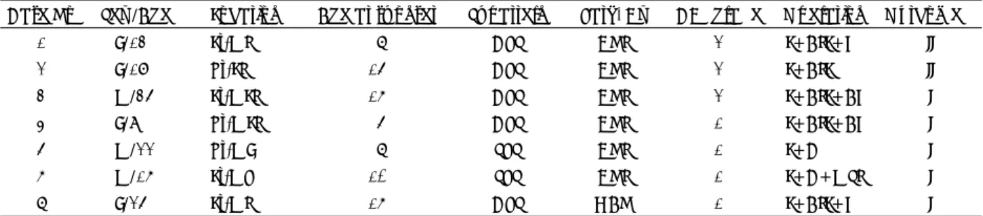
생기발육부전 신경상피종에서는 특징적인 핍지신경양 세포 (oligodendrocyte-like cell:OLC)가 나타나며(Fig. 2).
때로는 한 현미경시야에서 이중병리가 동시에 보이기도한다 (Fig. 3).
3. 수술후 추적 관찰 및 경과
수술후 모든 환자에서 술중검사 및 수술에 따른 합병증이 나 신경학적 문제는 없었으며 술후 간질 발작의 추적기간은 12개월에서 33개월까지 평균 21개월이었다. 이 중 5례에서 는 간질 발작이 없었으며(Engel’s class I), 2례에서는 현 저한 감소(Engel’s class II)를 보였다(Table 1).
고 찰
난치성 간질 환자에서 간질 조절을 목적으로 뇌 절제술을 시행한 후 병리조직을 분석해보면 정상조직으로부터 신경교 및 신경세포의 기형병변(neuronoglial malformative lesion) 과 종양, 신경교증(gliosis), 염증 등으로 다양하게 나타난다.
따라서 이런 병리조직이 간질수술후 예후에 미치는 영향 뿐 아니라, 술전에 미리 기저병리에 대해 알았을 경우와 이 중 병리(dual pathology) 소견이 동반된 경우 뇌엽 절제술 의 방법 및 범위에 대해서는 많은 연구와 논란들이 있었으
Fig. 1. Axial(T2 WI) and coronal(FLAIR) MRIs show cortical cystic mass with isosignal(to cortex) lesion suggesting cortical dysplasia which extend to ependymal layer, verified as dysembryoplastic neuroepithelial tumor(DNT).
Fig. 2. Characteristic microscopic finding of DNT(dysembryo-
plastic neuroepithelial tumor):many oligodendrocyte-
like cells(OLC) with prominent nucleoli and glial elements
in background(H & E stain, ×200).
나 1)3)7)9)11)16)19)20)24)27)
, 뇌종양과 피질 이형성이 동반된 경 우는 극히 드물었다. 저자들은 뇌종양이 피질 이형성과 동 반된 특이한 7례를 발견하여 문헌고찰을 해 본 결과 1988년 Daumas-Duport등이 피질 이형성이 동반된 태생기발육부 전 신경상피종(dysembryoplastic neuroepithelial tumor) 에 대한 보고가 있었으며 6) , 1993년 Prayson등이 보고한 13례중에는 신경절교종(ganglioglioma)이 8례로 가장 많았 고 태생기발육부전 신경상피종이 3례, 양성 신경교종이 2례 였다 24) . 그러나 본 연구 에서는 태생기발육부전 신경상피종 이 6례로 대부분이었고 신경절교종이 1례였다.
피질 이형성이란 용어는 1971년 Taylor가 처음 사용했 고 29) , 비정상적인 신경세포로 된 피질구조(cortical arch- itecture)의 분열된 상태로 특징 지어지는 신경세포 이주장 애(neuronal migrational disorder)의 하나이다 15)18)23) . 피 질이형성이 간질에 관여하는 것은 축색수지 접속이 감소하여 신경 연접(synaptogenesis)이 감소되는 것과 관계있다고 한 다 29) . 현미경적 병리 소견으로 cortical laminar disorga- nization, persistent subpial granular layer와 balloon cell change등 9가지 소견이 있다 15) . 자기공명영상검사로 대회
증(macrogyria), 소회증(microgyria) 그리고 피질두께의 증가등을 관찰하며 14)17)18) , 2mm 미만의 두께로 촬영한 고 해상 MRI를 이용하면 국소 피질이형성도 진단이 가능하다.
수술후 예후와 관계있는 것은 구조적 이상이 있는 부위의 절제여부가 중요하나 19) 피질 이형성의 병리학적 등급 자체 는 간질의 빈도와 관계 없다고 한다 15) .
신경절교종은 신경 조직과 신경교의 성분을 모두 가지는 종양으로 Courville가 처음 정의했다 5) . 임상적으로 성장속 도가 느리고 측두엽에 주로 위치하며 간질을 잘 동반하고 양성의 예후를 보인다. 병리학적으로 신생성의 성상세포와 비정상적인 신경절 세포가 혼재된 것을 특징적 소견으로 하 는데 신경절 세포(ganglion cell)는 Nissle 성분과 커다란 핵인을 가진다 21)31)32) . Synaptosin 같은 신경조직 표식자나 GFAP(glial fibrially acidic protein)같은 신경교의 표식자 를 이용한 병리조직검사가 진단에 도움이 된다 32) .
신경절교종이 피질 이형성의 범주내에서 종양성 형태(neo- plastic form)이거나 이형성 초점(dysplastic focus)에서 종 양으로 전환(neoplastic transformation)한 것이라는 주장 들이 있으며 20)24)25)32)
, Wolf등 32) 이 발견한 과오종(hamar-
Fig. 3. Dual pathologies in one microscopic field as dysorganized area of cortical dysplasia(A) and neoplastic area(B) of
oligodendrocyte-like cells(OLC)(H & E stain, ×50).
toma)과도 유사하다. 그러나 신경절 교종의 극소수에서는 악성으로 전환되거나 전이(metastasis) 되는 경우도 있다 고 한다 25) .
방사선학적검사상으로는 다양한 소견이 나타나서 진단의 특이성이 떨어지나 대체로 T2 강조영상에서 고신호 영상을 보이고 T1강조 영상에서는 저신호 소견을 보이며 조영제 증강효과는 잘 나타난다고 한다 2)21) . 신경절 교종의 수술적 치료방법에 대해서는 논란이 많으나 7)20)28) , 최근 Prayson 등 25) 의 경우를 보면 신경절교종의 완전 절제 및 간질성 병 소의 제거를 함께 하여 좋은 결과를 보이고 있다.
1988년 Daumaus-Duport는 소아에서 주로 발견되며 측두엽에 위치하고 수술후 동반되는 간질이 완치되는 일련 의 뇌종양을 태생기발육부전 신경상피종이라 명명하여 발표 했다 6) . 유점소의 기질내에 특이한 신경-교질성분(specific glioneural element within mucoid matrix), 피질 이형성과 동반된 다결절 성분(multinodular component associated with cortical dysplasia), 핍지신경양 세포(oligodendro- cyte-like cell)를 병리학적 특징으로 한다 6)22)30) . Cavanagh, Prayson 등은 과오종이라고 했고, Daumas-Duport등은 양성 종양으로 여기고 악성으로 전환될 수 있다고 생각했다 4)6)22)24) . 피질이형성과 동반되며 절제술만으로도 완치 될 수 있고 백 질(white matter)내에 위치하고 22)24)26) 또한 정상 신생아에 서 잔존 유막하 과립층이 잘 발견되는 위치가 태생기발육부 전 신경상피종의 호발 장소와 일치하는 등의 사실들은 태생 기발육부전 신경상피종이 과오종이라는 것을 잘 뒷받침 해
준다 6)9)22) . 그러나 최근 Raymond등은 세포분열의 소견과
다세포성, 저명한 세포의 다형태성과 PCNA(proliferating nucleolar organizing antigens)를 이용한 실험에서 강력한 유사분열의 소견이 나타나는 것을 미루어 과오종보다는 종 양에 가깝다고 했다 26) .
방사선학적으로 전산화단층촬영상 조영증강이 되지 않는 저밀도 영역으로 나타나고 자기공명영상의 T1 강조영상에 서 저신호 강도, T2 강조영상에서 고신호 강도를 나타내고 조영제에 의한 증강효과와 종괴효과(mass effect)는 없으
나 13)26) , 일부에서는 조영제 증강효과가 있다고 한다 12) (Ta-
ble 2). 본 연구의 태생기발육부전 신경상피종 6례의 MRI 소견을 보면, 국소 조영증강 소견과 낭성병변이 있는 경우 가 있으며 피질에 위치하거나 내측측두엽과 연관이 많았다.
수술적 치료법으로는 절제술만으로 충분하며 방사선 치료 나 항암제 치료같은 보강치료는 필요가 없다 6)12)13)26)
. 이상의 문헌고찰로 미루어 볼 때 신경절 교종이나 태생기 발육부전 신경상피종은 피질 이형성에서 발생 되거나 또는 이에 동반된 종양이다 25) . 그리고 간질성 유발병소는 동반된
피질 이형성이나 종양자체 또는 이차적 간질성 병소이다.
병리학적으로나 임상적으로 악성 전환을 완전히 배제하지는 못하나 대체로 양성의 종양이다. 그러면 간질을 주소로 한 이중 병소를 가진 환자의 수술은 어떻게 할것인가? 이런 문 제에 대한 의견은 다양하여 Falconer, Morris등 9)16) 은 뇌종 양 자체만 제거해서도 좋은 결과를 얻을 수 있다고 했고, Boon이나 Kirkpatrick등 3)11) 은 광범위 뇌엽 절제술로서 효 과적인 간질 조절을 할 수 있었다고 한다. 그러나 최근의 경 향들을 살펴보면 뇌종양의 제거 뿐만 아니라 간질성 병소의 제거도 함께 하는 것을 주장하는 보고 1)7)27) 들이 많다. 이들 중 Awad나 Pilcher등 1)16) 은 가능한 간질 병소의 제거를 위해 술중 뇌피질파검사를 강조했으며, Palmini, Prayson등 19)24) 은 피질 이형성이 간질의 주병소이므로 이를 최대한 제거해 야 한다고 했다. 이렇게 다양한 주장들이 나오게 된 계기는, 결국 간질의 주병소가 어디인지? 이를 제거함으로 간질이 충분히 조절될 것인지? 신경절교종이나 태생기 발육부전 신 경상피종 같은 뇌종양이 양성이기는 하나 악성으로 전환되 어 환자의 생존에 영향을 끼치지는 않는가? 하는 의문점 때 문이다. 저자들의 경우에서는 뇌종양에 인접한 부위에서 활 발한 간질양파가 술중 뇌피질파 검사상 감지되었고 환자에 게 신경학적 손상을 주지 않는 범위내에서 최대한 뇌종양을 절제(lesionectomy)한 후, 간질파가 나오는 인접 피질 부 위에 대한 추가 피질절제술(additional corticectomy)을 시 행하였으며, 이들 조직의 병리 검사소견상 피질 이형성이 발견되었다. 술후 추적 관찰에서도 5례에서는 간질이 소실 되었고 2례에서도 현저한 감소를 보이는 좋은 성적을 보였 다. 이상에서 저자들은 피질 이형성과 뇌종양이 동반되는 경우 간질의 조절을 위해서는 피질 이형성 부위를 최대한 제거해야하고 이의 감지 및 확인을 위해서 피질제거 전후에 뇌피질파 검사를 반드시 시행하고, 뇌종양에 대해서는 악성 으로 진행될 가능성이 희박하므로 신경학적 손상을 주지 않 는 범위내에서 최대한 절제하고 보강치료(adjuvant therapy)
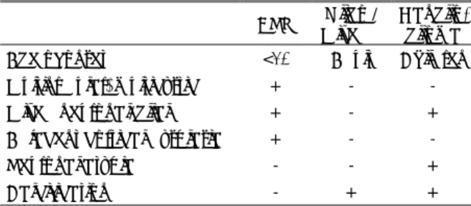
Table 2. Differential diagnosis of tumors coexistent with co- rtical dysplasia
†DNT* Oligo /
Mixed**
Ganglio- glioma Age of onset <20 Adult Children Multinodular, multicystic + - - Mixed neuronal/glial + - + Adjacent cortical dysplasia + - -
Neuronal atypia - - +
Calcification - + +
†
from ref. 21
*dysembryoplastic neuroepithelial tumor
**oligodendroglioma/mixed glioma
는 필요하지 않는 것으로 생각한다.
결 론
저자들은 1992년 12월부터 1996년 6월까지 난치성 간 질을 주소로 본원 신경외과에서 간질수술을 시행한 환자중 에서 술중 뇌지도화 및 뇌피질파를 이용하여 간질유발부위 를 최대한 제거한후 검사한 병리조직 소견상 7례에서 뇌종 양과 피질이형성의 공존이 확인되었으며, 술후 5례에서 간 질 발작이 소실되고 2례에서 호전되는 좋은 결과를 경험하 였다. 특히 술전 방사선학적 검사상 발견되지는 않았지만 술 중 뇌피질파 검사상 저명한 간질파가 나타나는 부위를 충분 히 제거함으로서 피질 이형성의 공존을 확인할 수 있었다.
아울러 문헌고찰에서 신경절 교종이나 태생기발육부전신경 상피종은 과오종으로 보는 경향이 높으며, 수술의 궁극적인 목표인 간질의 조절을 위해서는 간질유발부위로 추정되는 피질 이형성부위를 최대한 제거하여 철저한 병리검사를 시 행하는 것이 정확한 조직학적 규명과 함께 간질치료에 도움 이 될 것으로 사료된다.
•
논문접수일:1997년 12월 10일•
심사완료일:1998년 1월 12일•
교신저자:손 은 익700-712 대구광역시 중구 동산동 194 계명대학교 의과대학 신경외과학교실
전화:053) 250-7306, 전송:053) 250-7356
References
1) Awad IA, Rosenfeld J, Ahl J, et al:Intractable epilepsy and
structural lesions of the brain
:mapping, resection strategies, and seizure outcome. Epilepsia 32
:179-186, 1991
2) Bergen D, Bleck T, Ramsey R, et al:Magnetic resonance
imaging as a sensitive and specific predictor of neoplasms removed for intractable epilepsy. Epilepsia 30
:318-321, 1989
3) Boon PA, Williamson PD, Fried I, et al:Intracranial, intraaxial,space-occupying lesions in patients with intractable partial seizures
:an anatomoclinical, neuropsychological, and surgical correlation. Epilepsia 32
:467-476, 1991
4) Cavanagh JS:On certain tumors encountered in the temporal
lobe. Brain 81
:389-405, 1958
5) Courville CB:Ganglioglioma:
tumor of central nervous system, review of the literature and report of 2 cases. Arch Neurol Psychiatry 24
:439-491, 1930
6) Daumas-Duport C, Scheithauer BW, Chodkiewicz JP, et al:
Dysembryoplastic neuroepithelial tumor
:a surgically curable tumor of young patients with intractable partial seizures. Neu- rosurgery 23
:545-556, 1988
7) Drake J, Hoffman HJ, Kobayashi J, et al:Surgical manage-
ment of children with temporal lobe epilepsy and mass lesions.
Neurosurgery 21
:792-797, 1987
8) Engel J Jr:Outcome with respect to epileptic seizures, in
Engel J Jr
(ed
):Surgical Treatment of the Epilepsies. New York
:Raven Press, 1987. pp553-572
9) Falconer MA, Driver MV, Serafetinides EA:Temporal lobe
epilepsy due to distant lesions
:two cases relieved by operation.
Brain 85
:521-534, 1962
10) Hirose T, Scheithauer BW, Lopes BS, et al:Dysembryoplastic
neuroepithelial tumor
:an immunohistochemial and ultras- tructural study. J Neuropathol Exp Neurol 53
:184-195, 1994
11) Kirkpatrick PJ, Honavar M, Janota I, et al:Control of temporallobe epilepsy following en bloc resection of low-grade tumor.
J Neurosurg 78
:19-25, 1993
12) Koeller KK, Dillon WP:Dysembryoplastic neuroepithelial
tumors
:MR appearnace. AJNR 13
:1319-1325, 1992
13) Kuroiwa T, Kishikawa T, Kato A, et al:Dysembryoplasticneuroepithelial tumors
:MR Findings. J Comput Assist Tomogr 18
:352-356, 1994
14) Kuzniecky R, Garcia JH, Faught E, et al:Cortical dysplasia
in temporal lobe epilepsy
:magnetic resonance imaging cor- relations. Ann Neurol 29
:293-298, 1991
15) Mischel PS, Nguyen LP, Vinters HV, et al:Cerebral cortical
dysplasia associated with pediatric epilepsy. Review of neuro- pathologic features and proposal for a grading system. J Neu- ropathol Exp Neurol 54
:137-153, 1995
16) Morris HH, Estes ML, Gilmore R, et al:Chronic intractable
epilepsy as the only symptom of primary brain tumor. Epilepsia 34
:1038-1043, 1993
17) Osborn RE, Byrd SE, Naidich TP, et al:MR imaging of
neuronal migrational disorders. AJNR 9
:1101-1106, 1988
18) Palmini A, Andermann F, Olivier A, et al:Focal neuronalmigration disorders and intractable partial epilepsy
:a study of 30 patients. Ann Neurol 30
:741-749, 1991
19) Palmini A, Andermann F, Olivier A, et al:Focal neuronal
migration disorders and intractable partial epilepsy
:results of surgical treatment. Ann Neurol 30
:750-757, 1991
20) Pilcher WH, Silbergeld DL, Berger MS, et al:Intraoperativeelectrocorticography during tumor resection
:impact on seizure outcome in patients with gangliogliomas. J Neurosurg 78
:891- 902, 1993
21) Peretti-Viton P, Perez-Castillo AM, Raybaud C, et al:Magnetic
resonance imaging in gangliogliomas and gangliocytomas of the nervous system. J Neuroradiol 18
:189-199, 1991
22) Prayson RA, Estes ML:Dysembryoplastic neuroepithelialtumor. Am J Clin Pathol 97
:398-401, 1992
23) Prayson RA, Estes ML:Cortical dysplasia:
a histopathologic study of 52 cases of partial lobectomy in patients with epilepsy.
Hum Pathol 26
:493-500, 1995
24) Prayson RA, Estes ML, Morris HH:Coexistence of neoplasia
and cortical dysplasia in patients presenting with seizures.
Epilepsia 34
:609-615, 1993
25) Prayson RA, Khajavi K, Comair YG, et al:Cortical archite-
ctural abnormalities and MIB1 immunoreactivity in gangliog- liomas
:a study of 60 patients with intracranial tumors. J Neuropathol Exp Neurol 54
:513-520, 1995
26) Raymond AA, Halpin SFS, Alsanjari N, et al:Dysembryoplastic
neuroepithelial tumor
:Features in 16 patients. Brain 117
:461-475, 1994
27) Sperling MR, Cahan LD, Brown WJ, et al:Relief of seizures
from a predominantly posterior temporal tumor with anterior temporal lobectomy. Epilepsia 30
:559-563, 1989
28) Sutton LN, Packer RJ, Rorke LB, et al:Cerebral ganglio-
glioma during childhood. Neurosurgery 13
:124-128, 1983
29) Taylor DC, Falconer MA, Bruton CJ, et al:Focal dysplasia of
the cerebral cortex in epilepsy. J Neurol Neurosurg Psychiatry 34
:369-387, 1971
30) Theodore WH, Kuffa DK, et al:Pathology of temporal lobe
foci
:correlation with CT, MRI, and PET. Neurology 40
:797-803, 1990
31) Wolf HK, Birkholz T, Wellmer J, et al:Neurochemical profile