• 검색 결과가 없습니다.

Pulmonary Foreign Body Granulomatosis in Dental Technician

N/A
N/A
Protected

Academic year: 2021

Share "Pulmonary Foreign Body Granulomatosis in Dental Technician"

Copied!
5
0
0

로드 중.... (전체 텍스트 보기)

전체 글

(1)

Pulmonary Foreign Body Granulomatosis in Dental Technician

Sung Jun Chung, M.D.

1

, Gun Woo Koo, M.D.

1

, Dong Won Park, M.D.

1

, Hyun Jung Kwak, M.D.

1

, Ji Young Yhi, M.D.

1

, Ji-Yong Moon, M.D.

1

, Sang-Heon Kim, M.D.

1

, Jang Won Sohn, M.D.

1

, Ho Joo Yoon, M.D.

1

, Dong Ho Shin, M.D.

1

, Sung Soo Park, M.D.

1

, Ju Yeon Pyo, M.D.

2

, Young-Ha Oh, M.D.

2

and Tae-Hyung Kim, M.D.

1

Departments of

1

Internal Medicine and

2

Pathology, Hanyang University College of Medicine, Seoul, Korea

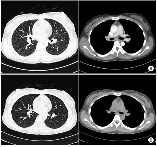
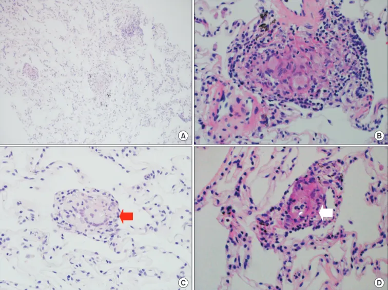
Occupational lung diseases are caused by several toxic substances including heavy metals; however, the exact pathologic mechanisms remain unknown. In the workplace, dental technicians are often exposed to heavy metals such as cobalt, nickel, or beryllium and occasionally develop occupational lung diseases. We described a case of occupational lung disease in a patient who was employed as a dental technician for over a decade. A 31-year-old, non-smoking woman presented with productive cough and shortness of breath of several weeks duration. Chest computed tomography revealed a large number of scattered, bilateral small pulmonary nodules throughout the lung field, and multiple mediastinal lymph nodes enlargement. Percutaneous needle biopsy showed multifocal small granulomas with foreign body type giant cells suggestive of heavy metals inhalation. The patient’s condition improved on simple avoidance strategy for several months. This case highlighted the importance of proper workplace safety.

Keywords: Lung Disease; Dental Technicians

Common occupational lung diseases include pneumoco- niosis, occupational asthma, lung granulomatosis, hypersensi- tivity pneumonitis, silicosis, asbestosis, contact dermatitis, and sick building syndrome

1-7

.

Various types of pneumoconioses have been reported in dental technicians

1,8

, which are named according to the re- lated toxic materials such as silicosis, asbestosis, hard metal disease, and the so-called dental technician’s pneumoconio- sis, in which Cr-Co-Mo alloys could play a role

1

. Dental techni- cians are, indeed, exposed to various dusts and other forms of chemicals when polishing and grinding prosthetics and dur- ing casting operations

1

. And, these occupational exposure can be different from person to person according to the working conditions and used materials

4

.

Therefore, dental technicians should be aware of the risk of occupational lung diseases

3,7

and also should be educated about proper protection and proper environment of work- place

3,4,7

. Once occupational lung diseases are suggested, they should visit hospital for confirmative diagnosis and proper management.

Because early radiologic findings of occupational lung dis- eases are non-specific, differential diagnosis might be very Copyright © 2015

The Korean Academy of Tuberculosis and Respiratory Diseases.

All rights reserved.

Introduction

There are many occupational lung diseases related to vari- ous toxic substances

1

, and respiratory system is a major route of occupational diseases

2

. Heavy metals, such as cobalt, nickel, or beryllium are one of the most common substances that is found in occupational lung diseases.

Address for correspondence: Tae-Hyung Kim, M.D.

Division of Pulmonary and Critical Care Medicine, Department of Internal Medicine, Hanyang University Guri Hospital, Hanyang University College of Medicine, 153 Gyeongchun-ro, Guri 11923, Korea

Phone: 82-31-560-2240, Fax: 82-31-553-7369 E-mail: [email protected]

Received: Aug. 24, 2014 Revised: Dec. 4, 2014 Accepted: Jan. 7, 2015

cc

It is identical to the Creative Commons Attribution Non-Commercial

License (http://creativecommons.org/licenses/by-nc/4.0/).

(2)

difficult without discrete occupational history and specific pathologic findings. Diagnosis of occupational lung disease should be based on the physician’s suspicion, and the miner- alogic studies, which had found that bronchoalveolar lavage and/or lung biopsies can be used to confirmative diagnosis of occupational lung disease

1

.

Case Report

A 31-year-old, non-smoking woman presented with mul- tiple symptoms, including productive cough, shortness of breath, and purulent sputum without fever or weight loss, and reported that she had been experiencing the symptoms for approximately 1 month. The patient’s medical history includ- ed diagnosis of rheumatoid arthritis 5 years prior to this visit at another hospital, where she was treated with non-steroidal anti-inflammatory drugs (prednisolone had been excluded).

A year later, the patient developed cough and hemoptysis that persisted for several months, and sputum culture results were positive for Mycobacterium tuberculosis. Therefore, the pa- tient was diagnosed with pulmonary tuberculosis (TB) of the right lower lobe and received treatment with anti-TB medica- tion for 9 months. There were no other significant events in

her medical history.

As part of our physical examination, pulmonary ausculta- tion during inspiration, we found out that breathing sound de- ceased lightly with mild end-inspiratory crackles at both lower lung fields. No other significant abnormalities were noted during physical examination. Laboratory tests results were as below: white blood count was 7,900/mm

3

, eosinophil count was 3.8% (300/mm

3

), hemoglobin level was 11.7 g/dL, and C- reactive protein level was 0.42 mg/dL. Sputum smear for acid- fast bacilli and bacterial culture of sputum were both negative.

Chest radiography revealed bronchial thickening in the right lower lobe and bilateral hilar lymph nodes enlargement, pos- sibly due to pneumonia or recurrent TB. For inspection of the hilar lymph nodes enlargement, the patient underwent com- puted tomography (CT). The chest CT revealed a large num- ber of small, scattered, bilateral pulmonary nodules through- out the lung field. A number of bilateral hilar and mediastinal lymph nodes enlargements were also observed. Based on these findings, the patient was suspected to have miliary TB with lymphadenopathies or sarcoidosis (Figure 1A).

The patient’s pulmonary function test results showed a forced expiratory volume in 1 second (FEV

1

) of 2.94 L (103%

of predicted value), forced vital capacity (FVC) of 3.26 L (99%

of predicted value), FEV

1

/FVC of 90.2%, and diffusing lung

A

B

Figure 1. Chest computed tomography

(CT) on admission (A) and at 3 months

after suitable precaution (B). (A) Chest

CT on admission showed multiple small

nodules in both lower lung fields. Both

hilar and mediastinal lymph nodes enlar-

gement are also seen. (B) After 3 months,

multiple small nodules are reduced in

size and number. Enlargement of both-

hilar and mediastinal lymph nodes are

absent.

(3)

capacity for carbon monoxide by alveolar volume of 74%. No other abnormal findings besides a slight decrease in diffusing capacity were noted.

Angiotensin converting enzyme level in serum was 62 U/L (normal range, 12–68 U/L).

To assist our differential diagnosis, we opted to perform a lung tissue biopsy, using fine needle aspiration biopsy (FNAB). Histologically, the biopsied tissue showed relatively well-preserved lung parenchyme except for multifocal non- caseating granulomas (Figure 2), and we assumed that they were located in the miliary nodules which were observed on the chest CT. These non-caseating granulomas contained a number of multinucleated foreign body type giant cells (Figure 2B), which appeared to have ingested metal debris (Figure

2C), forming needle-shaped particles, suspected as inorganic substances (Figure 2D). We assumed that granuloma forma- tion was the progression of inflammation caused by inhala- tion of inorganic substances such as heavy metals. Therefore, we concluded that these non-caseating granulomas were as- sociated with occupational exposure.

We then questioned the patient regarding her employment history and the work place environment. The patient revealed she had been employed as a dental technician for over 10 years, and did not usually wear protective equipments such as mask or glasses. In workplace, as dental technician, patient worked with polished and grinded prosthetics and casting operations. During the work, patient might be exposed and in- halated heavy metal such as cobalt, nickel, or beryllium. Since

A B

C D

Figure 2. Histologically, the lung biopsy reveals multifocal small and discrete non-caseating epithelioid granulomas containing foreign body-

type giant cells. (A) Multifocal granulomas shown in a low power field (H&E stain, ×100). (B) Representative non-caseating epithelioid

granulomas containing giant cells engulfing foreign bodies in a high power field (H&E stain, ×400). (C) Typical granuloma containing giant

cells engulfing refractile foreign bodies (arrow) (H&E stain, ×400). (D) Typical granulomas containing foreign body-type giant cells engulfing

refractile foreign bodies (arrow), which are conspicuous under polarized light microscopy (H&E stain, ×200).

(4)

CT had revealed a large number of tiny pulmonary nodules throughout the lung fields, which are not common findings in advanced cases of pneumoconiosis (e.g., in miners and glass factory workers), we presumed that these nodules were the result of acute and sub-acute inflammatory responses linked to occupational exposure. As acute response, we treated patient with simple avoidance therapy and education with- out any medication. We educated the patient regarding risk management in the workplace, and advised her to wear a dust mask and avoid any situation where dust could be present.

The patient was hospitalized for a week to facilitate further examination and stabilization. During the admission period, the patient discontinued all employment-related activity and following avoidance strategy, her symptoms much improved.

After discharge, outpatient observation and periodic examina- tions were conducted. After 3 months later, the patient’s symp- toms such as shortness of breath, productive cough, and puru- lent sputum expectoration had nearly disappeared. Follow-up chest CT confirmed that the size and number of nodules had significantly decreased, and that the consolidations had much improved (Figure 1B). The size of enlarged hilar lymph nodes and number of other enlarged mediastinal lymph nodes had also significantly decreased.

Discussion

Dental technicians are at high risk of occupational lung dis- eases

1,7,9

, because they are frequently exposed to dust, chemi- cal substances, toxic materials, and heavy metals used in the construction of dental prostheses and orthodontic appli- ances

1,9

. From those occupational lung diseases, the incidence of pneumoconiosis in dental technicians are higher than expected. And, the prevalence of pneumoconiosis in dental technician are from 9.8% to 24.2% in several reports

3,5,10

. Dur- ing construction of dental appliances or dental procedures, various toxic solvents and non-organic gases can be released into the air, leading to occupational lung disease

11

. The addi- tion of metal alloys, ceramics, acrylates, as well as finishing, grinding, and casting, may all increase the amount of heavy metals released.

Differential diagnosis based on clinical manifestations in- cluding subjective symptoms and radiological findings could be almost impossible due to their lack of specificity at earlier stage. In our case, the patient had experienced respiratory symptoms for one month, and various diseases were sus- pected based on her radiography and CT results, including infectious and non-infectious diseases. To reach a definitive diagnosis, we ran various laboratory tests and sputum culture to rule out various causes of infection.

As one study has reported that alveolar lavage and/or bi- opsy could be helpful in the diagnosis of similar conditions

1

, pathologic study might be necessary for exact diagnosis espe-

cially at early stage. For the confirmative diagnosis, we com- pared the usefulness and safety of FNAB and video-assisted thoracoscopy (VATS) for adequate pathologic confirmation, and chose FNAB in left upper lobe with the consideration of patient’s comfort and potential risk of VATS.

Based on the results of biopsy, we confirmed that the pul- monary nodules contained granulomatous inflammation including foreign bodies (Figure 1). Finally, considering the patient’s occupational history, we reached our final diagnosis of pulmonary granulomatosis, caused by occupational expo- sure and inhalation of heavy metals and inorganic dust

8,9,11

. The pathologic findings were different from TB which shows granulomas with caseous necrosis including Langhans’ giant cells. Although it is similar to sarcoidosis having non-caseating granulomas, inorganic substances within giant cells are spe- cific for foreign body granulomatosis by inorganic dust inhala- tion, common in dental technicians.

To protect dental technicians, several preventative mea- sures should be taken to reduce the incidence of occupational lung disease. Firstly, metal alloys that do not contain nickel or beryllium should be chosen to construct dental pros- thesis

4

. Secondly, local proper ventilation systems must be constructed to prevent respiratory and dermal exposure to airborne contaminants, and adequate general ventilation and enclosure systems must be provided

4,5,8

. Thirdly, eating, drink- ing, and smoking should all be prohibited in the workplace, as these activities can increase the technician’s exposure to toxic materials

4,12

. And lastly, dental technicians should be educated regarding workplace safety, early detection techniques, and the importance of proper personal protective equipments

4

.

As we have reported in this case, both education and avoid- ance therapy are important in the prevention and treatment of workers with occupational lung disease. Proper management can lead to improvement in the patient’s symptoms, even after the disease has already occurred. Since these diseases cause non-specific respiratory symptoms, we should evaluate more detailed working conditions including working hours, work- place environmental conditions, length of employment, and additionally we asked whether the patient takes the appropri- ate preventative measures during the working process

9

. A first level of treatment should consist of avoidance and prevention, and a second level of treatment should include the addition of symptomatic management. Together, these steps can stop the progression of disease, and potentially reverse existing pulmo- nary lesions.

In this case, the condition of the patient was improved in symptoms even during the admission period without expo- sure to the working environment. After 3 months of avoidance treatment, follow-up chest CT showed that both hilar and mediastinal lymph nodes enlargement and multiple small nodules were disappeared.

In Korea, this report is the first case report of pulmonary

granulomatosis occurred in dental technicians until now.

(5)

When considering the relatively high incidence of pneumoco- niosis in dental technicians from other reports, there could be many undetected cases of occupational lung disease due to lack of suspicion even in Korea. Also, with the development of more efficient ventilating systems and adequate precaution in the working place worldwide in the recent decades, thus the risk of occupational lung disease might be decreasing in the dental technicians.

In conclusion, the probability of occupational lung disease should be considered in dental technicians, especially in the case with chronic respiratory symptoms even before defini- tive radiologic abnormalities are noted. Detailed occupational history including the extent of exposure and the presence of adequate preventive management is the most important first step before the confirmative diagnostic procedure which could cause potential patients’ discomfort and complications.

And, even after diffuse radiologic abnormalities are found, avoidance treatment might improve symptoms and possibly, radiologic extent of the disease might as well.

Conflicts of Interest

No potential conflict of interest relevant to this article was reported.

References

1. Brancal eone P, Weynand B, De Vuyst P, Stanescu D, Pieters T. Lung granulomatosis in a dental technician. Am J Ind Med 1998;34:628-31.

2. Park SY , Kim HR, Song J. Workers’ compensation for occupa-

tional respiratory diseases. J Korean Med Sci 2014;29 Suppl:

S47-51.

3. Ozdemir Dogan D, Ozdemir AK, Polat NT, Dal U, Gumus C, Akkurt I. Prevalence of respiratory abnormalities and pneu- moconiosis in dental laboratory technicians. Tuberk Toraks 2010;58:135-41.

4. Torbica N, Krstev S. World at work: dental laboratory techni- cians. Occup Environ Med 2006;63:145-8.

5. Froudar akis ME, Voloudaki A, Bouros D, Drakonakis G, Hat- zakis K, Siafakas NM. Pneumoconiosis among Cretan dental technicians. Respiration 1999;66:338-42.

6. Thorett e C, Grigoriu B, Canut E, Sobaszek A, Tonnel AB, Tillie-Leblond I. Pulmonary disease in dental laboratory tech- nicians. Rev Mal Respir 2006;23 Suppl 2:4S7-4S16.

7. Abakay A, Atilgan S, Abakay O, Atalay Y, Guven S, Yaman F, et al. Frequency of respiratory function disorders among dental laboratory technicians working under conditions of high dust concentration. Eur Rev Med Pharmacol Sci 2013;17:809-14.

8. Kartalo glu Z, Ilvan A, Aydilek R, Cerrahoglu K, Tahaoglu K, Baloglu H, et al. Dental technician’s pneumoconiosis: miner- alogical analysis of two cases. Yonsei Med J 2003;44:169-73.

9. Cimrin A, Komus N, Karaman C, Tertemiz KC. Pneumoco- niosis and work-related health complaints in Turkish dental laboratory workers. Tuberk Toraks 2009;57:282-8.

10. Choudat D. Occupational lung diseases among dental techni- cians. Tuber Lung Dis 1994;75:99-104.

11. Choudat D, Triem S, Weill B, Vicrey C, Ameille J, Brochard P, et al. Respiratory symptoms, lung function, and pneumoco- niosis among self employed dental technicians. Br J Ind Med 1993;50:443-9.

12. Hu SW, Lin YY, Wu TC, Hong CC, Chan CC, Lung SC. Work-

place air quality and lung function among dental laboratory

technicians. Am J Ind Med 2006;49:85-92.

수치

Figure 1. Chest computed tomography  (CT) on admission (A) and at 3 months  after suitable precaution (B)
Figure 2. Histologically, the lung biopsy reveals multifocal small and discrete non-caseating epithelioid granulomas containing foreign body- body-type giant cells

참조

관련 문서

Percutaneous polymethylmethacrylate vertebroplasty in the treatment of osteoporotic vertebral body compression fractures: technical aspects. Pulmonary embolism caused

Conclusions : The mesiodens and the 3rd molar teeth are very similar to the inorganic component. These results provide reasonable rationale that mesiodens can be used

COL-I is widely distributed in the body, and synthesized by fibroblasts, odontoblasts, osteoblasts, and chondroblasts. It weakly interacted with

• Elastic body in equilibrium under the applied body forces and surface tractions undergoing compatible strain s whose displacement field is kinematically admissible, and for

 Assuming the materials exist as a continuum, meaning the matter in the body is continuously distributed and fills the entire region of space it occupies..  A continuum is

 The biological effect of ionizing radiation is a consequence of the energy transfer, by ionization and excitation, to cells in the body.  Factors of radiation effects

The aim of this Recommendation is, to request Member States, having regard to the assessments undertaken by the Performance Review Body and the results of contacts under

The 1990 American College of Rheumatology Classification Criteria for Wegener’s Granulomatosis and Churg-Strauss Syndrome (Data obtained from ref. 12, 13).. Wegener's