Introduction
Depression is one of the most common mental disorders and is characterized by numerous symptoms, including anhedonia, decreased attention, insomnia, loss of appetite, fatigue and lack of energy, feelings of worthlessness and guilt, and loss of libido, in addition to feeling dispirited and saddened. Depression can become severe and can result in suicide1). The World Health Organization(WHO) predicts that by 2020 depression will rank second among global diseases in terms of socioeconomic burden2).
Causes of depression have not yet been clearly identified, but seem to stem from an interaction of psychosocial, genetic, and biological factors. In particular, depression has been associated with a deficiency of monoamine neurotransmitters(such as serotonin, norepinephrine, and dopamine)3). Current treatments for depression aim to maintain levels of monoamine neurotransmitters in synapses at normal levels4). The major categories of depression treatments currently used in clinical practice include tricyclic antidepressants(TCA), monoamine oxidase inhibitors(MAOI), selective serotonin
reuptake inhibitors(SSRI), and serotonin and noradrenergic reuptake inhibitors(SNRIs)5). However, it has been reported that these antidepressants are effective in just 60–70% of depression patients and that, though they may be effective in treating depression, diverse side effects can occur, such as insomnia, headache, and sexual dysfunction. Further, they show efficacy levels similar to placebo or other antipsychotic drugs6,7). Hence, there is a need to develop new drugs that would replace the existing antidepressants or be used in combination with them to increase treatment effect.
Serotonin(5-hydroxytryptamine, 5-HT) is associated with the pathophysiology of multiple mental disorders, such as depression, anxiety, obsessive-compulsive disorder, and panic disorder8). There are 14 subtypes of 5-HT receptor.
Of those, the 5-HT6 receptor is the most recently discovered and is the major target of recent antipsychotic treatments. The 5-HT6 receptor belongs to the G-protein-coupled receptor(GPCR) family, is coupled to the Gs family of G proteins, and has been demonstrated to increase the production of cAMP by stimulating adenyl cyclase [9]. Recent interest in the 5-HT6 receptor began with research findings that the receptor has high affinity
Antidepressant Effects of Cynanchum wilfordii Hemsley, Phlomis umbrosa Turcz, and Angelica gigas Nakai via Inhibition of 5-HT6 Receptor-mediated
cyclic AMP Activity
Kyo-nyeo Oh, Dool-Ri Oh, Myung-A Jung, Yujin Kim, Eun Jin Choi, Ji Ae Hong, Jaeyong Kim, Chul-yung Choi*
Jeonnam Bioindustry Foundation, Jeonnam Institute of Natural Resources Research
A This study evaluated the antidepressant effects of the herbal mixture CPAE(Cynanchum wilfordii Hemsley, Phlomis umbrosa Turcz, and Angelica gigas Nakai) using several tests, including a test for serotonin 6(5-HT6) receptor activity, the forced swimming test(FST), and tests for corticosterone(CORT) and monoamine levels. CPAE showed antagonistic effects on the 5-HT6 receptor in a stable 5-HT6 receptor–expressing cell line. We subsequently confirmed the antidepressant effects of CPAE in chronic stress model in mice and explored the underlying mechanisms of its action. Specifically, we observed that CPAE treatment significantly reduced immobility time in the FST and effectively restored abnormal levels of CORT in plasma and of monoamines(serotonin, dopamine, and norepinephrine) in hippocampus and prefrontal cortex. These results suggest that CPAE has significant antidepressant effects.
keywords : Cynanchum wilfordii Hemsley, Phlomis umbrosa Turczaninow, Angelica gigas Nakai, Antidepressant, 5-HT6
receptor
* Corresponding author
Chul-yung Choi, Jeonnam Bioindustry Foundation, Center of Natural Resources Research, 288, Woodland-gil, Anyang-myeon, Jeollanamdo 59338, Korea
·E-mail : [email protected] ·Tel : +82-61-860-2620
·Received : 2018/04/12 ·Revised : 2018/08/27 ·Accepted : 2018/08/28
ⓒ The Society of Pathology in Korean Medicine, The Physiological Society of Korean Medicine pISSN 1738-7698 eISSN 2288-2529 http://dx.doi.org/10.15188/kjopp.2018.08.32.4.247 Available online at https://kmpath.jams.or.kr
for some antipsychotics and is widely distributed in the central nervous system (e.g., in the nucleus accumbens, hippocampus, striatum, and cerebral cortex)9). So far, it has been found that the 5-HT6 receptor is involved not only in cognitive processes and mood control, but also in depression and anxiety10-13). Studies examining the antidepressant and antianxiety effects of 5-HT6 receptor antagonists in preclinical models have shown that the 5-HT6
receptor is linked to depression and anxiety responses13-16). Cynanchum wilfordii Hemsley(CW), Phlomis umbrosa Turczaninow(PU), and Angelica gigas Nakai(AG) are herbal medicines that have long been used in Korea and other places in the Eastern Hemisphere to treat various illnesses.
So far, these plants have been reported to have physiological activities in diverse areas including anti-inflammation, antioxidation, anticancer, and anti-atherosclerosis17,18). A mixture extract based on CW, PU, and AG(CPAE, with a brand name of EstroG-100) has been evaluated in numerous animal experiments and in two randomized, double-blinded, placebo-controlled clinical trials. The CPAE has been shown to improve various menopausal symptoms, such as hot flashes, night sweats, paresthesia, insomnia, nervousness, depression, dizziness, fatigue, rheumatic pain and vaginal dryness. Additionally, it did not show any estrogenic effects in human breast and cervical cancer cells, demonstrating its potential to be a natural medicine effective in menopausal symptoms without side effects due to estrogenic activity19-22).
However, to our knowledge, neither in vitro nor animal studies have been conducted to determine the effects of CPAE on improving depression. Accordingly, the present study evaluated the antidepressant effects of CPAE. To do so, we assessed the effect of CPAE treatment on the 5-HT6
receptor activity using cells in which the 5-HT6 receptor was overexpressed. Furthermore, we measured levels of corticosterone(CORT), 5-HT, dopamine(DA), and norepinephrine(NE) in blood and brain tissue in animals after they were subjected to a forced swimming test(FST) in an animal model of restraint-induced chronic stress.
Materials and Methods
1. Chemicals
The chemicals 3-isobutyl-1-methylxantine (IBMX) and 4-(3-Butoxy-4-methoxybenzyl) imidazolidin-2-one(Ro 20-1724) were purchased from Sigma Chemical Co.(St.
Louis, MO, USA). The 5-HT, the selective 5-HT6 receptor antagonist(SB-399885) and escitalopram oxalate were
purchased from Tocris(Bristol, UK). DA, 5-HT, NE, and CORT ELISA kits were purchased from Abnova(Walnut, CA, USA).
2. Plant material and preparation of CPAE
To prepare CPAE, roots of CW and PU were purchased in July 2015 from the Jirisan Farming Association (Sancheong, Gyeongsangnam-do, Korea) and the Baocheng Chinese Herbal Med Co., Ltd.(Hunan, China), respectively, whereas root of AG was provided by the Yeongdong Herb Medicine Farming Association (Jecheon, Chungcheongbuk-do, Korea) in October 2015. CW, PU, and AG were mixed at a ratio of 1:1:1.08, extracted with distilled water(1:8 w/v) by boiling under reflux for 8 h, cooled to room temperature for 30 min, and filtered using Whatman #4 filter paper(Whatman, Maidstone, MA, USA). Subsequently, the filtrate was subsequently evaporated using a rotary evaporator (R-215, Buchi, Switzerland) under reduced pressure until 40° Brix and then lyophilized with a freeze drier(DW-86L728, Haier, China).
3. Measurement of intracellular cAMP levels in 1321N1 cells stably expressing human 5-HT6 receptors
The intracellular cAMP accumulation assay was performed according to the method described previously with some modifications37,38). Briefly, human astrocytoma 1321N1 cells stably express the human serotonin 5-HT6 receptor gene (cat. ES-316-CV, clone C1, Perkin-Elmer, Boston, MA, USA) were grown at 37℃ in a 5% CO2
incubator in Dulbecco Modified Eagle Medium(DMEM) supplemented with 10% fetal bovine serum, 1 mM sodium pyruvate, and 0.4 mg/mL Geneticin(Calbiochem, Darmstadt, Germany) for receptor expression selection. Briefly, 1321N1 cells were plated in a complete medium in 6-well plates at a concentration of 5 × 10⁵ cells/well. After 24 h, the cells were washed in induction buffer [1× phosphate-buffered saline(PBS, pH 7.4)] containing phosphodiesterase inhibitors IBMX(0.5 mM) and Ro 20-1724 (0.1 mM). Cells were treated with 5-HT or CPAE or SB-399885 alone for 30 min. The antagonistic effects were evaluated by exposing cels to CPAE or SB399885 for 15 min followed by treatment with 100 μM 5-HT for another 15 min. Cells were lysed with 300 μL of the ParameterTM cAMP 1X Cell Lysis Buffer.
Intracellular cAMP levels were measured using a ParameterTM cAMP Assay kit (R&D systems Europe Ltd, Abingdon, UK) according to the manufacturer’s instructions.
4. Animals
Adult SPF/VAF outbred male CrljOri:CD-1(ICR) mice(5 weeks old, 18-20) were purchased from ORIENT BIO(Sungnam, Korea). Animals were maintained at a constant room temperature of 22±2℃ with a humidity level of 50 ± 5% and with free access to water and food under a 12:12 h light : dark cycle(lights on at 8:00 am). The mice were randomly assigned to five groups (n=5 per group) : Group I received vehicle(saline) and served as the control;
Group II was subjected to chronic restraint-induced stress(CRS) and received vehicle (stress + saline); Group III was subjected to CRS and received escitalopram oxalate 10 mg/kg/day(stress + escitalopram 10 mg/kg); Group IV was subjected to CRS and received CPAE 100 mg/kg/day(stress + CPAE 100 mg/kg); Group V was subjected to CRS and received CPAE 300 mg/kg/day(stress + CPAE 300 mg/kg) The animals were acclimatized for 7 days before beginning the experiments. All experimental procedures were conducted in accordance with the relevant guidelines for the care of experimental animals and were approved by the Jeonnam Institute of Natural Resources Research(approval number JINR-1710). Every experimental group consisted of 10 animals and each mouse was used only once.
5. FST for chronic restraint-induced stress model
After acclimatization, mice in CPAE groups were administered either 100 or 300 mg/kg CPAE for 3 weeks.
The negative control group was administered the same volume of normal saline, and the positive control group was administered 10 mg/kg escitalopram. All drugs and vehicle were administered orally using a sonde needle at 10:30 a.m.
once per day for 21 consecutive days. Mice were under restraint stress continuously for 6 h(between 11:00 am and 5:00 pm) daily for 3 weeks while placed in an acrylic container with a height of 7 cm and a diameter of 3 cm.
FST was performed after the experimental materials and restraint stress were administered for 3 weeks. Each of the mice was placed for a duration of 6 min in a water tank(height of 40 cm and a diameter of 20 cm) filled with 25℃ water at a depth of 30 cm, forcing the mouse to swim.
The entire duration was videotaped, and animal behavior during the last 5 min(after discarding the first minute of video) was classified into 3 types for analysis. The 3 types of behavior were immobility behavior (the mouse floated near the surface of the water, slightly moving while only a part of the upper body including the face was above water), swimming behavior(the mouse moved horizontally around the cylinder moving the front and hind legs in a swimming motion), and climbing behavior(the mouse scratched the
wall, somewhat violently kicking the front extremities to move remain above water). The total duration of each behavior type was recorded.
6. Analysis of stress hormones and neurotransmitters Hippocampus and prefrontal cortex were homogenized in a 10-fold volume of an extract buffer [0.3 M sucrose, 10 mM HEPES(pH 7.9), 1.5 mM MgCl2, 10 mM KCl, 0.5 mM dithiothreitol (DTT), 0.2 mM phenylmethylsulfonyl fluoride(PMSF), 0.1% protease inhibitor, and 0.5% NP 40] on ice, and the filtrate was collected. The protein level in the filtrate was quantified by the Bradford assay using bovine serine albumin as the standard. The levels of CORT, DA, NE, and 5-HR in the serum or brain tissue(hippocampus and prefrontal cortex) of the experimental animals were determined using commercial ELISA kits(Abnova, Taiwan).
According to the manufacturer’s instructions for the ELISA kit, after adding the stop solution to each well, we read the optical density at 450 nm within 15 min using a microplate reader.
7. Statistical analysis
The results are expressed as the mean ± standard error of the mean(SEM). The data were statistically evaluated using Student’s t-tests or one-way analyses of variance(ANOVAs) followed by Duncan’s multiple range test to compare groups. The differences were considered significant at P<0.05.
Results
1. 5-HT6 receptor antagonism effects of CPAE in 5-HT6
receptor-expressing cell line
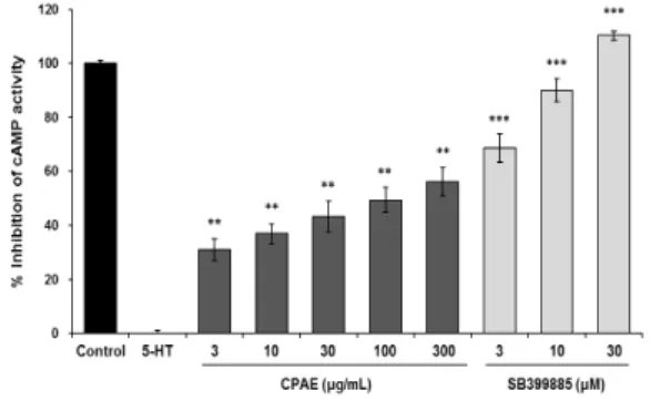
The 5-HT6 receptor belongs to the GPCR that when bound by its G-proteins ligand activates adenylyl cyclase to increase the production of cAMP9). Therefore, to evaluate the antidepressant effect of CPAE, we measured the effect of CPAE on 5-HT6 receptor activity via a cAMP immunoassay, using human astrocytoma 1321N1 cells stably expressing the 5-HT6 receptor. The results showed that 3, 10, 30, 100, and 300 μg/mL CPAE showed inhibitory effects on cAMP activity of 30.99 ± 4.00, 36.89 ± 3.66, 43.22 ± 5.75, 49.40 ± 4.74, and 56.00 ± 5.22%, respectively. Cell viability was determined by using MTT assay with various concentration of CPAE for 24h(Data not shown). There was a significant trend of CPAE inhibiting cAMP activity down to the lowest dose of CPAE, 3 μg/mL(P<0.01 or P<0.001), confirming antagonistic activity of CPAE on the 5-HT6
receptor(Fig. 1). These inhibitory effects were observed at doses of CPAE that did not affect cell survival (data not shown, P<0.05). The positive control group was administered SB-399885, a drug specifically known to be a 5-HT6
receptor antagonist, and in this group cAMP production was inhibited in a dose-dependent manner(P<0.001): 68.69 ± 5.22, 89.89 ± 4.30, and 110.27 ± 1.58% at doses of 3, 10, and 30 μM SB-399885, respectively. These findings suggest CPAE has 5-HT6 receptor antagonistic effect through the inhibition of cAMP activity.
The 5-HT6 receptor is distributed in the limbic system, a brain region involved in depression, and this receptor has a strong affinity for some antidepressants. Hence, in depression research this receptor is being actively studied as a major candidate gene23). Various 5-HT6 receptor antagonists have been developed so far24), and antidepressant- and antianxiety-like effects of several different selective 5-HT6 receptor antagonists have been reported in preclinical studies. Through a rodent study on a 5-HT6 receptor antagonist(SB-258585), Wesolowska et al.15) showed antidepressant-like effects of this drug in the FST and antianxiety-like effects in the conflict drinking test.
Other 5-HT6 receptor antagonists(SB-399885 and SB-271046) were also confirmed to have antidepressant-like activities in rats subjected to the FST13,14,16). These findings demonstrate that the 5-HT6 receptor is involved in depression and anxiety responses. Therefore, findings from the present study suggest that CPAE may be effective in treating depression via its antagonistic activity on the 5-HT6
receptor.
Fig. 1. The effect of CPAE on 5-HT6receptor–related cAMP activity in human astrocytoma 1321N1 cells stably expressing the human 5-HT6
receptor gene. Test extracts were co-treated with 100 μM 5-HT for 15 min. Intracellular cAMP levels were measured using a ParameterTM cAMP Assay according to the manufacture’s instructions. The data are expressed as the mean ± SEM. **P<0.01 and ***P<0.001, significantly different from the treated with 5-HT.
2. Antidepressant effect of CPAE on behavioral test performance in chronic stress model
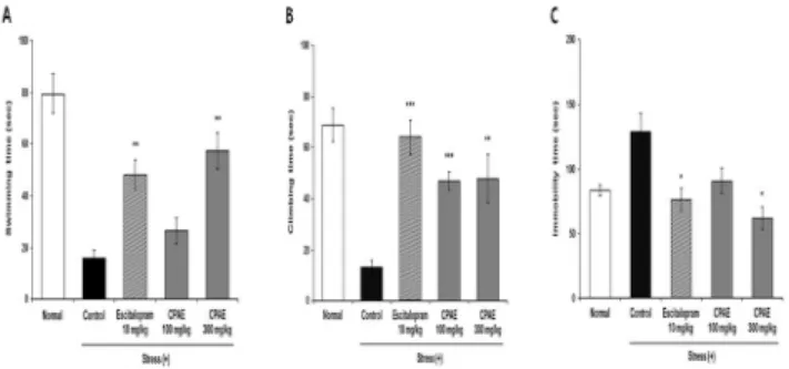
Fig. 2 shows the results of body weight in chronic restraint-stressed mice. There is no significant difference in the groups except for the control group. The reason for the decrease in body weight is will be caused by stress rather than toxicity. Fig. 3 shows the results in the FST in a mouse model of long-term restraint-induced stress. For the three types of behaviors, swimming, climbing, and immobility, durations in the control group was 16.0 ± 3.4, 13.4 ± 2.6, and 128.6 ± 14.49 s, respectively, and durations of first two behavioral types were shorter and the duration of immobility was longer than in the normal group(79.44 ± 7.60, 68.87 ± 6.65, and 83.63 ± 4.13 s, respectively). The duration of swimming in the group treated with 300 mg/kg CPAE was 57.4 ± 6.9 s, which was significantly higher compared with the control group(P<0.01; Fig. 3A). The duration of climbing was significantly higher in both the 100 mg/kg and the 300 mg/kg CPAE groups [47.2 ± 3.6 s(P<0.001) and 47.9 ± 9.5 s(P<0.01), respectively] compared with the control group(Fig. 3B). Regarding immobility, the duration in the group treated with 300 mg/kg CPAE was 61.6 ± 8.8 s, which was significantly lower than in the control group(P<0.05; Fig. 3C). The positive control group, which was administered escitalopram, showed significantly improved durations of swimming, climbing, and immobility versus the control group(P<0.01, P<0.001, and P<0.05, respectively).
FST is a model frequently used to evaluate the antidepressant activity of drugs. In the test, durations of swimming, climbing, and immobility are measured. Of the measured behaviors, immobility is regarded as behavioral despair, signaling that the animal has given up on attempting to get out of the water and, at the same time, is a behavioral manifestation of depression, and so it is used as an index to examine the antidepressant effects of a drug25,26). In the present study, the CPAE groups showed longer durations of swimming and climbing and a shorter duration of immobility in a chronic restraint-induced stress model, suggesting that CPAE may improve depression.
Fig. 2. The body weight of chronic restraint-stressed mice. Group I received vehicle(saline) and served as the control; Group II was subjected to chronic restraint-induced stress(CRS) and received vehicle (stress + saline); Group III was subjected to CRS and received escitalopram oxalate 10mg/kg/day(stress + escitalopram 10mg/kg); Group IV was subjected to CRS and received CPAE 100mg/kg/day(stress + CPAE 100mg/kg); Group V was subjected to CRS and received CPAE 300mg/kg/day(stress + CPAE 300mg/kg). Data are expressed as the mean ± SEM. *P<0.05, **P<0.01,
***P<0.001, significantly different from the control group (n=5).
Fig. 3. The effects of CPAE on the FST in chronic restraint-stressed mice(n=5). The mice were subjected to restraint-induced stress. The graphs show the duration(seconds) of the swimming(A), climbing(B) and immobility(C) in FST. Data are expressed as the mean ± SEM. *P<0.05,
**P<0.01, ***P<0.001, significantly different from the control group.
3. The effect of CPAE on CORT levels in chronic restraint-induced stress model
Changes in the level of a stress-related hormone, corticosterone(CORT), in the serum was measured in mice in which stress was induced by long-term restraint. CORT levels in the control group were 4326.10 ± 435.48 ng/mL, much higher than those of the normal group(1693.76 ± 602.64 ng/mL). In the group treated with 300 mg/kg CPAE, CORT levels were 3183.64 ± 220.17 ng/mL, a level significantly lower than in the control group(P<0.05). This effect was also observed in the positive control group administered with escitalopram compared with the control group(P<0.01; Fig. 4).
The restraint-induced stress model is commonly used in behavioral and biological research on depression, because it does not directly cause pain, hence the stress induced by the model is similar to mental stress in humans27). Repeated restraint-induced stress activates the hypothalamus-pituitary-adrenocortical(HPA) axis to facilitate the secretion of adrenocortical glucocorticoids. Normally, a negative feedback signal is sent to glucocorticoid receptors in the hippocampus and hypothalamus to return the activated HPA axis to normal, and the secretion of glucocorticoids thereby is inhibited25). In depression, however, glucocorticoids are excessively secreted because of impairment of this negative feedback, causing blood glucocorticoid levels to increase27). Several studies have reported that high blood levels of CORT, a type of
glucocorticoid, may induce depression-like behavior and impair hippocampal neurons28-30). In the present study, serum CORT levels significantly increased under restraint-induced stress, and this increase was significantly decreased because of daily oral administration of CPAE while restraint stress was delivered. These findings confirm a positive effect of CPAE on stress-induced depression.
Fig. 4. The effect of CPAE on corticosterone levels in the serum of chronic restraint-stressed mice(n=5). Data are expressed as the mean ± SEM. *P<0.05, **P<0.01, significantly different from the control group.
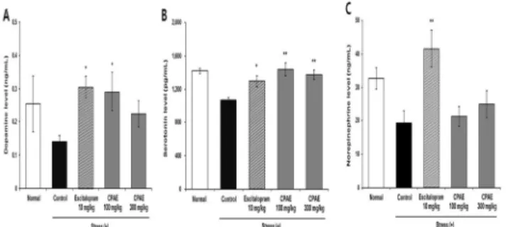
4. The effect of CPAE on changes in monoamine neurotransmitters in chronic restraint-induced stress model Brain tissues of mice subjected to the long-term restraint-induced stress model were examined to assess levels of neurotransmitters, specifically serotonin(5-HT), dopamine(DA), and norepinephrine(NE), in the hippocampus and prefrontal cortex. In the hippocampus, levels of DA, 5-HT, and NE were all lower in the stressed-control group(0.14±0.02 ng/mL, 1072.59±30.42 pg/mL, and 19.21±3.74 ng/mL, respectively) than in the normal group(0.25±0.08 ng/mL, 1417.51±32.09 pg/mL, and 32.66±3.13 ng/mL, respectively). Among the CPAE-treated groups was significantly higher in the group treated with 100 mg/kg CPAE(0.29±0.06 ng/mL) than in the stressed-control group(P<0.05; Fig. 5A), and 5-HT was significantly higher in the groups treated with 100 mg/kg and 300 mg/kg CPAE [1436.55 ± 78.43 pg/mL(P<0.01) and 1371.30 ± 57.17 pg/mL(P<0.01), respectively; Fig. 5B]. NE tended to be higher in CPAE-treated groups than in the stressed-control group, but the differences were not significant(Fig. 5C). All three neurotransmitters were significantly higher in the escitalopram group(P<0.05 for all three neurotransmitters).
In the prefrontal cortex, levels of DA, 5-HT, and NE were lower in the stressed-control group(113.88±6.63 ng/mL, 1417.23±45.01 pg/mL, and 27.07±2.68 ng/mL, respectively) than in the normal group(148.09±25.32 ng/mL, 1624.85±42.45 pg/mL, and 40.94±4.98 ng/mL, respectively).
In comparison with the stressed-control group, the group receiving 100 mg/kg CPAE showed significantly higher levels of DA(163.66±11.29 ng/mL; P<0.01) (Fig. 6A) and 5-HT(1675.73±53.58 pg/mL; P<0.05) (Fig. 6B). NE was somewhat higher in CPAE groups compared with the stressed-control group, but the differences were not significant(Fig. 6C). In contrast, levels of DA, 5-HT, and NE were significantly higher(P<0.01) in the escitalopram group.
Fig. 5. The effect of CPAE on monoamine levels in the hippocampus of chronic restraint-stressed mice(n=5). The graphs show the level of the dopamine(A), serotonin(B) and norepinephrine(C) in the hippocampus.
Data are expressed as the mean ± SEM. *P<0.05, **P<0.01, significantly different from the control group.
Fig. 6. The effect of CPAE on monoamine levels in the prefrontal cortex of chronic restraint-stressed mice (n=5). The graphs show the level of the dopamine(A), serotonin(B) and norepinephrine(C) in the prefrontal cortex. Data are expressed as the mean ± SEM. *P<0.05,
**P<0.01, ***P<0.001, significantly different from the control group.
Discussion
Monoamine neurotransmitters(5-HT, DA, and NE) are involved in depression and play an important role in modulating the effect of antidepressants. Several preclinical studies have demonstrated a clear association between chronic stress and depression via abnormal changes of neurotransmitters, their respective metabolites, turnover ratios and transporters31). Indeed, studies have found that monoamine neurotransmitters were markedly decreased in chronic stress models32,33). Decreased neurotransmitters can lead to depression, and many chemical compounds synthesized to increase neurotransmitters have been
empirically demonstrated to be effective as antidepressants23). Chronic stress factors increase the level of norepinephrine secretion in the brain. However norepinephrine resistance occurs when the same stress continues to be given in animals36). The present study has shown that in mice under chronic restraint-induced stress, the levels of dopamine and serotonin in the hippocampus and prefrontal cortex were reduced and this reduction was inhibited by administering CPAE. Also NE tended to be higher in CPAE-treated groups than in the stressed-control group, but the differences were not significant. Therefore, CPAE was confirmed to ameliorate abnormal changes in neurotransmitters due to chronic stress and to improve depression.
On the other hand, 5-HT6 receptor antagonists have been found to increase monoamine neurotransmitters in the extracellular space in the brain. In a study by Lacroix et al.34), a 5-HT6 receptor antagonist increased DA and NE in the prefrontal cortex of rats. Further, it was reported by Dawson et al.35) that another 5-HT6 receptor antagonist further increased DA and 5-HT in the prefrontal cortex and striatum of rats in which DA had already been increased by amphetamine. These findings suggest that 5-HT6 receptor antagonists function to modulate monoaminergic systems and, furthermore, may be involved in improving depression.
In the present study, we demonstrated the CPAE antagonistic activity against the human 5-HT6 receptor through intracellular cAMP signaling mechanism. In particular, 5-HT6 receptor is coupled to the Gαs protein, which results in the activation of intracellular cAMP signaling and this signaling activates the MAPK/ERK pathways. Further studies will be required to determine the antidepressant-like effects of these CPAE underlying mechanism remain unknown.
In conclusion, this study indicated that CPAE was observed to act as a 5-HT6 receptor antagonist and to increase three monoamines(5-HT, DA, and NE) in the hippocampus and prefrontal cortex, suggesting that the antagonistic activity of CPAE on the 5-HT6 receptor is involved in ameliorating depression via the modulation of neurotransmitters.
Conclusions
In conclusion, CPAE showed an antagonistic activity on the 5-HT6 receptor in the in vitro model. Also, it decreased the duration of immobility during the FST component of a chronic restraint-induced stress model and effectively
recovered levels of CORT and monoamine neurotransmitters which became abnormal because of restraint-induced stress. To conclude, CPAE improved stress-induced depression via its antagonistic activity on the 5-HT6 receptor, suggesting that CPAE can be developed into food and drug products to improve depression.
Acknowledgments
This research was supported by the Support Program for Creative Industry Institutes(Commercial Bio-technology Sophistication Platform Construction Program, R0003950) funded by the Ministry of Trade, Industry & Energy(MOTIE, Korea).
References
1. Nestler EJ, Barrot M, DiLeone RJ, Eisch AJ, Gold SJ, Monteggia LM. Neurobiology of depression. Neuron.
2002;34:13-25.
2. AKupfer DJ. Research in affective disorders comes of age. Am J Psychiatry. 1999;156:165-7.
3. Lanni C, Govoni S, Lucchelli A, Boselli C. Depression and antidepressants: molecular and cellular aspects. Cell Mol Life Sci. 2009;66:2985-3008.
4. Katz MM, Maas JW, Frazer A, Koslow SH, Bowden CL, Berman N, et al. Drug-induced actions on brain neurotransmitter systems and changes in the behaviors and emotions of depressed patients.
Neuropsychopharmacology. 1994;11:89-100.
5. Cleare AJ, Duncko R. Pharmacological management of depressive disorders. Medicine. 2016;44:756-60.
6. Möller HJ, Volz HP. Drug treatment of depression in the 1990s. Drugs. 1996;52:625-38.
7. Turner EH, Matthews AM, Linardatos E, Tell RA, Rosenthal R. Selective publication of antidepressant trials and its influence on apparent efficacy. N Engl J Med. 2008;358:252-60.
8. Sodhi M, SandersBush E. Serotonin and brain development. Int Rev Neurobiol. 2004;59:111-74.
9. Alcalde E, Mesquida N, López-Pérez S, Frigola J, Mercè
R. Indene-Based Scaffolds. 2. An Indole− Indene Switch:
Discovery of Novel Indenylsulfonamides as 5-HT6
Serotonin Receptor Agonists. J Med Chem. 2009;52:675-87.
10. Mitchell ES, Neumaie JF. 5-HT6 receptors: a novel target for cognitive enhancement. Pharmacol Ther.
2005;108:320-33.
11. Millan MJ. The neurobiology and control of anxious
states. Prog Neurobiol. 2003;70:83-244.
12. Svenningsson P, Tzavara ET, Qi H, Carruthers R, Witkin JM, Nomikos GG, et al. Biochemical and behavioral evidence for antidepressant-like effects of 5-HT6
receptor stimulation. J Neurosci. 2007;27:4201-9.
13. Wesołowska A, Nikiforuk A. Effects of the brain-penetrant and selective 5-HT6 receptor antagonist SB-399885 in animal models of anxiety and depression.
Neuropharmacology. 2007;52:1274-83.
14. Hirano K, Piers TM, Searle KL, Miller ND, Rutter AR, Chapman PF. Procognitive 5-HT6 antagonists in the rat forced swimming test: potential therapeutic utility in mood disorders associated with Alzheimer's disease. Life Sci. 2009;84:558-62.
15. Wesolowska A, Nikiforuk A, Stachowicz K. Anxiolytic-like and antidepressant-like effects produced by the selective 5-HT6 receptor antagonist SB-258585 after intrahippocampal administration to rats. Behav Pharmacol. 2007;18:439-46.
16. Wesołowska A, Nikiforuk A. The selective 5-HT6
receptor antagonist SB-399885 enhances anti-immobility action of antidepressants in rats. Eur J Pharmacol.
2008;582:88-93.
17. Kim SJ, Kang SM, Ko KH, Nam S. Physiological Activities and Inhibitory Effect of Extracts of Cynanchi wilfordii Radix and Perilla sikokiana against Cell Differentiation in 3T3-L1 Adipocytes. J Korean Soc Food Sci Nutr.
2016;45:642-50.
18. Ma YF, Jung JY, Jung YJ, Choi JH, Jeong WS, Song YS, et al. Anti-inflammatory activities of coumarins isolated from Angelica gigas Nakai on LPS-stimulated RAW 264.7 cells. Prev Nutr Food Sci. 2009;14:179-87.
19. Kim SJ, Jin SW, Lee GH, Kim YA, Jeong HG. Evaluation of Estrogenic Activity of Extract from the Herbal Mixture Cynanchum wilfordii Hemsley, Phlomis umbrosa Turczaninow, and Angelica gigas Nakai. Toxicol Res.
2017;33:71
20. Chang A, Kwak BY, Yi K, Kim JS. The Effect of Herbal Extract (EstroG‐100) on Pre‐, Peri‐and Post‐Menopausal Women: A Randomized Double‐blind, Placebo‐controlled Study. Phytother Res. 2012;26:510-6.
21. Kim SN, Li, YC, Xu HD, Yi DG, Kim MS, Lee SP, et al.
Phytoestrogenic effects of combined plant extracts on the change of bone metabolism of OVX rats. Korean J Food Sci Technol. 2008;40:316-20.
22. Lee KH, Lee DJ, Kim SM, Je SH, Kim EK, Han HS, et al.
Evaluation of effectiveness and safety of natural plants extract (Estromon®) on perimenopausal women for 1
year. J Korean Soc Menopause. 2005;11:16-26.
23. Bae D, Kim J, Oh DR, Kim Y, Choi EJ, Lee H, et al.
Multifunctional antistress effects of standardized aqueous extracts from Hippophae rhamnoides L. Anim Cells Syst.
2016;20:369-83.
24. Rosse G, Schaffhauser H. 5-HT6 receptor antagonists as potential therapeutics for cognitive impairment. Curr Top Med Chem. 2010;10:207-21.
25. Lee JW, Hong MC, Shin MK, Bae HS. Comparison of Nelumbinis semen extract with hypericum perforatum and fluoxetine in animal model of depression. Korean J Orient Physiol & Pathol. 2006;20:830-43.
26. Glavin GB, Paré WP, Sandbak T, Bakke HK, Murison R.
Restraint stress in biomedical research: an update.
Neurosci Biobehav Rev. 1994;18:223-49.
27. Gold PW, Drevets WC, Charney DS. New insights into the role of cortisol and the glucocorticoid receptor in severe depression. Biol Psychiatry. 2002;52:381-5.
28. Kalynchuk LE, Gregus A, Boudreau D, Perrot-Sinal TS.
Corticosterone increases depression-like behavior, with some effects on predator odor-induced defensive behavior, in male and female rats. Behav Neurosci.
2004;118:1365.
29. Johnson SA, Fournier NM, Kalynchuk LE. Effect of different doses of corticosterone on depression-like behavior and HPA axis responses to a novel stressor.
Behav Brain Res. 2006;168:280-8.
30. Murray F, Smith DW, Hutson PH. Chronic low dose corticosterone exposure decreased hippocampal cell proliferation, volume and induced anxiety and depression like behaviours in mice. Eur J Pharmacol.
2008;583:115-27.
31. O'mahony CM, Clarke G, Gibney S, Dinan TG, Cryan JF.
Strain differences in the neurochemical response to chronic restraint stress in the rat: relevance to depression. Pharmacol Biochem Behav. 2011;97:690-9.
32. Dang H, Chen Y, Liu X, Wang Q, Wang L, Jia W, et al.
Antidepressant effects of ginseng total saponins in the forced swimming test and chronic mild stress models of depression. Prog Neuro-Psychopharmacol Biol Psychiatry. 2009;33:1417-24.
33. Yu Y, Wang R, Chen C, Du X, Ruan, L, Sun, J, et al.
Antidepressant-like effect of trans-resveratrol in chronic stress model: behavioral and neurochemical evidences. J Psychiatr Res. 2013;47:315-22.
34. Lacroix LP, Dawson LA, Hagan JJ, Heidbrede, CA. 5‐HT6 receptor antagonist SB‐271046 enhances extracellular levels of monoamines in the rat medial prefrontal cortex. Synapse. 2004;51:158-64.
35. Dawson LA, Nguyen HQ, Li P. Potentiation of amphetamine-induced changes in dopamine and 5-HT by a 5-HT6 receptor antagonist. Brain Res Bul.
2003;59:513-21
36. Correlation between stress and biological indicators.
Epidemiology and health, 2003.
37. Bae D, Kim J, Oh DR, Kim Y, Choi EJ, Lee H, Jung MA, Lee SY, Jeong C, Lee M, Kang N, Lee J, Kim S.
Multifunctional antistress effects of standardized aqueous extracts from Hippophae rhamnoides L. Animal cells and systems. 2016;20:369-83.
38. Na JR, Oh DR, Han S, Kim YJ, Choi E, Bae D, Oh DH, Lee YH, Kim S, Jun W. Antistress Effects of Rosa rugosa Thunb. on Total Sleep Deprivation-Induced Anxiety-Like Behavior and Cognitive Dysfunction in Rat: Possible Mechanism of Action of 5-HT6 Receptor Antagonist. J Med Food. 2016;19:870-81.