수산화칼슘계 근관 충전제가 파골 세포의 분화 및 활성에 미치는 영향
장은영∙권 훈∙이장희*∙이창섭∙이상호 조선대학교 치과대학 소아치과학교실, 미생물학교실
본 실험은 수산화칼슘계 근관 충전제가 파골 세포의 분화 및 활성에 미치는 직접적인 억제 효과의 유무를 고찰하고자 chick embryo tibia의 골수로부터 추출한 파골 세포의 전구세포와 0.1, 0.01, 0.05㎍/ml로 희석된 네가지 실험 물질인 Ca(OH)2powder, Vitapex�, Metapaste�, pulpdent�를 사용하였다. 파골세포의 분화 및 활성에 미치는 억제 효과를 관찰 하고자 분화된 파골 세포의 수와 흡수와 면적이 측정되고 이들 약제의 효과가 세포독성에 의한 결과인지를 알아보고자 U2OS 골아세포에 대한 MTT assay를 시행하여 다음과 같은 결론을 얻었다.
1. 파골세포의 분화에 대한 억제 효과는 다음과 같은 순서로 분화된 파골세포의 수가 통계학적으로 유의한 증가를 보였다.:
Metapaste�, Ca(OH)2powder, Vitapex�. 하지만, 모든 농도에서 분화된 파골세포의 수가 증가한 pulpdent�군에서 는 통계학적으로 유의한 차이를 나타내지는 않았다.
2. 통계학적으로 분화된 파골세포 수의 감소가 유의한 결과를 나타낸 0.1㎍/ml로 희석된 세가지 실험 물질인 Ca(OH)2
powder, Vitapex�, Metapaste�가운데, Vitapex�군만이 대조군에 비해 유의한 세포독성을 나타내고 다른 두 집단은 유의한 결과를 보이지 않았다. 또한, 0.2% DMSO군은 통계학적으로 유의한 세포독성을 나타내었다.
3. 0.1㎍/ml로 희석된 세가지 실험 물질인 Ca(OH)2powder, Vitapex�, Metapaste�과 대조군에서 흡수와의 양상과 면 적을 관찰해 보면, Ca(OH)2powder군을 제외하고 대조군과 실험군사이에 유의한 차이를 나타내었고, 0.2% DMSO군 도 통계학적으로 유의한 감소를 보였다.
이상의 결과를 볼때, 수산화칼슘은 파골 세포 분화 및 활성의 직접적인 억제 작용에 기인한 경조직 흡수의 억제에 관여하는 것으로 사료된다.
주요어 : 수산화칼슘, 파골세포, 분화, 활성, 억제작용
Ⅰ. Introduction
Resorption is a condition associated with either a physiologic or a pathologic process resulting in a loss of dentin, cementum, or bone. Invariably, tooth resorption
results from injuries to or irrigation of the periodontal ligament and/or tooth pulp. It may arise as a sequela of traumatic luxation injuries, orthodontic tooth movement or chronic infections of the pulp or periondontal struc- ture1). It leads to the formation of multinucleated giant
A effect of calcium hydroxide endodontic materials on the differentiation and the activation of osteoclast
Eun-Young Jang, Hoon Kown, Chang-Hee Lee*, Chang-Seop Lee, Sang-Ho Lee
Department of Pediatric Dentistry, M icrobiology
*, College of Dentistry, Chosun University
국문초록
cells referred to as clasts2,3). The clastic cell is the key cell type responsible for all hard tissue resorptive processes. It is involved at elaborate interaction between inflammatory cells such as macrophages, monocytes and hard tissue structures4). Collectively, these cells orches- trate a complex interplay of molecular biologic events in- volving cytokines, enzymes, and hormones that influence the progression of resorption. Root resorption is mediat- ed by the odontoclast, a cell identical to the osteoclast in terms of its cytological features and its mineralized tis- sue resorpive function. Both cell types are identical as pleomorphic multinucleated cells, with many mitochon- dria and lysosomes, positive for tartrate-resistant acid phosphatase (TRAP) and calcitonin receptors, Because the two cell types appear to differ only in their resorp- tion substrates, one would assume that the differentia- tion process for the odontoclast would be the same as for the osteoclast and the process of tooth resorption be sim- ilar to that of bone resorption5).
In the many destructive hard tissue diseases, bacte- rias play an important role. Likewise, bacterias are inte- gral to the process of tooth resorption. The mechanism of bacteria-induced resorption is likely to be induction of osteolytic factors because of the effect of endotoxin, that is, lipopolysaccharides stimulate a number of molecular biologic events, including lysosomal enzyme release, col- lagenase release from macrophages and osteoblastic se- cretion of osteolytic factors IL-1, IL-6, M-CSF and PGE26). Together, these event result in the proliferation of osteoclast and enhanced bone resoption.
In previous study, Hammarstrom et al.7) had demon- strated that the bacterias in the pulp or dentinal tubules were the final targets for the resorbing cells because the resorption of the dentin did not spread laterally in the experimental cavites but seemed to follow the direction of the dentinal tubules toward the pulp. Therefore, the action mode of therapy for suppression of tooth resorp- tion was thought to relate to action on bacterial infec- tion. In clinical practice, calcium hydroxide is a well es- tablished root canal dressing material for treatment of progressive root resoption. Calcium hydroxide was first introduced as a pulp canal capping material and root canal sealer by Hellman at early nineteen century in the endodontic treatment area8).
Despite extensive research, the mechanism of action of calcium hydroxide is still not fully understood. But sev- eral theories9,10) have been postulated to explain its bio-
logical activity. One theory discusses its high alkaline pH, which is important in stimulating matrix formation by the formative cells11). Another theory postulates that a high pH neutralizes the acidic products of the resorp- tive cells, creating an unfavorable environment for them12). Furthermore, calcium hydroxide may promote healing because of its antibacterial properties. The high pH of calcium hydroxide, providing an unsuitable envi- ronment for the growth and activity of bacteria and re- sorbing cells appear to be the main reason for the thera- peutic effect of calcium hydroxide13). More recently, it has also been used in a number of specific endodontic treat- ment procedure, such as long-term pulpal dressing in teeth with large periapical lesions and temporary root canal filling to arrest root resorption12,14,15).
However, in spite of the widespread use of calcium hy- droxide in endodontic therapy, there seems to be no data concerning the experimental study on effect by interac- tion between this material and resorbing cell cultured in dircet contact with it.
The purpose of the present study is to investigate the direct inhibitory effect of some available calcium hydrox- ide materials on differentiation and activation of osteo- clast.
Ⅱ. Materials and Methods A. Experimental materials
Among bone marrow cells collected from tibia that was moved from 14 day-old chick embryo in 99.5℉, 80~82%
humidity, progenitor cells of osteoclast were isolated and incubated.
For this study, four commercially available calcium hydroxide endodontic materials, i.e. : Ca(OH)2 powder, Metapaste�(Meta Denta Co., LTD), Vitapex�(Neo Dental Chemical Products Co. Tokyo, Japan), Pulpdent� (PULPDENT Corp. U.S.A.) were used.
B. Experimental methods
1. Isolation of osteoclast progenitor cell
Tibia was dissected aseptically from 14 day-old chick embryo in 99.5℉, 80~82% humidity and placed into 60mm dish containing the HBSS media/antibiotics free of soft tissues. Therefore, cut across their epipyses and moved into 15ml cornical tube containing α-minimum es-
sential medium (α-MEM) and placed in -20℃ for 5min and later, put 60mm dish. In the state of holding of tibia by means of forcep, 1ml syringe needle fulfilled with 1ml medium inserted to bone and pushed the plunger. bone marrow cells flushed were collected in 50ml cornical tube and pipetted by 10ml pipet to increase single cell sus- pension. Clump was removed by let alone during 5~10min to sediment the cell clump or moving into 15ml cornical tube by pipet. After centrifugation, the cells suspended in αMEM containing 10% fetal bovine serum(FBS). These were plated at 2×10⁶total cells/well in 24-well plate. Cultures were fed at 4~6hr by replac- ing with fresh medium.
2. Differentiation into osteoclast
Isolated progenitor cells were plated in the medium at 500㎕/well having a similar number of cell. Four experi- mental groups (Ca(OH)2 powder, Metapast�, Vitapex�, Pulpdent�) were diluted at 0.1, 0.01, 0.05㎍/ml and all experimental materials and their respective controls were plated into three well. Also, Dimethyl sulfoxide, DMSO(SIGMA,CHEMICAL.CO) was diluted at 0.2%
concentration and used as a solvent of Vitapex�(Table 1).
Therefore, A total of 48 wells in series of four separa- tive trials were incubated during 36hr at 5% CO2
Incubator and therefore it was induced differentiation in- to osteoclast.
3. Measurement of the number of osteoclast
After incubation, each well was washed in a 37℃ pre- warm DDW, fixed with fixative consisted of citrate sol 25ml, acetone 65ml, 37% formaldehyde 8ml for 30sec.
Cytochemical staining for TRAP was conducted and mix- ture of it was consisted of the following: fast garnet GBC (wrap in hoil) 0.1ml, DDW 4.5ml, Naphthal AS-BI phosphate sol 0.05ml, acetate sol 0.2ml, tartrate sol 0.1ml(SIGMA DIAGNOSTICS. ST. Louis. Mo). Also, fast garnet GBC which was manufactured by mixing the fast garnet GBC base sol 0.5ml and sodium nitrate sol
0.5ml and was sensitive, thus covered by hoil. After TRAP staining, specimens were incubated for 1 hr at 37
℃ H2O bath and washed with distilled water. To exam- ine the osteoclast stained, GEL/MOUNT TM was dropped and put a cover glass. TRAP positive multinu- cleated cells were counted as osteoclast under the light microscope.
4. Cytotoxicity test
After measuring of number of osteoclast in all experi- mental materials and their respective controls, cytotoxic effect of calcium hydroxide materials was examined by MTT assay. U2OS osteoblastic cell was plated at 1×10⁴ cell/ well in 96-well plate In plate, 5columns were seed- ed: one was used for the control medium and three to evaluate the cytotoxicity of experimental groups, that is, Ca(OH)2powder, Metapaste�, Vitapex�at 0.1μg/ml dilu- tion and 0.2% DMSO was supplemented at remnant one. The 96-well dish was then placed into an incubator with an atmosphere of 5% CO2 for 24hr at 37℃. The medium was removed and immediately replaced with 20
㎕/well of a 0.5% solution of 3-(4,5-dimethylthiazol-2- yl)-2,5-dimethyltetrazolium bromide(MTT) prepared in the culture medium. After incubation for 4hr at 37℃, the supernatant was discarded and the intercellar for- mazan crystals were solubilized with 100㎕/well of 0.04 N HCl/isopropanol. The absorbance of 96-well plate was determined using an automatic microplate spectropho- tometer at 540nm.
5. Measurement of area of resorption lacunae Osteoclast progenitor cells isolated from bone marrow cell of chick embryo tibia were plated at artificial apatite crystal plate (OAASTM, OCT Inc. Dankook University School of Dentistry) in 36 well plate. In plate, 5columns were seeded :one was used for the control medium and three experimental groups, that is, Ca(OH)2 powder, Metapaste�, Vitapex� at 0.1㎍/ml dilution and 0.2%
DMSO was supplemented. After incubation for 96hr, 1M NH4OH was added for removal of osteoclast within re-
Table 1.Materials used in the present study
Materials Ca(OH)2Concentration(%) Manufacturer
Vitapex 30.3 Neo Dental Chemical Products Co. Tokyo, Japan
Metapaste �30 Meta Denta Co., LTD
Ca(OH)2powder 95 Meta Denta Co., LTD
Pulpdent - PULPDENT Corp. U.S.A
sorption lacunae. The resorbed area was measured using an image analysis system linked to the light microscope.
Ⅲ. Result
Isolated progenitor cells were plated in the medium at 500㎕/well having a similar number of cell. Four experi- mental groups [Ca(OH)2powder, Metapaste�, Vitapex�, Pulpdent�] were diluted at 0.1, 0.01, 0.05㎍/ml and each was plated into three well. Dimethyl sulfoxide, (DMSO) (SIGMA, CHEMICAL CO.)was diluted at 0.2%
concentration and used as a solvent of Vitapex�. All ex- perimental materials and their respective controls were incubated during 36hr at 5% CO2 Incubator and then measured the number of differentiated osteoclast.
In three experimental groups, that is, Ca(OH)2 pow- der, Metapaste�, Vitapex�at 0.1㎍/ml dilution that were statistically significant in reduction of the number of dif- ferentiated osteoclast and 0.2% DMSO, MTT assay on U2OS osteoblast were conducted to examine cytotoxic ef- fect. Also, pit forming assay was done to prove the effect of calcium hydroxide materials on resorptive activity of osetoclast. Result were as follow.
A. Measurement of the number of osteoclast
The results of the inhibitory effect upon osteoclast dif- ferentiation in all experimental groups and their respec- tive controls were shown in Table 2 and Fig. 1. In Vitapex group, it displayed significantly less number at all concentration : These presented a decrease rate at 0.01㎍/ml(20.4%), 0.05㎍/ml(55.8%), 0.1㎍/ml(74.3%).
Also, there were statistically significant difference among diluted concentration (p<0.05).
Table 2.Number of osteoclast in the solution of four experimental groups
Vitapex Metapaste Ca(OH)2powder Pulpdent
control 112.5± 2.12 343 ±96.17 275±35.36 83.67± 5.51
0.01㎍/ml 90 ± 1.41* 309.5±10.6 220±39.60 100 ±12.49
0.05㎍/ml 50 ±11.31* 365 ± 1.41 185± 5.66* 100.3 ± 6.66
0.10㎍/ml 28.5± 7.78* 91 ±36.77* 97±21.21* 106.3 ± 9.45
Fig. 1. Relative decrease percentage of the number of osteoclast in the solution of four experimental materials compared to control.
Fig. 3.Area of resorption lacunae on solution of four exper- imental groups.
Fig. 2.Absorbance of U2OS osteoblast cell on solution of four experimental groups.
There were shown a significant difference in Metapaste group at 0.1㎍/ml dilution (73.4%) and Ca(OH)2 powder group at 0.05㎍/ml(32.8%), 0.1㎍/ml (64.7%) dilution and no significant difference was found among different concentrations except 0.1μg/ml concen- tration(p<0.05).
In Pulpdent group, they were exhibited that the num- ber of differentiated osteoclast was increased at all con- centration but no significant difference was found(Fig.
4~7).
B. Cytotoxicity test
In three experimental groups, that is, Ca(OH)2
Powder, Metapaste�, Vitapex�at 0.1㎍/ml dilution that were statistically significant in reduction of the differen- tiated osteoclast, Vitapex�group showed significant dif- ference compared to control and another two groups ex- hibited no significant difference(p<0.05).
0.2% DMSO group was shown statistically significant cytotoxicity(Fig. 2).
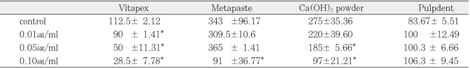
C. Measurement of area of resorption lacunae
Pattern and measured area of resorption lacunae in the control and the three experimental groups, that is, Ca(OH)2powder, Metapaste�, Vitapex�at 0.1μg/ml dilu- tion were presented(Fig. 3).
Except Ca(OH)2powder group, statistically significant differences were found between experimental groups and control group.: These presented a decreased rate at Vitapex�(79.1%), Metapaste�(73.8%) (p<0.05). Also, DMSO group showed statistically significant decrease (68.9%)(Fig. 8~12).
Ⅳ. Discussion
Osteoclasts are highly polarized cells that form ruffled borders and clear zones toward the bone surface and de- rived from hemopoietic cells of the monocyte-macrophage lineage16). These are subject to extensive regulatory mechanism that are facilliated to a large degree by os- teoblasts mediating the effects of osteotropic hormones and local mediator on these cells. Osteoclastic formation, regulation, activation are dependent on several local fac- tors including M-CSF, IL-1, IL-6, IL-11, TNF-αand systemic regulatory factors such as PTH, 1,25(OH)2D3,
calcitonin17-20).
The resorptive process itself can be described as being bimodal, involving the degradation of the inorganic crys- tal structure of hydroxyapatite and the organic structure of collagen. The unique structural arrangement of the osteoclasts to hard tissues allows the cell to establish a microenvironment between the ruffled border and the bone in which resorption takes place. Therefore, osteo- clastic bone resorption consists of several complicated processes : osteoclast development, attachment of osteo- clast to calcified tissues, development of a ruffled border and clear zone, followed by the secretion of acids and lysosomal enzymes into the space beneath the ruffled border21).
Considering osteoclast function to be studied biochemi- cally during the past decades, several different thera- peutic approaches have been taken to reduction of osteo- clastic resorption. A previous report demonstrated that intrapulpal application of an antibiotic/corticosteriod combination effectively eliminates experimentally in- duced external inflammatory resorpion in monkey teeth through directly inhibited the spreading of odontoclast, suggesting that this medicament acts the detaching re- sorbing cells from the root surface22-24).
Another alternatives, bisphosphonates are widely used as anti-bone-resorbing agents in various disease associ- ated with stimulated bone resorption25). The mechanism by which bisphosphonates inhibit bone resorption has not been established but proposed that it is achieved by inhibition of differentiation or recruitment of osteo- clast26,27), direct or indirect stimulation of osteoclast apoptosis28), suppression of the resorption activitiy of mature osteoclast29), morphologic change of osteoclast.
Pierce et al30). demonstrated the potent effects on in- flammatory resorption of intra-canal insertion of calci- tonin into reimplanted monkey teeth. Residual inflam- mation found within the periodontal membrane was re- duced and this reduction was notably greater than that achieved in earlier studies using alternative therapies such as antibiotics alone or calcium hydroxide. The mode of action of calcitonin in deactivation of resorbing cell is thought to relate both specific and direct inhibition of root-resorbing cell and suppression of Inflammation31). According to SUDA et al.6), calcitonin inhibited osteoclast function by calcitonin-induced morphological changes of the cytoskeleton of osteoclast and by disrupted actin ring and inhibition of pit formation .
Nevertheless, with regard to the management of den- tal resorption, intracanal application of calcium hydrox- ide is a popular treatment. Its high pH has been shown to be effective in destroying bacteria in dentinal tubules and thus reducing the inflammatory stimulus of resorp- tion and also ascribed the superfical necrosis of adjacent tissues1,32). Hammarstrom et al.7)found that calcium hy- droxide applied intrapulpally caused limited necrosis of both dentin-resorbing cells and cells forming reparative cementum in an experimental groove in the root surface.
Thus, a temporary necrotizing effect on reparative cells could encourage increased areas of ankylosis at the ex- pense of the re-establishment of new periodontal attach- ment33). Likelywise, ankylosis was attributed to the po- tential toxicitiy of the medicament in high concentration on reparative cells34). Early investigations showed that calcium hydroxide was highly toxic for HeLa cell or hu- man fibroblast35-37). In present study, MTT assay on os- teoblast was conducted to examine whether or not effect of calcium hydroxide on inhibition of differentiation of os- teoclast is due to cytotoxicity. No statistically significant difference was found between control group and experi- mental substrate. But in the Vitapex group, it was sta- tistically significant difference that might be resulted from DMSO toxicity.
Zmener and Cabrini38,39) studied the adverse effects of three calcium hydroxide-based materials on the behavior of a mixed cell population of human blood monocytes and lympocytes and demonstrated that the adverse ef- fects of the test materials would lead to cell detachment from the substratum or to produce alterations in cell morphology. According to their results, it seems that the degrees of inhibition of cell attachment correlated with one of the most undesirable properties of endodontic ma- terial, i.e toxicity. In the present study, inhibitory effect of commercially available calcium hyroxide endodontic materials on differentiation of osteoclast increased in the following order : Metapaste , Ca(OH)2powder, Vitapex . These results may be attributed that difference of con- centration of Ca(OH)2 and other components from each material adversly affect differentiation of cells cultured into osteoclast. Although Ca(OH)2Powder is very higher concentration than that of others, it was shown the low- er effect than that of Vitapex because of it’s relative in- solubility. As another possibility, effect of Dimethyl sul- foxide, DMSO was used as a solvent of vitapex might be considered.
Previously, some researcheres40-42)have been described the inhibition of cell functions and the suppression of their vital activity when they are cultured in contact with calcium hydroxide or when this material is placed in direct contact with pulpal or periapical tissue.
In present stuty, to examine effect of calcium hydrox- ide materials on activity of osteoclast, pit forming assay using artificial apatite crystal plate was conducted.
Except Ca(OH)2powder group, calcium hydroxide mate- rials inhibited the formation of resoption lacunae com- pared to control group. These result may be due to lack of complete solution of Ca(OH)2 powder group.
Therefore, there are possible differences in the ability of calcium and hydroxyl ions to egress from Ca(OH)2pow- der material, producing different levels of alkalinity in the culture medium. Through this report, it can be pro- posed that calcium hydroxide is responsible for suppres- sion of hard tissue resorption by a direct inhibition of differentiation and activation of osteoclast. However, on the basis of the molecular mechanism of the regulation of osteoclastic bone resorption, further studies will be es- tablished to elucidate the action of calcium hydroxide on formation, regulation, activation of osteoclast.
Ⅴ. Conclusion
Using the osteoclast progenitor cells isolated from bone marrow cell of chick embryo tibia and four experimental materials(Ca(OH)2 powder, Metapaste�, Vitapex�, Pulpdent�) diluted at 0.1, 0.01, 0.05㎍/ml, it was evalu- ated the direct effect of all experimental materials and their respective controls on differentiation and resorptive activity of osetoclast. Also, MTT assay on U2OS os- teoblast was conducted to examine cytotoxic effect.
Results were as follow.
1. Considering the result of the inhibitory effects upon osteoclast differentiation, there were shown a signif- icantly different increase in the following order:
Metapaste�, Ca(OH)2 powder, Vitapex�. But, no significant difference was found in Pulpdent group that the number of differentiated osteoclast was in- creased at all concentration(p<0.05).
2. Among three experimental groups, that is, Ca(OH)2
powder, Metapaste�, Vitapex� at 0.1㎍/ml dilution that were statistically significant in reduction of the number of differentiated osteoclast, Vitapex group
showed significant difference compared to control and another two groups exhibited no significant dif- ference. Also, 0.2% DMSO group was shown statis- tically siginificant cytotoxicity (p<0.05).
3. Examining pattern and measured area of resorption lacunae in the control and the three experimental groups, that is, Ca(OH)2 powder, Metapaste�, Vitapex� at 0.1㎍/ml dilution, statistically signifi- cant inhibitory effect were found between experi- mental groups and control group except Ca(OH)2
powder group. Also, DMSO group showed statisti- cally significant decrease(p<0.05).
Through this report, it can be proposed that calcium hydroxide is responsible for suppression of hard tissue resorption by a direct inhibition of differentiation and ac- tivation of osteoclast. However, on the basis of the mole- cular mechanism of the regulation of osteoclastic bone resorpion, further studies will be established to elucidate the action of calcium hydroxide on formation, regulation, activation of osteoclast.
References
1. Ne RF, Witherspoon DE, Gutmann JL : Tooth re- sorption. Quintessence international 30:9-25, 1999.
2. Anan H, Akamine A, Maeda K : An enzyme histo- chemical study of the behavior of rat bone cells dur- ing experimental apical periodontitis. J Endod 19:83-86, 1993.
3. Boyde A, Ali NN, Jones SJ : Resorption of dentine by isolated osteoclasts in vitro. Br Dent J 156:216- 220, 1984.
4. Akamine A, Anan H, Hamachi T, et al. : A histo- chemical study of the behavior of macrophage during experimental apical periodontitis. J Endod 20(10):474-478, 1994.
5. Wu SM, Richards DW, Rowe DJ : Production of ma- trix-degrading enzymes & Inhibition of osteoclast- like cell differentiation by fibroblast-like cells from the periodontal ligament of human primary teeth. J Dent Res 78(2):681-689, 1999.
6. Suda T, Nakamura I, Jimi E : Regulation of osteo- clast function. Journal of Bone and Mineral Research 12(6):869-879, 1997.
7. Hammarstrom LE, Blomlof LB, Feiglin B, et al. : Effect of calcium hydroxide treatment on periodontal
repair and root resorption. Endod Dent Traumatol 2:184-189, 1986.
8. Rheu E, Jeon SM : PH changes at the surface of root dentin when using root canal sealers containing calcium hydroxide. J of Korean Academy of Conservative Dentistry 23(2):710-717, 1998.
9. Calts S, Serper A, Ozcelik B : PH changes & Ca2+
ion diffusion from calcium hydroxide dressing mate- rials through root dentin. J Endod 25(5):329-331, 1999.
10. Foreman PC, Barnes IE : A review of calcium hy- droxide. Int Endod J 23:283-297, 1990.
11. Yousef saad A : Calcium hydroxide in the treatment of external root resorption. JADA 118:579-581, 1989.
12. Stock CJR : Calcium hydroxide ; Root resorption and perio-endo lesions. Br Dent J 158:325-334, 1985.
13. Staehle HJ, Pioch T, Hoppe W : The alkalizing properties of calcium hydroxide compound. Endod Dent Traumatol 5:147-152, 1989,
14. Caliskan MK, Sen BH : Endodontic treatment of teeth with apical periodontitis using calcium hydrox- ide : A long-term study. Endod Dent Traumatol 12:215-221, 1996.
15. Holland R, Valle GF, Taintor JF, et al. : Influence of bony resorption on endodontic treatment. Oral Surg 2:191-203, 1983.
16. Wedenberg C, Sumita S : Evidence for an inhibitor of osteoclast attachment. Endod Dent Traumatol 6:255-259, 1990.
17. Kim TS, Yu YJ, Gwak WA, et al. : Formation of os- teoclasts by cytokine released from periodontal liga- ment cells in response to interleukin -1β. J of Korean Academy of Pediatric Dentistry 23(1):189- 205, 1996.
18. Martin T, Udagawa N : Hormonal regulation of os- teclast function. Trends Endocrinol Metab 9:6-12, 1998.
19. Suda T, Takahashi N, Udagawa N, et al. : Modulation of osteclast differentiation and function by the new members of the tumor necrosis factor re- ceptor and ligand families. Endocrine Reviews 20(3):345-357, 1999.
20. Suda T, Jimi E, Nakamura I, et al. : Role of 1α,25- dihyroxyvitamin D3 in osteoclast differentiation and function. Methods Enzymol 282:223-235, 1997.
21. Gillies JA, Carnes DL, Stewart Windeler A : Development of an in vitro culture system for the study of osteoclast activity and function. J Endod 20(7):327-331, 1994.
22. Hammarstrom LE, Blomlof LB, Feiglin B, et al. : Replantation of teeth and antibiotics treatment.
Endod Dent Traumatol 2:51-57, 1986.
23. Pierce A, Lindskog S : The effect of an antibiotic/
corticosteriod paste on inflammatory root resorption in vivo. Oral Surg Oral Med Oral Pathol 64:216- 220, 1987.
24. Pierce A, Heithersay G, Lindskog S : Evidence for direct inhibition of dentinoclasts by a corticosteriod/
antibiotics endodontic paste. Endod Dent Traumatol 4:44-45, 1988.
25. Lee IS, Kim HJ, Ryoo HM, et al. : Effect of bisphos- phonate on osteoclast differentiation. J of Korean Academy of Pediatric Dentistry 27(2):309-317, 2000.
26. Cocchini MG, Fleisch H : Bisphosphonate in vitro specifically inhibit among the hem atopoietic series, the development of the mouse mononuclear phago- cyte lineage. J Bone Miner Res 5:1019-1027, 1990.
27. Hughes DW, MacDonald BR, Russel RGG, et al. : Inhibition of osteoclast-like cell formation by bis- phosphonate in long-term cultures of human bone marrow. J Clin Invest 83:1930-1935, 1989.
28. Hughes DE, Wright KR, Sasake A, et al. : Bisphosphonates promote apoptosis in murine osteo- clasts in vitro and in vivo. J Bone Miner Res 10:1478-1487, 1995.
29. Sato M, Grasser W : Effect of bisphosphonate on isolated rat osteoclasts as examined by reflected light microscopy. J Bone Miner Res 5:31-40, 1990.
30. Pierce A, Berg GO, Lindskog S : Calcitonin as an al- ternative therapy in the treatment of root resorp- tion. J Endod 14:459-464, 1988.
31. Wiebkin OW, Cardari SC, Heithersay GS : Therapeutic delivery of calcitonin to inhibit external inflammatory root resorption. Endod Dent Traumatol 12:265-271, 1996.
32. Tronstad L : Root resorption- etiology, terminology and clinical manifestations. Endod Dent Traumatol 4:241-252, 1988.
33. Cotton WR : Bacterial contamination as a factor in healing of pulp exposure. Oral Surg 38:441-450, 1974.
34. Pierce AM : Experimental basis for the management of dental resorption. Endod Dent Traumatol 5:255- 265, 1989.
35. Im MK, Lee CS : A study on the cytotoxicity of root canal sealers to several cell lines, J of Korean Academy of Conservative Dentistry 17(2)263-282, 1992.
36. Spangberg L : Biological effects of root canal filling materials toxic effect in vitro of root canal filling ma- terials on HeLa cells and human skin fibroblasts.
Odontol Revy 20:1-10, 1969.
37. Torneck CD, Moe H, Howley TP : The effect of calci- um hydroxide on porcine pulp fibroblast in vitro. J Endod 9(4):131-136, 1983.
38. Zmener O, Cabrini RL : Effects of three calcium hy- droxide-based material on human blood monocytes and lympocytes. Endod Dent Traumatol 3:28-32, 1987.
39. Zmener O, Cabrini RL : Adhesion of human blood monocytes and lympocytes to different endodontic cements. A methodological in vitro study. J Endod 2(4):150-155, 1986.
40. Holland R, Mello WD, Nery MJ, et al. : Reaction of human periapical tissue to pulp extirpation and im- mediate root canal filling with calcium hydroxide. J Endod 3:63-67, 1977.
41. Schroeder V, Granath L : Early reaction of intact human teeth to calcium hyroxide following experi- mental pulpotomy and its significance to the devel- opment of a hard tissue barrier. Odontol Revy 22:379-396, 1971.
42. Lengheden A, Blomlof L, Lindskog S : Effect of de- layed calcium hydroxide treatment on periodontal healing in contaminated replanted teeth. Scand J Dent Res 99:147-153, 1991.
Explanation of figures
Fig. 14. Light micrograph of differentiated osteoclast in respective control and at 0.1㎍/ml dilution of vitapex group(×
40)
Fig. 15. Light micrograph of differentiated osteoclast in respective control at 0.1㎍/ml dilution of Metapaste group(×
40)
Fig. 16. Light micrograph of differentiated osteoclast in respective control at 0.1㎍/ml dilution of Ca(OH)2group(×40) Fig. 17. Light micrograph of differentiatedosteoclast in respective control at 0.1㎍/ml dilution of Pulpdent group(×40) Fig. 18. Light micrograph of resorption lacunae in Control group(×40)
Fig. 19. Light micrograph of resorption lacunae in vitapex group(×40) Fig. 10. Light micrograph of resorption lacunae in Metapaste group(×40) Fig. 11. Light micrograph of resorption lacunae in Ca(OH)2group(×40) Fig. 12. Light micrograph of resorption lacunae in DMSO group(×40)
Fig. 4
Fig. 5
Fig. 6
Fig. 7
Explanation of figures
Fig. 8 Fig. 9
Fig. 10 Fig. 11
Fig. 12
Explanation of figures
Abstract
A EFFECT OF CALCIUM HYDROXIDE ENDODONTIC MATERIALS ON THE DIFFERENTIATION AND THE ACTIVATION OF OSTEOCLAST
Eun-Young Jang, Hoon Kwon, Chang-Hee Lee*, Chang-Seop Lee, Sang-Ho Lee
Department of Pediatric Dentistry, Microbiology*, College of Dentistry, Chosun University
The purpose of this study was to investigate the direct inhibitory effect of calcium hydroxide materials on dif- ferentiation and activation of osteoclast. we used the osteoclast progenitor cells isolated from bone marrow cell of chick embryo tibia and four experimental materials [Ca(OH)2 powder, Metapaste�, Vitapex�, Pulpdent�] di- luted at 0.1, 0.01, 0.05㎍/ml. There were measured both the number of differentiated osteoclast and the area of resorption lacunae. Also, we conducted MTT assay on U2OS osteoblast to examine of cytotoxic effect and ob- tained following result.
1. Considering the result of the inhibitory effects upon osteoclast differentiation, There were shown a signifi- cant difference increased in the following order: Metapaste�, Ca(OH)2powder, Vitapex�. But no significant difference was found in pulpdent group that the number of differentiated osteoclast was increased at all concentrations(p<0.05).
2. Among the three experimental groups, that is, Ca(OH)2powder, Metapaste�, Vitapex�at 0.1㎍/ml dilution that were statistically significant in reduction of the number of differentiated osteoclast. Vitapex group showed significant cytotoxic effect compared to control and another two groups exhibited no significant dif- ference. Also, 0.2% DMSO group was shown statistically siginificant cytotoxicity (p<0.05).
3. Examining pattern and measured area of resorption lacunae in the control and the three experimental groups ,that is, Ca(OH)2 powder, Metapaste�, Vitapex� at 0.1㎍/ml dilution, except Ca(OH)2 powder group, statistically significant differences were found between experimental groups and control group. Also, DMSO group showed statistically significant decrease (p<0.05).
From these results, we think that calcium hydroxide is responsible for suppression of hard tissue resorption by a direct inhibition of dfferentiation and activation of osteoclast.
Key words : Calcium hydroxide, Osteoclast, Differentiation, Activation, Inhibitory effect.