약학회 지 제 48 권 제 3 호 197~201 (2004)
Yakhak Hoeji Vol. 48, No. 3 藥 學 舍 확
Phosphodiesterase 저해제 Pentoxifylline 이 피골세포 분화에 미치는 영향
김 민 혜•전 윤 나• 임미정# 숙명여자대학교 약학대학
(Received May 12,2004; Revised June 11,2004)
Effect of Pentoxifylline, a Phosphodiesterase Inhibitor, on Osteoclast Formation
Minhye Kim, Yunna Chun and M ijung Yim#
College of Pharmacy, Sookmyung Womens University, Seoul 140-742, Korea
Abstract — Phosphodiesterases (PDEs) are enzymes that degrade intracellular cAMR In the present study, pentoxifylline, a PDE inhibitor, induced osteoclast formation in co-cultures of mouse bone marrow cells and calvarial osteoblasts. To address the involvement of the osteoclast differentiation factor TNF-related activation-induced cytokine (TRANCE, identical to RANKL, ODF; and OPGL), mouse bone marrow cells and calvarial osteoblasts were co-cultured with pentoxifylline in the presence of OPG, a decoy receptor for TRANCE. The osteoclastogenic effect of pentoxifylline was completely blocked by addition of OPG, suggesting that TRANCE is involved in the osteoclast formation induced by pentoxifylline. Northern blot analysis revealed that pentoxifylline significantly induced TRANCE mRNA expression in calvarial osteoblasts. These results suggests that pentoxifylline regulates TRANCE expression in osteoblasts, which in turn controls osteoclast for
mation.
Keywords □ osteoblast, osteoclast, phosphodiesterase, TRANCE, pentoxifylline
인체에서뼈는신체지지의 골격을제공하고, 각종면역세포의 발달과성숙의 장소가되며, 칼슘과같은무기질의 저장소역할 을 하는 중추적 기관이다. 건강한 뼈는 일생동안 조골세포 (osteoblast)와파골세포(osteoclast)의작용에 의해뼈가지속적 으로형성되고파괴되는뼈의 재형성 과정을거치게 된다.
파골세포는조혈모세포(hematopoietic cell)에서분화한것으 로, 파골세포의 분화는뼈형성을담당하는조골세포에 의해 엄 격하게 조절되고 있다^ 2> 조골세포는 1,25-dihydroxyvitamin D3[l,25(ᄋH)2D3],부갑상선 호르몬(PTH),Prostaglandin E2 (PGE2) 등의자극에의해파골세포분화인자인 TRANCE(TNF- related activation-induced cytokine, OPGL, 이) ^ or RANKL) 를 생성하는 한편, decoy receptor인 OPG(osteoprotogenin)를
분비하여 TRANCE를억제함으로써 파골세포의 분화를 조절
한 다 . ^
이러한조골세포■파골세포상호작용에 있어서, cAMP는중요 한내부신호전달자•로작용한다. PTH나 PGE2 등의 자극에 의
#본 논문에 관한 문의는 저자에게로 (전화) 02-710-9572 (팩스) 02-?15-9498 (E-mail) [email protected]
해 세포내 cAMP의 농도가 상승하면, protein kinase A(PKA)가 활성화되고 이를 통해 TRANCE의 발현이 증가된다고 알려져 있다.7^
세포내 cAMP의농도는 2가지효소에 의해 항상일정하게유 지된다. adenylyl cyclase는 ATP를 기질로 cAMP를 합성하고,
phosphodiesterase(PDE)는 cAMP를 5’-AMP로분해시킨다.10’u) 지금까지 PDE family는 PDE1 에서 11까지의 isozyme이존재하 는것으로밝혀졌으며, 각각의 isozyme은 PDE1A, IB , 1C와같 은 subtype을가진다.u)
Pentoxifylline은 폭 넓게 사용되는 PDE 저해제로,cAMP의 분해를 억제함으로써 세포내 CAMP 농도를 상승시킨다. 마우스 를 사용한 in vivo 실험에서 pentoxifylline 및 타 PDE 억제제의 투여는 골밀도를 증가시킨다고 보고된 바 있고,12-14) 이로 인해 PDE 저해제를 골다공증 치료제로 개발히•려는 다양한 시도가 이 루어지고 있다. 그러나 조골세포 및 파골세포에 대한 pentoxifylline의 효과와 이에 대한 기전 연구는 중분히 수행되지 않은 실정이다. 따라서 본 연구에서는 pentoxifylline의 효과를 초 기 배양한 마우스 조골세포와 골수세포의 공배양계 (co-culture) 를 이용하여 검토하였다. 본 연구 결과 pentoxifylline은 공배양 계에서 파골세포의 분화를 유도하였다. 이러한 pentoxifylline의
198 김민혜 . 전윤나 • 임미정
파골세포유도반응은 OPG의첨가로억제되어 TRANCE 분자 의관여를 시사하였다. 실제로 pentoxifylline은 TRANCE의발 현을 전사수준에서 조절하고있음이 Northern blot 방법에 의 해명확하게 밝혀졌다.
실험방법
口I무스 조골세포의 초기 배양
생후 0~1일의신생아 ddy mouse로부터 두개골 피부를벗긴 후두개골을 적출하였다. 이때후두골근육부착부분은 제거하 였다. 부착된 근육, 혈구 둥을 제거한 후 minimum essential medium, alpha modification(oc-MEM)으로 가볍게 세척하였다. 0.1% collagenase + 0.2% dispase 효소용액에넣어 37°C에서 5 분간진탕시킨후상등액을버리고 새로운효소용액을가하였 다. 37°C에서 약 10분간진탕하여 상등액을모으는조작을 4회 반복하였다. 원심분리한후 10% FBS 함유 ot-MEM으로약 3~
5X 10⁴ 세포/100 mm plastic dish가되도록접종하였다. 5% C02, 37°C에서 3~4일간배양한세포를이후조골세포로실험에사용 하였다.
口[우스 골수세포의 배양
ddy mouse(6~9주, 수컷)를경추탈골한후 70% 에탄올로소 독하였다. 경골부분의 피부를 절개하여부착근육을떼어냈다. 경골원심부를절단하고슬개골을탈골시켜 경골을적출하였다. 뼈양끝을조금잘라한쪽끝에 25G의주시비늘을꽂고 a-MEM 을흘려보내 골수세포를시험관에 모았다. 원심 분리한후 0C- MEM에현탁하고 2배의 Gey's solution을가해 적혈구를 제거 했다. 원심 분리한후 10% FBS가함유된 ct-MEM으로 재현탁 했다.
공배양(co-culture)법을 이용한 파골세포의 분화유도
96 well plate에초기 배양한조골세포 5X 10³ cells/well, 골수 세포 1 X 10⁵ cells/well을 일정 농도의 pentoxifylline(Sigma- Aldrich) 존재하에서 10% FBS가함유된 ct-MEM으로공배양했 다. 배지는 3일에 한번씩 교환했다. 배양이 끝난세포는 10%
formalin으로 10분간고정한 후 ethanol-acetone(l: 1)로 1분간 재고정하여 TRAP(tartrate-resistant acid phosphatase) staining 을 했다. 3개 이상의 핵을 가진 TRAP + 세포를 다핵 파골세포 로판정했다.
RT-PCR 분석
Total RNA 1 n g i: Superscript II(Invitrogen, CA, USA)로역 전사하였다. 얻어진 cDNA의일부를 Go Taq DNA polymerase (Promega)로 PCR 증폭하였다. PCR 증폭에사용한 primer의서
열은다음과 같다. calcitonin
receptor(CTR): 5'-tttcaagaaccttagctgccagag-3' (forward) 5'-caaggcacggacaatgttgagaag-3' (reverse) cathepsin K: 5'-cttccaatacgtgcagcaga-3' (forward)
5'-acgcaccaatatcttgcacc-3' (reverse) GAPDH: 5'-gaaggtcggtgtgaacggatttggc-3' (forward)
5'-catgtaggccatgaggtccaccac-3' (reverse) 반응조건은 940C에서 3분초기반응후, 940C에서 30초, 58°C 에서 45초, 그리고 720C에서 1분간격으로 총 32주기(CTR과 cathepsin K) 또는 28주기 (GAPDH)로이루어졌다. 중폭된 PCR 산물들은 1% agarose gel에서 전기영동하여 분리하였다.
Northern blot 분■스i
초기배양마우스조골세포를 600|iM pentoxifylline0-?- 처리 하여 3시간 배양한후, total RNA를 Trizol reagent(Invitrogen) 로희수하였다. Total RNA 20 (ig을■ 1.2% agarose-formaldehyde gel로 전기영동한 후 nylon membrane filter에 이동시켰다 (Hybond N+, Amersham Biosciences), membrane을 32P로표 지한 cDNA probe와반응시킨후세척액으로세척하고,-70oC에 서 x-ray 필름에노출시켰다.
실험 결과 및 고찰
Pentoxifylline의 파골세포 분화 유도
TRAP+ 다핵 파골세포는초기 배양한마우스조골세포와골
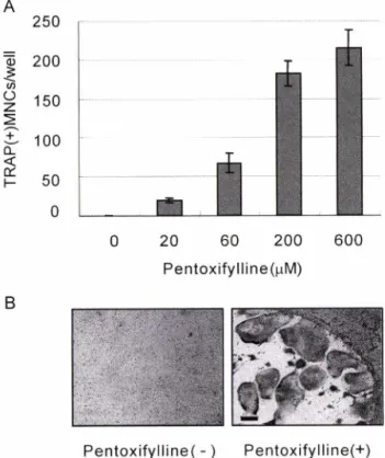
수세포의 공배양계(co-culture)에 1,25-dihydroxyvitamin D3 [l,25(OH)2D3], 부갑상선호르몬(PTH), Prostaglandin E2(PGE2), Interleukin- 1(IL-1), lipopolysaccharide(LPS) 등의 자극제를 처 리함으로써분화유도할수있다.1) 이러한공배양계를이용하여 pentoxifylline이파골세포분화에미치는억제또는유도효과를 조사하였다. 먼저 pentoxifylline은공배양계에서 1,25-dihydroxy- vitamin D3[l,25(OH)2D3]의 처리로유도된파골세포분화에아 무런억제효과를보이지 않았다(미발표결과). 반면공배양계에 pentoxifylline을단독 처리하였을 때 TRAP+ 다핵세포의 수가 농도의존적으로증가되어, pentoxifylline이파골세포의분화를 촉진함을시사하였다(Fig. 1A). Fig. IB에서 pentoxifylline으로
유도된 TRAP+ 다핵파골세포의 현미경사진을확인할수있다.
Pentoxifylline의처리로유도된 TRAP+ 다핵세포가파골세포 특이적유전자를발현하는지 RT-PCR 방법으로확인하였다(Fig.
2). pentoxifylline 무처리에 비해 pentoxifylline 처리시파골세포 특이적유전자인 calcitonin receptor(CTR), cathepsin K의발현 이증가하여,pentoxifylline의처리로유도된 TRAP+ 다핵세포 는파골세포임이 입증되었다.
J. Pharm. Soc. Korea
Phosphodiesterase 저해제 Pentoxifylline 이 파골세포 분화에 미치는 영향 199
P e n to x ify llin e - - + +
O P G + - +
Fig. 3 - Requirement of TRANCE for osteoclast formation induced by pentoxifylline. Mouse bone marrow cells and calvarial osteoblasts were cultured with (+) or without (-) 600 jiM of pentoxifylline on the presence (+) or absence (-) of OPG (100 (ig/ml) for 6 days. The cells were then fixed and stained for TRAR and the number of TRAP-positive multinucleated cells (MNCs) was counted. Data are expressed as the mean 土 SD of triplicate cultures.
P e n to x ify llin e
허 ^ g |
TRANCE
GAPDH
Fig. 4 - Effect of pentoxifylline on TRANCE mRNA expression induced by pentoxifylline in calvarial osteoblasts. Calvarial osteoblasts were treated with (+) or without (-) 600 (j_M of pentoxifylline for 3 hours. Total RNA was then extracted from the osteoblasts and the expression of TRANCE mRNA was examined by Northern blot analysis.
TRANCE의 발현 증가와 연관되어 있다고 알려져 있다.7#
pentoxifylline^: phosphodiesterase(PDE) 저해제로, cAMP의분
해를억제하여 세포내 cAMP의농도를증가시킬 것으로기대된
다. 따라서 pentoxifylline의 파골세포 분화 유도가 TRANCE 발현증가와 관련되어 있는지 조사하였다. 먼저 TRAN CE와의 관련성을 알아보기 위해 공배양계에 pentoxifylline과 함께 TRANCE의 decoy receptor인 OPG를 처리하였다. pentoxifyl
line 의처리로유도된 파골 세포분화는 OPG의첨가로 완벽하 게억제되어, pentoxifyllinePl TRANCE 발현에 작용함을나타 내었다(Fig. 3). pentoxifylline이전사수준에서 TRANCE 발현 을조절하는지 northern blotting으로 조사하였다. 대조군에 비 해 pentoxifylline 처리시 TRANCE의 mRNA 발현이 크게 증가 하는것을볼수있다(Fig. 4). 따라서 pentoxifylline^: TRANCE 의발현을전사수준에서 조절하고있음이 밝혀졌다.
0 20 60 2 0ᄋ 600
P e n to x ify llin e (fiM )
P e n to x ify llin e ( - ) P e n to x ify llin e (+ ) Fig. 1 - Effect of pentoxifylline on osteoclast formation. A, Dose-
dependent effect of pentoxifylline on osteoclast formation in co-cultures. Mouse bone marrow cells and calvarial osteoblasts were co-cultured in the presence of the indicated concentrations of pentoxifylline for 6 days. Cells were then fixed and stained for TRAP TRAP-positive (+) multinucleated cells (MNCs) were counted. Data are expressed as the mean 土 SD of triplicate cultures. B, Photographs of co-cultures in which cells were co-cultured with (+) or without (-) 600 |iM of pentoxifylline for 6 days.
Cells were fixed and stained for TRAFj which appears as dark-stained cells. Bar = 200 jiM.
RT:
C a lcito n in re c e p to r C a th e p sin K
G APD H
P e n to x ify llin e (-) P e n tᄋx ify llin e (+ ) Fig. 2 - Expression of osteoclast marker genes in co-cultures. Bone
marrow cells and calvarial osteoblasts were co-cultured for 6 days with (+) or without (-) 600 (iM of pentoxifylline.
Total RNA was then isolated from the cells and cDNA templates prepared with (+) or without (-) reverse transcriptase (RT). mRNA expression was determinrd by RT-PCR using specific primers designed for each gene.
Pentoxifylline의 TRANCE 발현증가
조골세포내 cAMP의증가는 파골 세포 분화 유도 인자인
PA/vsoNlAK+
Q rv c ri
Tx- —
> ^불
:C !
AlA/vsozIAK+dv
lrJ.
200 김민혜 ᅵ전윤나 • 임미정
이는 지금까지 보고된 부갑상선 호르몬(PTH), Prostaglandin E2(PGE2) 등 세포내 cAMP 상승 물질의 TRANCE 발현 증가와 일치하는 결과로,1) pentoxifylline0] 이들과 동일한 신호전달 경 로를 매개하여 TRANCE 발현을 유도하는지에 대해 현재 연구 가 진행 중이다.
기존의 동물 모델을 이용한 실험에서 pentoxifylline 및 기타 PDE 억제제들은 골밀도를 중가시킨다고 보고된 바 있다.12-14) Pentoxifylline은 조골세포의 분화를 촉진시킨다는 V.TIT. 있어15>
뼈 형성에 촉진적으로 작용하는 것으로 생각되어져 왔다. 그러 나 주지한 바와 같이 본 실험에서 pentoxifylline은 파골세포의 분화를 촉진시키는 효과도 가지는 것으로 나타났다. 이는 부갑 상선 호르몬이 골 형성과 골 파괴의 양쪽 효과를 모두 가지는 것 과 매우 유사하다.16) 부갑상선 호르몬은 실험동물에 간헐적으로 투여시 골밀도 증가 효과를,연속적으로 투여시 골밀도 감소 효 과를 나타낸다고 알려져 있다.16) 부갑상선 호르몬이 투여조건에 따라 골밀도에 대해 상반된 효과를 나타내는 원인은 아직 명확 히 규명되지 않았으나, 간헐적인 부갑상선 호르몬의 투여는 현 재 골다공중 치료제의 일환으로 널리 이용되고 있다. 이는 pentoxifylline 또한 골다공증 치료제로서 이용될 수 있는 가능성 을 시사하는 것.0■로, pentoxifylline이 부갑상선 호르몬과 동일하 게 골다공증 치료제로 개발되기 위해 향후 pentoxifylline의 효과 및 기전 연구가 지속적으로 이루어져야 할 것이다.
감사의 말씀
본 연구는 보건 장학회 연구지원으로 수행되었으므로 이에 감 사드립니다.
문 헌
1) Takahashi, N., Akatsu, T, Udagawa, N., Sasaki, T., Yamaguchi, A., Moseley, J. M.,Martin, T. J. and Suda, T. : Osteoblastic cells are involved in osteoclast formation. Endocrinology 123,2600 (1988).
2) Suda, T., Takahashi, N., Udagawa, N., Jimi, E., Gillespie, M. T.
and Martin, T. J. : Modulation of osteoclast differentiation and function by the new members of the tumor necrosis factor receptor and ligand families. Endocr. Rev. 20,345 (1999).
3) Wong, B. R., Rho, J., Arron, J., Robinson, E., Orlinick, J., Chao, M.,Kalachikov, S., Cayani, E.,Bartlett, E S. 3rd, Frankel, W N., Lee, S. Y. and Choi, Y. : TRANCE is a novel ligand of the tumor necrosis factor receptor family that activates c-Jun N- terminal kinase in T cells. J. B iol Chem. 272, 25190 (1997).
4) Yasuda, H.,Shima, N., Nakagawa, N., Yamaguchi, K.,Kinosaki, M., Mochizuki, S., Tomoyasu, A., Yano, K.,Goto, M .,
Murakami, A., Tsuda, E.’ Morinaga, T., Higashio, K., Udagawa, N., Takahashi, N. and Suda, T. : Osteoclast differentiation factor is a ligand for osteoprotegerin/osteoclastogenesis- inhibitory factor and is identical to TRANCE/RANKL. Proc.
N atl Acad. Sci USA 95, 3597 (1998).
5) Lacey, D. L., Timms, E.,Tan, H. L., Kelley, M. J., Dunstan, C.
R., Burgess, T., Elliott, R., Colombero, A., Elliott, G., Scully, S., Hsu, H.’ Sullivan, J., Hawkins, N., Davy, E.’ Capparelli, C., Eli, A., Qian, Y. X., Kaufman, S., Sarosi, I., Shalhoub, V, Senaldi, G., Guo, J., Delaney, J. and Boyle, W. J. : Osteoprotegerin ligand is a cytokine that regulates osteoclast differentiation and activation. Cell 93,165 (1998).
6) Simonet, W. S., Lacey, D. L., Dunstan, C. R.,Kelley, M., Chang, M. S., Luthy, R., Nguyen, H. Q., Wooden, S., Bennett, L.,
Boone, T., Shimamoto, G.,DeRose, M., Elliott, R.’ Colombero, A., Tan, H. L., Trail, G., Sullivan, J., Davy, E.’ Bucay, N., Renshaw-Gegg, L.,Hughes, T. M.,Hill, D.,Pattison, W, Campbell, R and Boyle, W J. : Osteoprotegerin: a novel secreted protein involved in the regulation of bone density. Cell 89, 309 (1997).
7) Kondo, H. Guo, J. and Bringhurst, F. R. : Cyclic adenosine monophosphate/protein kinase A mediates parathyroid hormone/
parathyroid hormone-related protein receptor regulation of osteoclastogenesis and expression of RANKL and osteopro - tegerin mRNAs by marrow stromal cells. ]. Bone Miner. Res.
17,1667 (2002).
8) Kaji, H., Sugimoto, T., Kanatani, M., Fukase, M., Kumegawa, M. and Chihara, K. : Prostaglandin E 2 stimulates osteoclast
like cell formation and bone-resorbing activity via osteoblasts:
role of cAMP-dependent protein kinase. J. BoneMiner. Res. 11, 62 (1996).
9) Fu, Q., Jilka, R. L., Manolagas, S. C. and O'Brien, C. A. : Parathyroid hormone stimulates receptor activator of NF- kB ligand and inhibits osteoprotegerin expression via protein kinase A activation of cAMP-response element-binding pro - tein. J. B iol Chem. 277,48868 (2002).
10) Antoni, F. A. : Molecular diversity of cyclic AMP signaling.
Front Neuroendocrinol. 21,103 (2000).
11) Essayan, D. M. : Cyclic nucleotide phosphodiesterases. J.
Allergy Clin. Immunol. 108,671 (2001).
12) Kinoshita, T., Kobayashi, S., Ebara, S., Yoshimura, Y., Horiuchi, H.,Tsutsumimoto, T, Wakabayashi, S. and Takaoka, K. : Phosphodiesterase inhibitors, pentoxifylline and rolipram, increase bone mass mainly by promoting bone formation in normal mice. Bone 27,811 (2000).
13) Waki, Y., Horita, T, Miyamoto, K., Ohya, K. and Kasugai, S. : Effects of XT-44, a phosphodiesterase 4 inhibitor, in osteoblastgenesis and osteoclastgenesis in culture and its therapeutic effects in rat osteopenia models. Jpn. J. Pharmacol
J. Pharm. Soc. Korea
Phosphodiesterase 저해제 Pentoxifylline 이 피골세포 분화에 미치는 영향 201
79,477 (1999).
14) Miyamoto, K., Waki, Y., Horita, T, Kasugai, S. and Ohya, K. : Reduction of bone loss by denbufylline, an inhibitor of phosphodiesterase 4. Biochem. Pharmacol. 54, 613 (1997).
15) Rawadi, G.,Ferrer, C., Spinella-Jaegle, S., Roman-Roman, S., Bouali, Y. and Baron, R. : l-(5-oxohexyl)-3,7-Dimethylxanthine, a phosphodiesterase inhibitor, activates MAPK cascades and
promotes osteoblast differentiation by a mechanism independent of PKA activation. Endocrinology 142, 4673 (2001).
16) Tam, C. S., Heersche, J. N.,Murray, T. M. and Parsons, J. A. : Parathyroid hormone stimulates the bone apposition rate independently of its resorptive action: differential effects of intermittent and continuous administration. Endocrinology 110,506 (1982).