DOI 10.17480/psk.2015.59.5.207
P2Y 6 수용체 길항제의 파골세포 분화 촉진 효과 규명
노아롱새미 · 문미란 · 임미정
# 숙명여자대학교 약학대학(Received May 7, 2015; Revised September 9, 2015; Accepted September 9, 2015)
The Stimulatory Effect of P2Y
6Receptor Antagonist on RANKL-induced Osteoclastogenesis
A Long Sae Mi Noh, Miran Moon and Mijung Yim
# College of Pharmacy, Sookmyung Women’s University, Seoul 140-742, KoreaAbstract — P2Y receptors, a type of P2 receptor family, are G-protein coupled receptors and 8 subtypes have been char- acterized (P2Y1, P2Y2, P2Y4, P2Y6, P2Y11-14). Recently, several studies have shed light on the role of P2Y receptors in bone biology. Among them, little is known on the role of P2Y6 receptor on osteoclast differentiation. Thus, we investigated the role of P2Y6 receptor on osteoclastogenesis using P2Y6 receptor selective antagonist, MRS 2578. When osteoblasts and bone marrow cells were co-cultured in the presence of VitD3 and PGE2, P2Y6 antagonist increased the formation of TRAP positive osteoclasts. To elucidate the target cells of P2Y6 antagonist, we first checked the effect of MRS 2578 on osteoblasts.
Treatment of MRS 2578 did not affect OPG : RANKL mRNA ratio in osteoblasts. Next, we checked the effects of P2Y6 antagonist on osteoclast precursors using mouse bone marrow macrophages (BMMs). Addition of MRS 2578 increased the number of osteoclasts in RANKL-treated BMMs. Although P2Y6 antagonist had no effect on RANKL-induced NFATc1, c- Fos and MafB expression levels, it significantly stimulated RANKL-induced Blimp1 mRNA expression in BMMs. Taken together, these data indicate that P2Y6 antagonist increases osteoclast formation by upregulation of Blimp1 expression.
Keywords □ P2Y6 receptor, osteoclast
건강한 골은 일생 동안 골이 지속적으로 형성되고 파괴되며, 이 를 골의 재형성이라 한다. 골을 구성하는 두 가지 세포 중 조골세 포는 골을 구성하는 기질성분을 분비하고 합성하며 골 표면 사이 의 무기질 이동에 관여하는 골세포로 중요한 역할을 수행 한다.
한편, 파골세포는 골의 기질 성분을 용해시켜 칼슘염과 인산염을 유리시키는 골 흡수 분해를 담당함으로써 골의 농도를 유지시키 는 기능을 수행 한다.1-5)체내 조골세포와 파골세포는 밀접하게 상 호 작용하고 있으며, 파골세포의 분화는 조골세포에 의해 엄격하 게 조절된다.6) 즉, 조골세포는 파골세포 분화인자인 M-CSF (macrophage-colony stimulating factor) 및 RANKL (receptor activator of nuclear factor-kappaB ligand)을 통해 파골세포의 분
화를 조절하며 이로써 체내 골 형성과 골 흡수의 동적인 평형을 유지한다. 또한 OPG(osteoprotegerin)는 RANKL의 decoy receptor로서 파골세포 분화의 중요한 억제인자로 알려져 있다.7-10) ATP나 UTP 같은 nucleotide는 세포내에서 일차적인 에너지 원으로 이용될 뿐만 아니라 세포외에도 미량 존재하며 세포내 다 양한 생리적 기능에 관여하는 것으로 잘 알려져 있다.11)세포외 액의 nucleotide의 작용은 세포 외막에 존재하는 수용체를 매개 로 이루어지는데, 이는 크게 P1과 P2 수용체로 구분한다. P1 수 용체는 adenosine과 AMP를 인식하며 A1, A2, A3로 나뉘고, P2 수용체는 ATP, ADP, UTP와 UDP를 인식하며 리간드-의존성 이 온채널인 P2X와 G-단백과 연결된 P2Y로 나누어진다.12,13)현재 까지 알려진 P2X 수용체 subtype은 최소 8개 이상이며(P2X1~8), P2Y 수용체에도 P2Y1, P2Y2, P2Y4, P2Y6, P2Y11, P2Y12, P2Y13, P2Y14등의 subtype들이 보고되어 있다.13) P2Y 수용체 subtype들 중 P2Y1, P2Y2, P2Y4, P2Y6, P2Y11수용체는 사람에 게서 클로닝 되어왔고 기능적으로 활성화된 형태라고 알려져 있
다.12-15)일반적으로 세포 외액에서의 nucleotide는 포유동물의
#
Corresponding Author Mijung Yim
College of Pharmacy, Sookmyung Women’s University, Seoul 140- 742, Korea
Tel.: 02-710-9572 Fax.: 02-710-9871
E-mail: [email protected]
Short Report
종설눈, 뇌, 뼈, 태반, 골격근, 폐, 비장, 신장, 심장 등과 같은 조직에 광범위하게 분포하고 있으면서 다양한 생리적 기능을 조절하고, 세포내 신호전달 조절을 통한 세포의 성장, 분화, 증식, 사멸 등의 기전을 매개하며, 면역계 질환, 염증, 통증, 신경퇴행성 질환, 골다 공증 등의 다양한 병리적 기전에도 관여한다. 뿐만 아니라 관련 수용체는 암이나 허혈성 손상, 약물 및 방사선 독성, 외상성 조직 손상, 뼈의 재흡수, 스트레스 및 출혈성 쇼크 등과 같은 병태생리 학적 상황에서 보호기전을 활성화시키는데도 관여하는 것으로 알 려져 있다.15-17)최근 파골세포에서 P2 수용체에 관한 연구가 다 방면으로 수행되어 사람 파골세포는 P2X1, P2X4, P2X7, P2Y1, P2Y2, P2Y4, P2Y6, P2Y11수용체를, 마우스는 P2X6와 P2Y4를 제 외한 모든 P2 수용체가 발현되고 있음이 보고되었다.18,19)그 중 P2Y6수용체는 파골세포의 apoptosis를 억제하여 생존을 증가시키 며 골 흡수 기능에 관여함이 보고되었다.20-23)그러나 파골세포 분 화에 대한 P2Y6수용체의 기능은 거의 연구된 바 없다. 따라서 본 연구에서는 P2Y6길항제를 사용하여 P2Y6수용체의 파골세포 분 화 조절 기능 및 작용 기전을 조사하였기에 보고하는 바이다.
실험방법
시약
MRS 2578은 TOCRIS Bioscience(Missouri, USA), UDP는 Santa Cruze Biotechnology, 모든 항체는 Cell Signaling Technology(Beverly, MA, USA)에서 구입하였으며, 그 외 실험에 사용한 모든 시약은 Sigma Aldrich(St. Louis, MO)에서 구입하였다.
마우스 조골세포의 초기 배양
생후 0~1일의 신생아 ICR mouse로부터 두개골 피부를 벗긴 후 두개골을 적출하였다. 이때 후두골 근육 부착부분은 제거하 였다. 부착된 근육, 혈구 등을 제거한 후 α-MEM(minimum essential medium)으로 가볍게 세척하였다. 0.1% collagenase와 0.2% dispase 효소 용액에 넣어 37oC에서 5분간 진탕시킨 후 상 등액을 버리고 새로운 효소 용액을 가하였다. 37oC에서 약 15분 간 진탕하여 상등액을 모으는 조작을 4회 반복하였다. 원심 분 리한 후 10% FBS(fetal bovine serum) 함유 α-MEM으로 약 3- 5×10⁴cells/100 mm plastic dish가 되도록 접종하였다. 5%
CO2, 37oC에서 3~4일간 배양한 세포를 이후 조골세포로 실험 에 사용하였다.
마우스 파골전구세포의 배양
ICR mouse(6~9주, 수컷)를 경추 탈골한 후 70% 에탄올로 소 독하였다. 경골 부분의 피부를 절개하여 부착 근육을 떼어냈다.
경골 원심부를 절단하고 슬개골을 탈골시켜 경골을 적출하였다.
뼈 양끝을 조금 잘라 한 쪽 끝에 25G의 주사바늘을 꽂고 α-MEM
을 흘려보내 골수세포를 시험관에 모았다. 1200 rpm에서 5분 원 심 분리한 후 10% FBS가 함유된 α-MEM으로 재현탁하고 M- CSF 10 ng/ml로 하룻밤 배양하여 부유세포를 얻은 후 M-CSF 30 ng/ml로 3일간 추가 배양했다. 이를 회수하여 파골전구세포로 사용하였다. 모든 실험은 숙명여자대학교 동물윤리심의 위원회의 승인 하에 수행되었다(승인번호 SMU-IACUC-2011-0915-019호).
MTT 분석
MTT(3-(4,5-dimethylthiazol-2-yl)-2,5-diphenyltetazolium bromide) 정량은 Mosmann의 방법을 변형하여 실시하였다. 1×
104 cells/well 농도로 96 well plate에 분주한 세포에 시료를 처 리하고 일정 시간 동안 배양하였다. 배양액을 버리고 PBS로 세 척한 후, MTT 용액(0.5 mg/ml)을 100 μl/well 첨가하여 호일로 싼 상태에서 5시간 동안 배양하고, Solubilization buffer(10%
SDS in 0.01 M HCl)를 100 μl/well 첨가하고 다시 호일로 쌌다.
16~17시간 동안 배양한 후, 570 nm에서 흡광도를 측정하였다.
파골세포의 분화유도
초기 배양한 조골세포 5×10³ cells/well와 골수세포 1×10⁵ cells/well을 10% FBS가 함유된 α-MEM으로 공배양 했다. 또는 파골전구세포를 RANKL 200 ng/ml과 M-CSF 30 ng/ml 존재 하 에서 4일간 배양했다. 배양이 끝난 세포는 10% formalin으로 10 분간 고정한 후 ethanol-aceton(1 : 1)로 1분간 재고정하여 TRAP (tartrate-resistant acid phosphatase) 염색을 했다. 3개 이상의 핵을 가진 TRAP 양성 세포를 다핵 파골세포로 판정했다.
RNA 분리 및 RT-PCR 분석
Total RNA 추출은 Easy-blue(Intron, Biochemistry. INC.)를 이용하였다. cDNA는 1 μg의 total RNA를 oligodT primer, 10 mM dNTP, 1 unit RNase inhibitor 그리고 4 unit Script reverse transcriptase(Fermentas, Life science)로 42oC에서 60분 처리하 여 합성한 후, 70oC에서 10분 가열함으로써 반응을 중지시켰다.
Polymerase chain reaction(PCR)의 조건과 사용한 primer의 서 열은 아래와 같다.
Primer 서열
P2Y
6R (F) 5 '-CCTGGCACTGGCGGACCTGAT-3' (R) 5'-GGCGGGCCATGCGACAATAAC-3' Calcitonin
receptor (CTR)
(F) 5'-TTTCAAGAACCTTAGCTGCCAGAG-3' (R) 5'-CAAGGCACGGACAATGTTGAGAAG-3' Cathepsin K
(CTK)
(F) 5 '-CTTCCAATACGTGCAGCAGA-3' (R) 5'-ACGCACCAATATCTTGCACC-3' β-Actin (F) 5'-TGTGATGGTGGGAATGGGTCAG-3'
(R) 5'-TTTGATGTCACGCACGATTTCC-3'
Real-Time PCR 분석
추출된 cDNA 2 μl에 PCR 혼합액 18 μl를 넣은 후 기포가 생 기지 않도록 혼합하였다. PCR 혼합액은 SYBR® Green PCR Master Mix(Applied Biosystems, CA, USA)와 10 pmol의 primer(Cosmogenetech, Korea), 그리고 멸균 증류수를 포함한 다. Real time PCR 반응은 7500 Real Time PCR System (Applied Biosystems, CA, USA) 장비를 사용하여 시행하였다.
Cycle threshold(Ct) value는 GAPDH를 기준으로 자동적으로 계 산되었으며, 실험결과 분석은 7500 System Sequence Detection Software version(Applied Biosystems, CA, USA) 프로그램을 사 용하였다. Thermocycling은 먼저 95°C에서 10분간 유지하였다.
이후 95oC에서 15초, 57oC에서 1분, 60oC에서 1분을 40 cycle 반 복하였다. 분석을 위해 사용된 primer의 서열은 다음과 같다.
Western blot 분석
시료를 처리한 세포를 lysis buffer로 용해하고 원심분리 하였 다. 여기서 얻은 상등액을 10% SDS-PAGE를 이용해 전기영동하 고 이를 nitrocellulose membrane으로 이전시켰다. 이를 5% skim milk가 함유 된 PBST 용액으로 blocking 하고, 1차 항체로서 anti- NFATc1(1 : 200), anti-c-Fos(1 : 1000), anti-β-actin(1 : 4000) 항 체와 각각 반응 시켰다. PBST로 5회 세척하고 HRP(horseradish peroxidase)가 결합된 2차 항체와 반응시킨 후 ECL Select (Amersham. CO.)로 발색시켜 LAS-3000으로 측정 한 후 Scion image 프로그램을 사용하여 분석하였다.
통계처리
실험결과는 평균±표준편차로 표기하였고, Student’s t-test로 분 석하여 p 값이 0.05 미만일 때 통계적으로 유의하다고 판단하였다.
실험결과 및 고찰
P2Y6길항제가 공배양계에 미치는 영향 규명
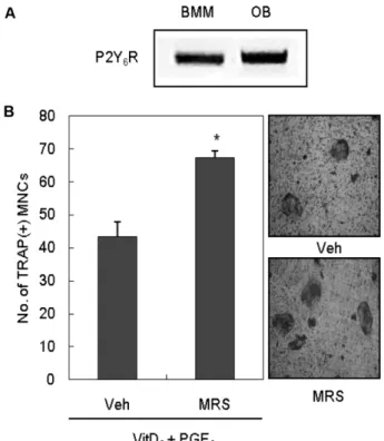
먼저 조골세포와 파골전구세포에서 P2Y6 mRNA가 발현
되는지의 여부를 P2Y6특이적 서열을 사용한 RT-PCR 방법으 로 확인하였다. 마우스 두 개관에서 초기배양한 조골세포(OB) 와 마우스 골수유래의 파골전구세포(Bone marrow-derived macrophages, BMMs)는 모두 P2Y6수용체 mRNA를 발현하 는 것으로 확인되었다(Fig. 1A). PGE2(prostaglandin E2)와 VitD3(1α,25-Dihydroxyvitamin D3)의 처리는 초기 배양한 마우 스 조골세포와 골수세포의 공배양계에서 TRAP 양성 다핵 파골 세포의 분화를 유도하는 것으로 알려져 있다.24)이 실험계를 이 용하여 P2Y6수용체의 길항제인 MRS 2578이 파골세포 분화에 미치는 효과를 조사한 결과, P2Y6 길항제(0.3 μM)는 공배양계에 서 PGE2와 VitD3에 의한 파골세포 형성을 증가시키는 것으로 나타났다(Fig. 1B). 공배양계에는 조골세포와 파골전구세포가 함 께 존재하므로, P2Y6 길항제가 어느 세포에 작용하여 파골세포 분화를 증가시켰는지 규명하기 위해 각각의 세포에 P2Y6길항 제가 미치는 영향을 조사하였다. 먼저 P2Y6길항제에 의한 파골
Primer 서열
RANKL (F) 5'-CCAAGATCTCTAACATGACG-3' (R) 5'-CACCATCAGCTGAAGATAGT-3' OPG (F) 5'-ACGGACAGCTGGCACACCAG-3'
(R) 5 '-CTCACACACTCGGTTGTGGG-3' MafB (F) 5'-AACGGTAGTGTGGAGGAC-3'
(R) 5'-TCACAGAAAGAACTCAGGA-3' Blimp1 (F) 5'-TTCTTGTGTGGTATTGTCGGGACTT-3'
(R) 5 '-TTGGGGACACTCTTTGGGTAG AGTT-3' GAPDH (F) 5'-TCGACCACCAACTGCTTAGC-3'
(R) 5'-GGCATGGACTGTGGTCATGAG-3'
Fig. 1 − The effect of P2Y
6R antagonist on osteoclast formation in coculture. (A) The mRNA expression of P2Y
6R on mouse BMMs and osteoblasts (OB). mRNA expression was determined by RT-PCR using specific primers designed for P2Y
6R. (B) Mouse osteoblasts were cultured with bone marrow cells for 7 days with or without 0.3 μM MRS 2578 in the presence of VitD
3(2×10
−8M) and PGE
2(0.3 μM).
Cells were then fixed and stained for TRAP activity. TRAP-
positive multinucleated cells containing more than three
nuclei were counted as osteoclasts. Veh : Vehicle, MRS :
MRS 2578, BMM : Bone marrow macrophage, OB :
Osteoblast, VitD
3: 1α,25-Dihydroxyvitamin D
3, PGE
2:
Prostaglandin E
2; * : p<0.05.
세포 분화 증가 효과가 세포 수 증가에 기인하는 것인지 확인하 기 위하여 조골세포와 파골전구세포에서 MTT 분석을 수행하였 다. 그 결과 P2Y6길항제는 조골세포와 파골전구세포 수를 모두 변화시키지 않았다(Fig. 2A, B). 이 결과는 P2Y6 길항제가 조골 세포나 파골전구세포의 수를 변화시킴으로써 파골세포 분화를 촉진하는 것은 아니라는 점을 시사한다.
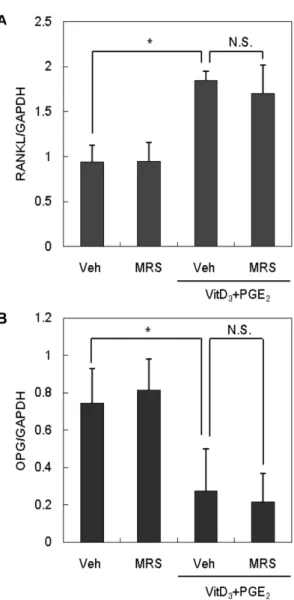
조골세포에 대한 P2Y6저해제의 영향 규명
조골세포에 PGE2와 VitD3를 처리하면 파골세포 분화 유발인
자인 RANKL의 발현이 증가하는 한편, 분화 억제인자인 OPG 의 발현은 감소하는 것으로 알려져 있다.25,26) RANKL은 전구파 골세포의 표면에 있는 수용체인 RANK와 상호작용을 하여 파골 전구세포가 성숙한 파골세포로 분화되게 유도한다. OPG는 교란 수용체로서 RANKL에 결합함으로서 RANKL-RANK 상호작용 을 방해 한다.27)앞의 공배양계 실험에서 P2Y6길항제가 파골세 포의 분화를 촉진하였으므로 그 효과가 조골세포에서 RANKL 또는 OPG의 발현을 조절함으로써 나타난 것인지 확인하고자 하 였다. 이를 위해 조골세포를 P2Y6길항제 존재 하에서 PGE2와
Fig. 2 − The effect of P2Y
6R antagonist on the proliferation of BMMs
and osteoblasts. Mouse BMM cells (A) or osteoblasts (B) were treated with MRS 2578 (0.3 μM) for 6 days. Then, cells were treated with MTT solution for 5 hrs and added Solubilization buffer. The level of cell proliferation was measured at 570 nm. All values are the mean±SD of three experiments. Veh : Vehicle, MRS : MRS 2578, BMM : Bone marrow macrophage, OB : Osteoblast, N.S. : Not significant.
Fig. 3 − The effect of P2Y
6R antagonist on RANKL and OPG mRNA expression. Primary osteoblasts were cultured with VitD
3and PGE
2for 24 hrs with or without 0.3 μM MRS 2578.
mRNA expressions were determined by Real-Time PCR
using specific primers designed for RANKL or OPG. All
values are the mean±SD of three experiments. Veh :
Vehicle, MRS : MRS 2578, VitD
3: 1 α,25-Dihydroxyvitamin
D
3, PGE
2: Prostaglandin E
2; N.S. : Not significant, * :
p<0.05.
VitD3를 처리한 후 RANKL과 OPG의 mRNA 발현을 Realtime PCR을 통해 조사하였다. 기존에 보고된 바와 동일하게 PGE2와 VitD3의 처리는 RANKL의 mRNA 발현을 증가시키는 한편 OPG 의 발현은 감소시켰다. 한편 P2Y6길항제는 PGE2와 VitD3의 효 과에 아무런 영향을 미치지 않는 것으로 나타났다(Fig. 3). 이상 의 결과로부터 P2Y6길항제에 의한 파골세포의 분화 촉진은 조 골세포의 RANKL 또는 OPG 발현 변화에 기인하지 않는 것으 로 추정되었다.
파골전구세포에 대한 P2Y6길항제의 영향 규명
P2Y6길항제가 조골세포를 표적하지 않는 것으로 나타났으므 로 다음은 파골전구세포에 대한 효과를 조사하였다. 파골전구세 포로는 마우스 골수세포를 M-CSF로 3일간 배양하여 얻은 BMM
세포를 사용하였다. 파골전구세포를 RANKL과 M-CSF 존재 하 에서 4일간 배양하면 TRAP 양성의 파골세포로 분화하며(Fig.
4A), 파골세포 특이적 표지 유전자인 CTR(calcitonin receptor) 와 CTK(cathepsin K)의 mRNA 발현도 증가 한다(Fig. 4B). 파 골전구세포를 RANKL과 M-CSF 존재 하에서 P2Y6길항제를 처 리하였을 때 파골세포로의 분화가 촉진되었으며(Fig. 4A), 이에 따라 CTR과 CTK의 mRNA 발현 역시 증가하는 것으로 나타났 다(Fig. 4B). 반면 P2Y6수용체의 선택적 리간드로 작용하는 UDP 를 파골전구세포에 처리하였을 때 파골세포로의 분화가 억제되 는 것을 발견하였다(Fig. 4C). P2Y6길항제가 파골세포의 분화 과정 중 어느 단계에 작용하는지 조사하기 위해 P2Y6길항제를 배양 기간의 초기(day 0~2) 또는 후기(day 2~4)에 처리하여 파 골세포 분화 변화를 살펴보았다(Fig. 4D). 파골전구세포를 배양
Fig. 4 − The effect of P2Y
6R antagonist on RANKL-induced osteoclast formation. BMMs were cultured with 200 ng/ml RANKL and 30 ng/ml
M-CSF in the presence or absence of MRS 2578 (0.3 μM). Cells were then fixed and stained for TRAP activity. (A) TRAP-positive
multinucleated cells containing more than three nuclei were counted as osteoclasts. (B) mRNA expressions were determined by RT-
PCR using specific primers designed for CTR, CTK, and β-actin. (C) BMMs were cultured with RANKL and M-CSF in the presence
or absence of UDP (100 μM). (D) MRS 2578 was added during the indicated culture time. All values are the mean±SD of three
experiments. Veh : Vehicle, R : RANKL, MRS : MRS 2578, UDP : Uridine 5'-diphosphate, * : p<0.05.
초기에 P2Y6길항제로 처리하였을 때 파골세포로의 분화가 촉 진되었으며, 이러한 분화 촉진 효과는 전체 배양 기간(day 0~4) 동안 처리하였을 때보다 오히려 증가되었다. 한편 배양 후기에 P2Y6길항제를 처리하였을 때는 분화 촉진 효과가 나타나지 않 았다.
이상의 결과를 종합하면 P2Y6길항제는 파골전구세포에 직접 적으로 작용하여 파골세포로의 분화를 촉진하며, 특히 분화의 초 기 단계에 작용하는 것으로 추정할 수 있다.
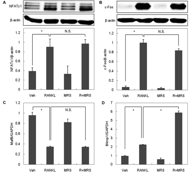
P2Y6길항제의 파골세포 분화 촉진 기전 규명
P2Y6길항제가 파골세포 분화를 촉진하는 작용기전을 규명하 기 위해 먼저 파골세포 분화를 결정짓는 중요 전사인자인 NFATcl 의 발현을 조사하였다. 파골전구세포에 RANKL을 처리하면 NFATc1의 발현이 증가함을 확인하였으며, P2Y6길항제는 예상
과 달리 NFATc1의 발현 유도에 영향을 미치지 않는 것으로 나 타났다(Fig. 5A). 또 다른 전사인자인 c-Fos는 NFATc1의 발현에 촉진적으로, MafB(v-maf musculoaponeurotic fibrosarcoma oncogene family, protein B)는 억제적으로 작용한다고 알려져 있
다.28-30)실제로 RANKL 처리 시 c-Fos의 발현이 증가되었으며
(Fig. 5B), MafB의 발현은 감소하였다(Fig. 5C). NFATc1에 대한 결과와 일치하게 P2Y6길항제는 RANKL에 의한 c-Fos의 발현 증가 및 MafB의 발현 감소에 아무런 영향을 미치지 않았다. 이 는 P2Y6길항제가 NFATc1 경로와 무관하게 파골세포 분화를 조 절한다는 것을 의미한다.
최근 파골세포 분화를 조절하는 새로운 기전으로 Blimp1(B- lymphocyte induced maturation protein 1) 분자가 제시되었다.
파골세포 특이적 Blimp1 결손 마우스는 파골세포 분화가 원활 하지 않으며 Blimp1은 궁극적으로 파골세포 분화를 촉진하는 전
Fig. 5 − The effect of P2Y
6R antagonist on RANKL-induced NFATc1, c-Fos, MafB and Blimp1 expression. BMMs were treated with M-CSF
and RANKL in the presence or absence of MRS 2578 (0.3 μM). After 24 hrs incubation, whole-cell extracts were harvested and
analyzed by Western blotting (A and B) or Real-Time PCR (C and D). All values are the mean±SD of three experiments. Veh : Vehicle,
R : RANKL, MRS : MRS 2578, N.S : Not significant, * : p<0.05.
사인자임이 밝혀졌다.31,32) P2Y6길항제의 파골세포 분화 촉진 효과가 NFATc1 경로와 무관한 것으로 나타났으므로, Blimp1과 의 연관성을 조사하였다. 파골전구세포에 RANKL을 처리하면 보고된 바와 동일하게 Blimp1의 mRNA 발현이 증가하며, P2Y6 길항제의 처리는 그 발현을 증강시키는 것으로 나타났다(Fig.
5D). 이상의 결과를 종합할 때 P2Y6길항제는 파골전구세포에서 RANKL에 의한 Blimp1의 전사를 촉진함으로써 파골세포로의 분 화를 증가시키는 것으로 사료되었다.
결 론
P2Y6길항제가 파골세포 분화에 미치는 영향을 조사한 결과 조골세포와 골수세포의 공배양계에서 파골세포로의 분화를 증가 시키는 것으로 나타났다. P2Y6길항제는 조골세포내 RANKL 및 OPG 발현을 변화시키지 않았으며, 파골전구세포의 분화 초기에 직접적으로 작용하여 분화를 촉진하는 것으로 밝혀졌다. P2Y6 길항제의 파골세포 분화 촉진 효과는 RANKL에 의해 유도되는 NFATc1 경로와는 무관하였으며, P2Y6길항제가 Blimp1의 전사 를 증가시키는 것에 기인할 것으로 사료된다. 본 연구 결과는 P2Y6수용체에 의한 파골세포 분화 조절 가능성을 처음으로 제 시한 것으로, 이는 향후 P2Y6수용체와 골대사와의 연관성을 밝 히는데 기여할 수 있을 것으로 기대된다.
References
1) Godring, S. R. and Gravallese, E. M. : Mechanisms of bone loss in inflammatory arthritis: Diagnosis and therapeutic implications. Arthritis Res. 2, 33 (2000).
2) Rodan, G. A. and Martin, T. J. : Role of osteoblasts in hormonal control of bone resorption-a hypothesis. Calcif. Tissue Int. 33, 349 (1981).
3) Suda, T., Takahashi, N., Udagawa, N., Jimi, E., Gillespie, M. T.
and Martin, T. J. : Modulation of osteoclast differentiation and function by the new members of the tumor necrosis factor receptor and ligand families. Endocr. Rev. 20, 345 (1999).
4) Takahasi, N., Akatsu, T., Udagawa, N., Sasaki, T., Yamaguchi, A., Moseley, J. M., Martin, T. J. and Suda, T. : Osteoblastic cells are involved in osteoclast formation. Endocrinology 123, 2600 (1988).
5) Takayanagi, H., Ogasawara, K., Hida, S., Chiba, T., Murata, S., Sato, K., Takaoka, A., Yokochi, T., Oda, H., Tanaka, K., Nakamur, K. and Taniguchi, T. : T-cell-mediated regulation of osteoclastogenesis by signaling cross-talk between RANKL and IFN-γ. Nature 408, 600 (2000).
6) Phan, T. C. A., Xu, J. and Zheng, M. H. : Interaction between osteoblast and osteoclast : impact in bone disease. Histol.
Histo. Phatol. 19, 1325 (2004).
7) Asagiri, M. and Takayanagi, H. : The molecular understanding of osteoclast differentiation. Bone 40, 251 (2007).
8) Kong, Y. Y., Feige, U., Sarosi, I., Bolon, B., Tafuri, A., Morony, S., Capparelli, C., Li, J., Elliott, R., McCabe, S., Wong, T., Campagnuolo, G., Moran, E., Bogoch, E. R., Van, G., Nguyen, L. T., Ohashi, P. S., Lacey, D. L., Fish, E., Boyle, W. J. and Penningeral, J. M. : Activated T cells regulate bone loss and joint destruction in adjuvant arthritis through osteoprotegerin ligand. Nature 402, 304 (1999).
9) Simonet, W. S., Lacey, D. L., Dunstan, C. R., Kelly, M., Chang, M. S., Luthy, R., Nguyen, H. Q., Wooden, S., Bennett, L., Boone, T., Shimamoto, G., DeRose, M., Elliott, R., Colombero, A., Tan, H. L., Trail, G., Sullivan, J., Davy, E., Bucay, N., Renshaw-Gegg, L., Hughes, T. M., Hill, D., Pattison, W., Campbell, P. and Boyle, W. J. : Osteoprotegerin : A novel secreted protein involved in the regulation of bone density. Cell 89, 309 (1997).
10) Yasuda, H., Shima, N., Nakagawa, N., Mochizuki, S. I., Yano, K., Fujise, N., Sato, Y., Goto, M., Ymaguchi, K., Kuriyama, M., Kanno, T., Murakami, A., Tsuda, E., Morinaga, T. and Higashio, K. : Identity of osteoclastogenesis inhibitory factor (ocif) and osteoprotegerin (opg): A mechanism by which opg/ocif inhibits osteoclastogenesis in vitro. Endocrinology 139, 1329 (1998).
11) Buckley, K. A., Hipskind, R. A., Gartland, A., Bowler, W. B. and Gallagher, J. A. : Adenosine triphosphate stimulates human osteoclast activity via upregulation of osteoblast-expressed receptor activator of nuclear factor-kappa B ligand. Bone 31, 582 (2002).
12) Lazarowski, E. R., Boucher, R. C. and Harden, T. K. : Mechanisms of release of nucleotides and integration of their action as P2X- and P2Y-receptor activating molecules. Mol.
Pharmacol. 64, 785 (2003).
13) Ralevic, V. and Brunstock, G. : Receptors for purines and pyrimidines. Pharmacol. Rev. 50, 413 (1998).
14) Jacobson, K. A., Jarvis, M. F. and Williams, M. : Purines and pyrimidine (P2) receptors as drug targets. J. Med. Chem. 45, 4057 (2002).
15) Von Kügelgen, I. : Pharmacological profiles of cloned mammalin P2Y-receptor subtypes. Pharmacol. Ther. 110, 415 (2006).
16) Communi, D., Janssens, R., Suraex-Huerta, N., Robaye, B. and Boeynaems, J. M. : Advances in signaling by extracellular nucleotides. the role and transduction mechanisms of P2Y receptor. Cell Signal. 12, 351 (2000).
17) Jorgensen, N. R., Solgaard, M. and Schwarz, P. : Role of P2 purinergic receptors in bone metabolism and their therapeutic potential. Med. Hypothesis Res. 3, 615 (2006).
18) Burnstock, G. and Knight, G. E. : Cellular distribution and functions of P2 receptor subtypes in different system. Int. Rev.
Cytol. 240, 31 (2004).
19) Steinberg, T. H. and Hiken, J. F. : P2 receptors in macrophage fusion and osteoclast formation. Purinergic Signaling 3, 53 (2007).
20) Orriss, I. R., Wang, N., Burnstock, G., Arnett, T. R., Gartland, A., Robaye, B. and Noeynaems, J. : The P2Y6 receptor stimulates bone resorption by osteoclasts. Endocrinology 152, 3706 (2011).
21) Korcok, J., Raimundo, L. N., Du, X., Sims, S. M. and Dixon, S. J. : P2Y6 nucleotide receptors activate NFκB and increase survival of osteocalsts. J. Biol. .Chem. 280, 16909 (2005).
22) Zhang, Z., Wang, Z., Ren, H., Yue, M., Huang, K., Gu, H., Liu, M., Du, B. and Qian, M. : P2Y6 agonist uridine 5'-diphosphate promotes host defense against bacterial infection via monocyte chemoattractant protein-1-mediated monocytes/macrophages recruitment. J. Immunol. 186, 5376 (2011).
23) Orriss, I. R., Wang, N., Burnstock, G., Arnett, T. R., Gartland, A., Robaye, B. and Boeynaems, J. M. : The P2Y(6) receptor stimulates bone resorption by osteoclasts. Endocrinology 152, 3706 (2011).
24) Murillo, A., Guerrero, C. A., Acosta, O. and Cardozo, C. A. : Bone resorptive activity of osteoclast-like cells generated in vitro by PEG-induced macrophage fusion. Biol. Res. 43, 205 (2010).
25) Lee, J. H., Jin, H., Shim, H. E., Kim, H. N., Ha, H. and Lee, Z. H. : Epigallocatechin-3-gallate inhibits osteoclastogenesis by down-regulating c-Fos expression and suppressing the NF- κB signal. Mol. Pharmacol. 77, 17 (2010).
26) Takayanagi, H. : Osteoimmunology: shared mechanisms and
crosstalk between the immun and bone systems. Nat. Rev.
Immunol. 7, 292 (2007).
27) Hsu, H., Lacey, D. L., Dunstan, C. R., Solovyev, I., Colombero, A., Timms, E., Tan, H. L., Ellioti, G., Kelly, M. J., Sarosi, I., Wang, L., Xia, X. Z., Elliott, R., Chiu, L., Black, T., Scully, S., Capparelli, C., Morony, S., Shimamoto, G., Bass, M. N. and Boyle, W. J. : Tumor necrosis factor receptor family member rank mediates osteoclast differentiation and actiation induced by osteoprotegerin ligand. Proc. Natl. Acad. Sci. USA 96, 3540 (1999).
28) Takayanagi, H., Kim, S., Koga, T., Nishina, H., Isshiki, M., Yoshida, H., Saiura, A., Isobe, M., Yokochi, T., Inoue, J., Wagner, E. F., Mak, T. W., Kodama, T. and Taniguchi, T. : Induction and activation of the transcription factor NFATc1 (NFAT2) integrate RANKL signaling in terminal differentiation of osteoclasts. Dev. Cell. 3, 889 (2002).
29) Takayanagi, H. : The role of NFAT in osteoclast formation.
Ann. NY. Acad. Sci. 1116, 227 (2007).
30) Kim, K., Kim, J. H., Lee, J., Jin, H. M., Kook, H., Kim, K. K., Lee, S. Y. and Kim, N. : MafB negatively regulates RANKL- mediated osteoclast differentiation. Blood 109, 3253 (2007).
31) Martins, G. and Calame, K. : Regulation and functions of Blimp-1 in T and B lymphocytes. Annu. Rev. Immunol. 26, 133 (2008).
32) Nishikawa, K., Nakashima, T., Hayashi, M., Fukunaga, T., Kato, S., Kodama, T., Takahashi, S., Calame, K. and Takayanagi, H. : Blimp1-mediated repression of negative regulators is required for osteoclast differentiation. Proc. Natl. Acad. Sci. USA 107, 3117 (2010).