Effect of microgrooves and fibronectin
conjugation on the osteoblast marker gene expression and differentiation
Su-Jung Park1, Richard Leesungbok1, Su-Jin Ahn1, Byung-Jin Im2, Do Yun Lee3, Yu-Jin Jee4, Joon-Ho Yoon5, Taixing Cui2, Sang Cheon Lee6*, Suk Won Lee1*
1Department of Biomaterials & Prosthodontics, Kyung Hee University Hospital at Gangdong, Institute of Oral Biology, School of Dentistry, Kyung Hee University, Seoul, Republic of Korea
2Department of Dentistry, Graduate School of Dentistry, Kyung Hee University, Seoul, Republic of Korea
3ED Dental Clinic, Seoul, Republic of Korea
4Department of Oral and Maxillofacial Surgery, Kyung Hee University Hospital at Gangdong, Institute of Oral Biology, School of Dentistry, Kyung Hee University, Seoul, Republic of Korea
5Department of Prosthodontics, National Health Insurance Medical Center Ilsan Hospital, Goyang, Gyeonggi, Republic of Korea
6Department of Maxillofacial Biomedical Engineering and Institute of Oral Biology, School of Dentistry, Kyung Hee University, Seoul, Republic of Korea
PURPOSE. To determine the effect of fibronectin (FN)-conjugated, microgrooved titanium (Ti) on osteoblast differentiation and gene expression in human bone marrow−derived mesenchymal stem cells (MSCs).
MATERIALS AND METHODS. Photolithography was used to fabricate the microgrooved Ti, and amine functionalization (silanization) was used to immobilize fibronectin on the titanium surfaces. Osteoblast differentiation and osteoblast marker gene expression were analyzed by means of alkaline phosphatase activity assay, extracellular calcium deposition assay, and quantitative real-time PCR. RESULTS. The conjugation of fibronectin on Ti significantly increased osteoblast differentiation in MSCs compared with non-conjugated Ti substrates. On the extracellular calcium deposition assays of MSCs at 21 days, an approximately two-fold increase in calcium concentration was observed on the etched 60-μm-wide/10-μm-deep microgrooved surface with fibronectin (E60/10FN) compared with the same surface without fibronectin (E60/10), and a more than four- fold increase in calcium concentration was observed on E60/10FN compared with the non-etched control (NE0) and etched control (E0) surfaces. Through a series of analyses to determine the expression of osteoblast marker genes, a significant increase in all the marker genes except type I collagen α1 mRNA was seen with E60/10FN more than with any of the other groups, as compared with NE0. CONCLUSION. The FN-conjugated,
microgrooved Ti substrate can provide an effective surface to promote osteoblast differentiation and osteoblast marker gene expression in MSCs. [J Adv Prosthodont 2015;7:496-505]
KEY WORDS: Titanium; Microgrooves; Fibronectin conjugation; Gene expression; Osteoblast differentiation
Corresponding author:
Sang Cheon Lee
Department of Maxillofacial Biomedical Engineering and Institute of Oral Biology, School of Dentistry, Kyung Hee University, 26 Kyungheedae-ro, Dongdaemun-gu, Seoul 02447, Rep. of Korea
Tel. 82 2 961 2317: e-mail, [email protected] Suk Won Lee
Department of Biomaterials & Prosthodontics, Kyung Hee University Hospital at Gangdong, Institute of Oral Biology, School of Dentistry, Kyung Hee University, 892 Dongnam-ro, Gangdong-gu, Seoul 05278, Rep. of Korea
Tel. 82 2 440 7518: e-mail, [email protected]
Received November 25, 2015 / Last Revision December 18, 2015 / Accepted December 22, 2015
© 2015 The Korean Academy of Prosthodontics
This is an Open Access article distributed under the terms of the Creative Commons Attribution Non-Commercial License (http://creativecommons.org/licenses/
by-nc/3.0) which permits unrestricted non-commercial use, distribution, and reproduction in any medium, provided the original work is properly cited.
This research was supported by Basic Science Research Program through the National Research Foundation of Korea (NRF) funded by the Ministry of Education (2010-0021268).
This work was supported by the National Research Foundation of Korea (NRF) grant funded by the Korean government (MSIP) (NO. 2013R1A2A2A01009239).
INTRODUCTION
Titanium (Ti) has been used as a practical biomaterial for dental implants in edentulous areas. Many studies have demonstrated that microgrooved rather than smooth titani- um surfaces promote better adhesion of gingival fibroblasts and osteoblastic cells, enhancing soft tissue sealing and an osteogenesis reaction.1-3 In our previous study, we showed that 60-μm-wide and 10-μm-deep etched microgrooves were the most effective surface topography for promoting osteo- blast maturation and inducing differential gene expression in human bone marrow-derived mesenchymal stem cells (MSCs).1
Because undifferentiated MSCs are osteoblastic precur- sors cells producing bone marrow, their capability for form- ing bone and undergoing differentiation into osteoblasts is important for estimating the interaction between the bone and implant surface during osseointegration. MSCs cultured on nanoscale topography of Ti surfaces have been demon- strated to express significantly higher levels of alkaline phosphatase (ALP) activity and type I collagen, extracellular matrix mineralization, and bone matrix protein production than those cultured on smooth Ti surfaces.4,5 In our previous study using osteogenic cultures of primary human cells at day 14, it was found that the osteoblast marker gene expres- sion, except for type I collagen α1 (COL1A1), was significant- ly higher on 60-µm-wide and 10-µm–deep etched micro- grooves than it was on smooth surface.6
Fibronectin, a plasma protein that is one of the major molecules mediating cell adhesion, was found to bind spe- cifically to collagen and gelatin.7 In vitro, fibronectin is pri- marily adsorbed to titanium surfaces and promotes fibro- blast adhesion and activity8 through increased focal contact formation9; however, in vivo, dermal attachment around transcutaneous devices is not affected by adsorbed fibronec- tin.10 It is suggested that adsorbed fibronectin can be dissi- pated during implant insertion due to physical abrasion or the presence of other competing serum proteins. To enhance dermal attachment of fibronectin for intraosseous transcutaneous titanium implants in vivo, a more predictable chemical attachment other than simple adsorption to Ti is required. Silanization causes covalent bonds between pro- teins and metals with a silane complex, improving cell adhe- sion and activity.11 The bioactivity of fibronectin conjugat- ed on silanized Ti alloy was determined by analysis on cell morphology, area, immunolocalization of focal contacts, and metabolism.12 Several studies have reported that micro- grooved Ti surfaces precisely modulate endogenous fibro- nectin expression at the transcriptional and post-transcrip- tional levels, as well as the amount of fibronectin assem- bled in the extracellular matrix.13,14
In our previous study, etched microgrooves on Ti sub- strates also enhanced cell proliferation and altered the expression of various genes including fibronectin.4 Such surface treatment appears to increase the degree to which exogenous human plasma fibronectin is adsorbed onto the Ti substrates.2 These data suggest a need to determine
whether an organic coating of fibronectin (such as conju- gated fibronectin) on silanized microgrooved Ti12 would further enhance the overall cell response.
In this study, our aim was to determine the effect of fibronectin (FN)-conjugated microgrooved Ti on the osteo- blast differentiation and osteoblast marker gene expression in MSCs. For this, we performed multiple tests, including the ALP activity assay, extracellular calcium deposition assay, and quantitative real-time PCR.
MATERIALS AND METHODS
Grade 2 commercially pure Ti sheets (TSM-TECH, Ulsan, Korea) with 0.14-mm thickness were polished using a cloth wheel (Yougar Enterprise, Incheon, Korea) at 1,800 rpm on an angle grinder (GWS 20-230) (Robert Bosch, Stuttgart, Germany) to produce a polished surface with a roughness average (Ra) of 0.1 μm. These sheets were washed using acetone and acted as the smooth Ti surface control (non- etched control, NE0). Subsequent acid etching was per- formed for 10 seconds using 1% hydrofluoric acid (HF), and these constituted a second control group (etched con- trol, E0). As described in our previous study,15 we used photolithography to create truncated, V-shaped micro- grooves 15-, 30-, or 60-μm wide and 3.5- or 10-μm deep on Ti surfaces. Subsequent acid etching was performed for 2 seconds using 1% HF on the overall surface of the micro- grooved Ti substrates (E15/3.5, E30/10, and E60/10) (Table 1). In every experiment, the fabricated Ti substrates were washed three times for 30 minutes in an ultrasonic device with sterile distilled water. After another triple wash using distilled water, Ti substrates were dried at room tempera- ture overnight before use. The surfaces of the five Ti sub- strates (NE0, E0, E15/3.5, E30/10, and E60/10) were examined using field emission scanning electron microsco- py (S-800 FESEM) (Hitachi, Tokyo, Japan) (Fig. 1).
Human serum fibronectin was conjugated on Ti surfac- es that had been functionalized (silanized) with 3-amino- propyltriethoxysilane (APTES) (Sigma-Aldrich, St. Louis, MO, USA) (Fig. 2). Functionalized amine was produced in anhydrous toluene by adding 1 mL of APTES to 9 mL tol- uene solution per well of Ti substrates (silanization). The reaction was maintained at 120°C in a nitrogen atmosphere with a reflux condenser for 24 hours. After the reaction, the Ti substrates were washed three times with methanol to eliminate silane remnant. The silanized Ti substrates were dried with a vacuum pump for 24 hours and then washed three times for 20 minutes each time using distilled water in an ultrasonic device.
Human plasma fibronectin (human pFN) (Sigma-Aldrich) was conjugated on the surface of the silanized Ti substrates in a 10 µg/mL concentration and maintained for 24 hours at 4°C. After that, the pFN-conjugated Ti substrates were washed with distilled water to remove residual pFN and were dried at room temperature for 45 minutes. As a result, the FN-conjugated non-etched control Ti substrates (NE0FN),
Fig. 1. Images taken of A- NE0 (×500), B- E0(×500), C- NE15/3.5 (×200), D- E15/3.5 (×200), E- E30/10 (×200), and F- E60/10 (×200) using field emission scanning electron microscopy.
A C
E B
D F
Table 1. Titanium (Ti) substrates used in this study (μm)
Groove (ridge) width Groove depth Bottom width Acid-etching FN conjugation
Ti-series NE0 0 0 0 Non-etched N/A
E0 0 0 0 Acid-etched N/A
E15/3.5 15 3.5 8 Acid-etched N/A
E30/10 30 10 10 Acid-etched N/A
E60/10 60 10 40 Acid-etched N/A
FN-series NE0FN 0 0 0 Non-etched FN-conjugated
E0FN 0 0 0 Acid-etched FN-conjugated
E15/3.5FN 15 3.5 8 Acid-etched FN-conjugated
E30/10FN 30 10 10 Acid-etched FN-conjugated
E60/10FN 60 10 40 Acid-etched FN-conjugated
NE0: polished Ti substrate, E0: NE0 with subsequent hydrofluoric (HF) acid etching, Eα/β: Ti substrata containing α-μm wide and β-μm deep surface microgrooves with subsequent HF acid etching, γFN: fibronectin-conjugated Ti substrata γ, FN: fibronectin, N/A: non-applicable.
the FN-conjugated acid-etched control Ti substrates (E0FN), and the FN-conjugated microgrooved Ti sub- strates (E15/3.5FN, E30/10FN, and E60/10FN) were pro- duced (Table 1).
Human bone marrow-derived MSCs were purchased from Lonza (Walkersville, MD, USA). The cells were cul- tured in specific growth medium (MSC GMTM) (Lonza) at 37°C in 5% CO2. Cells were maintained in Dulbecco’s mod- ified Eagle’s medium (DMEM; WelGene, Daegu, Korea), containing 10% fetal bovine serum (FBS; Sigma-Aldrich, St.
Louis, MO, USA) and 1% antibiotic-antimycotic solution at 37°C in 5% CO2. MSCs at passages 3 to 5 were used.
MSCs were seeded on 24-well Ti substrates (NE0, E0, E15/3.5, E30/10, and E60/10) and FN-conjugated Ti sub- strates (NE0FN, E0FN, E15/3.5FN, E30/10FN, and E60/10FN) at 4×10⁴ cells/well and were cultured for 2 days for confluence. Cells were then cultured in osteogenic media, which consisted of DMEM (WelGene) supplemented
with 10% FBS (Sigma-Aldrich), with 50 μg/mL of α-ascorbic acid (Sigma-Aldrich), 10 mM of β-glycerophosphate (Sigma- Aldrich), and 100 mM of dexamethasone (Sigma-Aldrich), at 37°C in 5% CO2 for 7 and 14 days to determine ALP activity. The ALP activity was assayed according to the pro- cedure described previously.2 The reaction suspensions were transferred to 96-well plates to be monitored at 405 nm by a microplate reader (Bio-Rad, Hercules, CA, USA).
Measurements were compared using p-nitrophenol stan- dards to be normalized to total protein.
MSCs were seeded on 24-well Ti substrates of all 10 groups at 4×10⁴ cells/well and were cultured for 2 days for confluence. Cells were cultured in osteogenic media at 37°C in 5% CO2 for 21 days and were washed in phosphate buff- ered solution (PBS) (Gibco BRL, Grand Island, NY, USA).
Ti substrates with remaining calcium deposits were incubat- ed with 0.5 N hydrogen chloride at 4oC overnight. After centrifugation, the resultant amount of calcium was quanti- Fig. 2. Schematic illustration of the fabrication of FN-conjugated microgrooved titanium substrata and the expected enhancement of cellular activity.
fied by Calcium LiquiColor (Stanbio Laboratory, Boerne, TX, USA). The reaction suspensions were transferred to 96-well plates to be monitored at 650 nm by a microplate reader (Bio-Rad, Hercules, CA, USA).
The expression of osteoblast marker genes in MSCs was analyzed using quantitative real-time PCR. The genes were COL1A1 (Hs00164004_m1), ALP (Hs01029144_m1), C B FA 1 / RU N X 2 ( H s 0 1 0 4 7 9 7 8 _ m 1 ) , S P 7 / O S X (Hs01866874_s1), BGLAP/OC (Hs00609452_g1), SPP1/
OPN (Hs00959010_m1), bone sialoprotein II (IBSP/BSP2;
Hs00173720_m1), and osteonectin (SPARC/ON, Hs00234160_m1). MSCs were seeded on the 24-well Ti substrates of all groups at 4×10⁴ cells/well and cultured for 2 days for confluence and were subsequently cultured for 14 days in osteogenic media at 37°C in 5% CO2. The quan- titative real-time PCR was performed as in our previous study.6 The relative-fold-change expression levels were ana- lyzed by normalizing the values to the internal control, glyceraldehyde 3-phosphate dehydrogenase.
The ALP activity assay, extracellular calcium deposition assay, and quantitative real-time PCR analyses of MSCs were repeated independently and simultaneously five times.
One-way analysis of variance (ANOVA) was used to com- pare the mean values of the results for the substrate groups
under Tukey’s multiple comparison tests. Pearson’s correla- tion analysis was performed to determine the correlations.
Multiple regression analysis was used to determine the influential factor genes on osteoblastic differentiation. The SPSS 18.0 was used for statistical analyses.
RESULTS
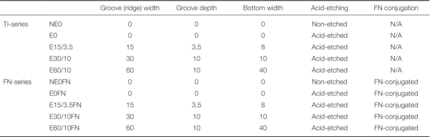
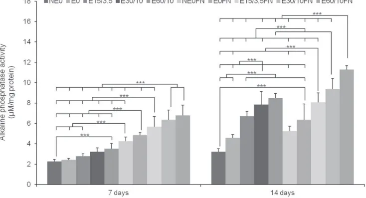
MSCs’ ALP activities were tested after 7 and 14 days of osteogenic culturing. These time points were selected from previously reported of MSCs’ peak ALP activity after 14 days of osteogenic culturing.16 The extracellular calcium deposition assay was conducted after 21 days of osteogenic culturing. MSCs showed the highest level of ALP activity on both day 7 and day 14 (Fig. 3), with the highest calcium level found to be on E60/10FN relative to the other groups of Ti surfaces (Fig. 4). On the extracellular calcium deposition assay, every Ti surface group with conjugated fibronectin had significantly higher levels of calcium com- pared with the Ti groups that had the same microstructure but without conjugated fibronectin at 21 days. When com- pared with NE0 or E0, E60/10FN showed a significant increase (up to more than four-fold) in calcium deposition at 21 days (Fig. 4)
Fig. 3. The multiple comparison result of alkaline phosphatase (ALP) activity in human bone marrow-derived mesenchymal stem cells (MSCs) after 7 and 14 days of osteogenic culture on titanium substrata with specific surface topographies and fibronectin conjugation signaled by the ALP activity assay. One-way ANOVA (n = 5). ***: significant difference (P < .001).
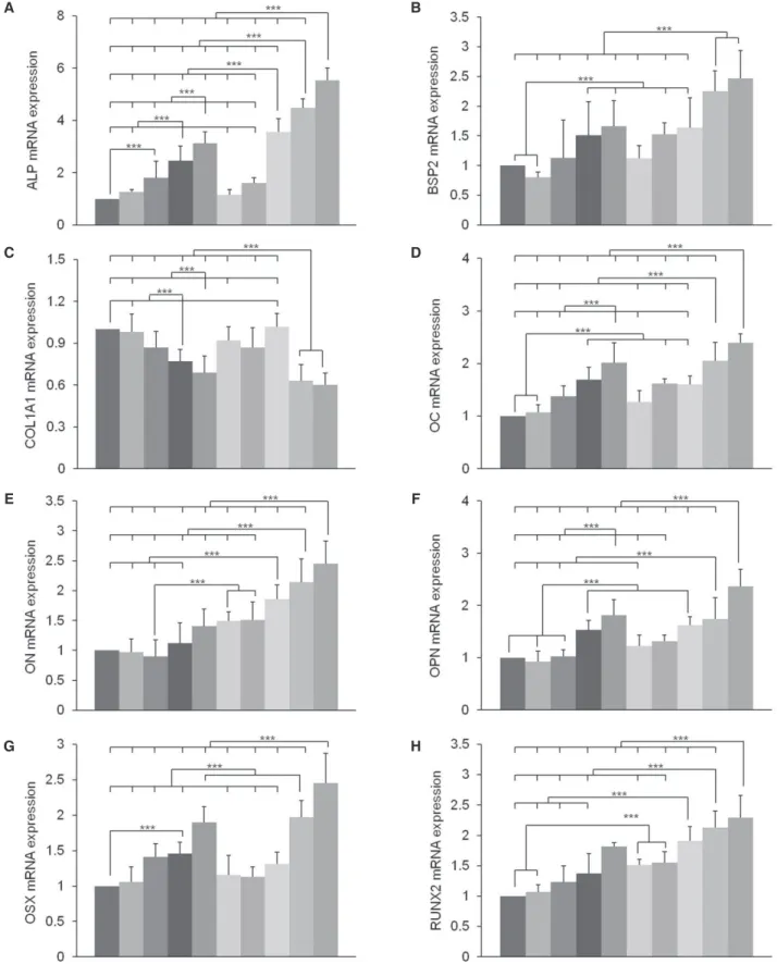
In our previous study, it was shown that the relative COL1A1 gene expression was distinctly patterned when compared to other osteoblast marker genes analyzed at day 14.6 Thus, we chose day 14 as the critical time point during MSCs’ osteoblast differentiation on microgrooved Ti sub- strates. The expression levels of BSP2, ALP, RUNX2, OSX, OC, OPN, and ON were higher on E60/10FN than on any other Ti substrate (Fig. 5). However, the difference in the expression levels of BSP2 mRNA was not statistical- ly significant between E60/10FN and E30/10FN, whereas the levels of ALP, RUNX2, OSX, OC, OPN, and ON were significantly different compared to other groups. Only COL1A1 showed a lower level of gene expressed on E60/10FN versus NE0. This result corresponds to those in our previous study, in which E60/10FN caused significant- ly lower expression levels of COL1A mRNA than any of the Ti substrates.6 Fig. 5 provides detailed results of the rel- ative protein levels in MSCs.
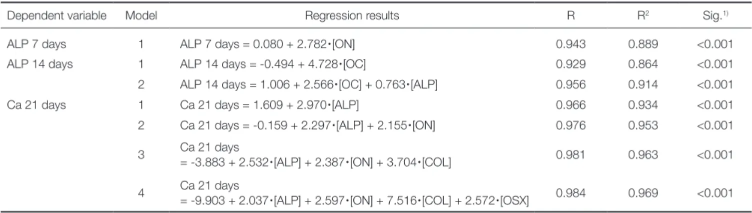
Using the representative gene expression as indepen- dent variables, we performed a Pearson’s correlation analy- sis. Correlations were significant between the expression levels of each of the eight mRNAs and MSC differentia- tion. Multiple regression analyses showed that expression of ON, OC, ALP, COL, and OSX had significant influence on MSC differentiation at 21 days on the Ti substrates with various topographies and fibronectin conjugation (Table 2).
These were determined as the influential factor genes that enhanced MSCs’ osteoblast differentiation in response to FN-conjugated microgrooved Ti substrates, with ALP
apparently being the most influential one. From the step- wise method, ON, OC, and ALP were determined as the most influential factor for ALP 7 days, ALP 14 days, and Ca 21 days, respectively, and the scatter plots for each cor- relation result are presented in Fig. 6.
DISCUSSION
The results of a series of tests, including the ALP and extracellular calcium deposition assays, quantitative real- time PCR, Pearson’s correlation, and multiple regression analysis, suggest that FN-conjugated microgrooved Ti sub- strates promote osteoblast differentiation in MSCs. Indeed, more than three-fold increase in ALP activity at 14 days and more than four-fold increase in calcium concentration at 21 days were observed in the MSCs on E60/10FN com- pared with those on NE0 or E0 (Fig. 3 and Fig. 4). The conjugation of fibronectin on Ti significantly increased osteoblast differentiation in MSCs when compared with the non-conjugated Ti substrates. Also, the extracellular calci- um deposition assay revealed an approximately two-fold increase in calcium concentration in MSCs on E60/10FN compared with E60/10. The addition of both conjugated fibronectin and microgrooves on Ti substrates enhanced osteoblast differentiation in MSCs synergistically when compared with that achieved on the control substrates. The E60/10FN substrates used in this study provided optimal environment for the promotion of a variety of cellular responses in vitro; therefore, an optimal microgroove struc- Fig. 4. The Multiple comparison result of osteoblast differentiation in human bone marrow-derived mesenchymal stem cells after 21 days of osteogenic culture on titanium substrata with specific surface topographies and fibronectin conjugation signaled by the extracellular calcium (Ca) deposition assay. One-way ANOVA (n = 5). ***: significant difference (P < .001).
Fig. 5. The relative fold change osteoblast-marker-gene expression in human bone marrow-derived mesenchymal stem cells (MSCs) after 2-day confluence and 14 days of osteogenic culture on titanium substrata with specific surface topographies and fibronectin conjugation by quantitative real-time PCR. Note that the results are presented as a ratio to the mRNA expression levels of the reference GAPDH gene, followed by a standardization of the NE0’s Ct (threshold cycle) expression as 1. One-way ANOVA (n = 5). ***: significant difference (P < .001).
A
C
E
G
B
D
F
H
ture and the secondary submicroscale of etched topogra- phy with conjugated fibronectin on Ti substrates can exert the strongest impact on osteoblast differentiation in MSCs.
Earlier studies indicated that microgrooved Ti regulates the amount of fibronectin that assembles in the extracellu- lar matrix.14 In addition, etched microgrooves and ridges on the Ti surface exclusively control endogenous fibronectin expression and appear to increase the degree to which exogenous human plasma fibronectin is adsorbed onto Ti substrates.2 Other studies suggest that a fibronectin coating on Ti improves a longevity of the outcome of transcutane- ous implants by promoting soft-tissue attachment and pre- venting infection or epithelial downgrowth.17 MSCs were allowed for limited spreading but enhanced osteoblast dif- ferentiation on microgrooved substrates.18 Moreover, they displayed high sensitivity to substrate differences, and in appropriate-for-surface topography studies, differentiated
more rapidly into osteoblasts, and required less time for matrix maturation and mineralization, as compared with human periodontal ligament cells.4 Microgrooves that are narrower than 10 μm induce changes in cell morphology and alter specific gene expression.14,19 On the other hand, sufficient surface area of truncated, V-shaped, etched microgrooves with 30- or 60- μm width induce fibroblast crawling or migration, and the proliferation of cells is sig- nificantly enhanced.4 However in this study, ALP activity and calcium concentration were higher on E60/10 than on E30/10 and on any other surface groups in the MSC cul- tures (Fig. 3 and Fig. 4). Taken together with our result in the present study, cells on FN-conjugated microgrooved Ti substrates with microgrooves of sufficient dimension are expected to secure the peri-implant environment and regu- late strong and rapid osseointegration.
Based on the osteoblast marker gene expression in Table 2. The influential genes of the osteoblastic differentiation in human bone marrow-derived mesenchymal stem cells after 7, 14 and 21 days of osteogenic culture, as determined by multiple stepwise regression analysis
Dependent variable Model Regression results R R2 Sig.1)
ALP 7 days 1 ALP 7 days = 0.080 + 2.782
∙
[ON] 0.943 0.889 <0.001ALP 14 days 1 ALP 14 days = -0.494 + 4.728
∙
[OC] 0.929 0.864 <0.0012 ALP 14 days = 1.006 + 2.566
∙
[OC] + 0.763∙
[ALP] 0.956 0.914 <0.001Ca 21 days 1 Ca 21 days = 1.609 + 2.970
∙
[ALP] 0.966 0.934 <0.0012 Ca 21 days = -0.159 + 2.297
∙
[ALP] + 2.155∙
[ON] 0.976 0.953 <0.0013 Ca 21 days
= -3.883 + 2.532
∙
[ALP] + 2.387∙
[ON] + 3.704∙
[COL] 0.981 0.963 <0.0014 Ca 21 days
= -9.903 + 2.037
∙
[ALP] + 2.597∙
[ON] + 7.516∙
[COL] + 2.572∙
[OSX] 0.984 0.969 <0.0011) Significances of each regression model were tested by analysis of variance (n = 50).
R: coefficient of multiple correlations, R2: coefficient of determination, ALP α days: the result of alkaline phosphatase activity assay in human bone marrow-derived mesenchymal stem cells after α days of osteogenic culture, Ca 21 days: the result of extracellular calcium deposition assay in human bone marrow-derived mesenchymal stem cells after 21 days of osteogenic culture.
Fig. 6. The scatter-plot of correlation results between the expressed genes determined as greatest influential factors for osteoblastic differentiation and relevant assays. Significant correlations were present for A, B, and C (P < .01) (n = 30).
A B C
human bone marrow MSCs after 14 days of osteogenic cul- ture on Ti substrates having various surface topographies and fibronectin conjugation by quantitative real-time PCR, we observed significant upregulation of ALP, RUNX2, OSX, OC, and OPN genes in MSCs at day 14 of osteogen- ic culture on E60/10FN. We also observed significant downregulation of COL1A1 gene expression in MSCs on E60/10FN. This result corresponds with our previous study,6 and indicates a presence of mechanism for prevent- ing excessive collagen deposition and for inducing effective mineralization.20 The 14-day time point culture (Fig. 5) was chosen for gene expression analysis based on previous reports showing that the nanotopography upregulates the expression of genes responsible for the sequential osteo- blast differentiation progress in MSCs on Ti.21-23 Several studies suggest that MSCs at day 14 represent an immature osteoblast-dominant phase between pre-osteoblast and mature osteoblast.24,25 Although our results contradict pre- vious reports that showed a decrease in OPN on dual acid- etched Ti substrates,26 they coincide with the findings in other studies showing that upregulation of ON, RUNX2, and OPN gene expression correlated with the increase in adhe- sion and proliferation on Ti grooved surfaces.27-29 Expression patterns of RUNX2 and OPN were similar during osteoblast differentiation and de novo bone formation,29 due to the known fact that RUNX2 is a transcription factor activating the expression of OPN,28,30 and also to the reported possi- bility that RUNX2 may maintain the expression levels of OPN in immature osteoblasts with ultimate downregula- tion.31 The multiple stepwise regression analysis showed that expression of ON, ALP, COL, and OSX had the most significant influence on MSC differentiation at 21 days. OC and ALP were influential genes at 14 days, and ON was important at 7 days on Ti substrates with various topogra- phies and fibronectin conjugation (Table 2). These results demonstrated the influence of several marker genes on MSC osteoblast differentiation. Scatter-plot results showed significant correlations between the expressed genes which are found to be the factors with the greatest influence on osteoblast differentiation and relevant assays (Fig. 6).
CONCLUSION
The conjugation of FN by silanization on Ti with 60-μm-wide /10-μm-deep etched microgrooves significantly enhanced the ALP activity and osteoblast differentiation in human MSCs. Likewise, induction of osteoblast marker gene expression caused by etched microgrooves with conjugated FN is indicative of the positive effect of such surface mod- ification on the acceleration of the osseointegration for oral and orthopedic Ti implants. Further study is essential to explore the application of other nanoscale and submi- croscale topographies in various methods of cellular assays and to perform a various timelines of analysis on osteoblast marker gene expression.
ORCID
Su-Jung Park http://orcid.org/0000-0002-4111-2231 Richard Leesungbok http://orcid.org/0000-0002-8381-723X Su-Jin Ahn http://orcid.org/0000-0003-2128-1561
Byung-Jin Im http://orcid.org/0000-0001-5661-2258 Do Yun Lee http://orcid.org/0000-0002-8638-7423 Yu-Jin Jee http://orcid.org/0000-0003-2526-4005 Joon-Ho Yoon http://orcid.org/0000-0002-4571-7342 Taixing Cui http://orcid.org/0000-0002-9528-7299 Sang Cheon Lee http://orcid.org/0000-0002-8560-0173 Suk Won Lee http://orcid.org/0000-0003-2726-3567 REFERENCES
1. Lee SW, Kim SY, Lee MH, Lee KW, Leesungbok R, Oh N.
Influence of etched microgrooves of uniform dimension on in vitro responses of human gingival fibroblasts. Clin Oral Implants Res 2009;20:458-66.
2. Lee MH, Oh N, Lee SW, Leesungbok R, Kim SE, Yun YP, Kang JH. Factors influencing osteoblast maturation on mi- crogrooved titanium substrata. Biomaterials 2010;31:3804-15.
3. Park JA, Leesungbok R, Ahn SJ, Lee SW. Effect of etched microgrooves on hydrophilicity of titanium and osteoblast responses: A pilot study. J Adv Prosthodont 2010;2:18-24.
4. Kim SY, Oh N, Lee MH, Kim SE, Leesungbok R, Lee SW.
Surface microgrooves and acid etching on titanium substrata alter various cell behaviors of cultured human gingival fibro- blasts. Clin Oral Implants Res 2009;20:262-72.
5. Guida L, Annunziata M, Rocci A, Contaldo M, Rullo R, Oliva A. Biological response of human bone marrow mesen- chymal stem cells to fluoride-modified titanium surfaces. Clin Oral Implants Res 2010;21:1234-41.
6. Lee MH, Kang JH, Lee SW. The significance of differential expression of genes and proteins in human primary cells caused by microgrooved biomaterial substrata. Biomaterials 2012;33:3216-34.
7. Engvall E, Ruoslahti E. Binding of soluble form of fibro- blast surface protein, fibronectin, to collagen. Int J Cancer 1977;20:1-5.
8. Dean JW, 3rd, Culbertson KC, D’Angelo AM. Fibronectin and laminin enhance gingival cell attachment to dental im- plant surfaces in vitro. Int J Oral Maxillofac Implants 1995;
10:721-8.
9. Gallant ND, Michael KE, Garcia AJ. Cell adhesion strength- ening: contributions of adhesive area, integrin binding, and focal adhesion assembly. Mol Biol Cell 2005;16:4329-40.
10. Fini M, Savarino L, Nicoli Aldini N, Martin L, Giavaresi G, Rizzi G, Martini D, Ruggeri A, Giunti A, Giardino R.
Biomechanical and histomorphometric investigations on two morphologically differing titanium surfaces with and without fluorohydroxyapatite coating: an experimental study in sheep tibiae. Biomaterials 2003;24:3183-92.
11. Rezania A, Thomas CH, Healy KE. A probabilistic approach to measure the strength of bone cell adhesion to chemically modified surfaces. Ann Biomed Eng 1997;25:190-203.
12. Middleton CA, Pendegrass CJ, Gordon D, Jacob J, Blunn
GW. Fibronectin silanized titanium alloy: a bioinductive and durable coating to enhance fibroblast attachment in vitro. J Biomed Mater Res A 2007;83:1032-8.
13. Pendegrass CJ, Middleton CA, Gordon D, Jacob J, Blunn GW. Measuring the strength of dermal fibroblast attachment to functionalized titanium alloys in vitro. J Biomed Mater Res A 2010;92:1028-37.
14. Chou L, Firth JD, Uitto VJ, Brunette DM. Substratum sur- face topography alters cell shape and regulates fibronectin mRNA level, mRNA stability, secretion and assembly in hu- man fibroblasts. J Cell Sci 1995;108 (Pt 4):1563-73.
15. Lee SW, Kim SY, Rhyu IC, Chung WY, Leesungbok R, Lee KW. Influence of microgroove dimension on cell behavior of human gingival fibroblasts cultured on titanium substrata.
Clin Oral Implants Res 2009;20:56-66.
16. Jaiswal N, Haynesworth SE, Caplan AI, Bruder SP.
Osteogenic differentiation of purified, culture-expanded hu- man mesenchymal stem cells in vitro. J Cell Biochem 1997;
64:295-312.
17. Branemark PI, Albrektsson T. Titanium implants permanent- ly penetrating human skin. Scand J Plast Reconstr Surg 1982;16:17-21.
18. Dalby MJ, McCloy D, Robertson M, Wilkinson CD, Oreffo RO. Osteoprogenitor response to defined topographies with nanoscale depths. Biomaterials 2006;27:1306-15.
19. den Braber ET, de Ruijter JE, Ginsel LA, von Recum AF, Jansen JA. Quantitative analysis of fibroblast morphology on microgrooved surfaces with various groove and ridge dimen- sions. Biomaterials 1996;17:2037-44.
20. Li Z, Hassan MQ, Jafferji M, Aqeilan RI, Garzon R, Croce CM, van Wijnen AJ, Stein JL, Stein GS, Lian JB. Biological functions of miR-29b contribute to positive regulation of os- teoblast differentiation. J Biol Chem 2009;284:15676-84.
21. Valencia S, Gretzer C, Cooper LF. Surface nanofeature ef- fects on titanium-adherent human mesenchymal stem cells.
Int J Oral Maxillofac Implants 2009;24:38-46.
22. Mendonca G, Mendonca DB, Simoes LG, Araujo AL, Leite ER, Duarte WR, Aragao FJ, Cooper LF. The effects of im- plant surface nanoscale features on osteoblast-specific gene expression. Biomaterials 2009;30:4053-62.
23. Mendonca G, Mendonca DB, Aragao FJ, Cooper LF. The combination of micron and nanotopography by H(2)SO(4)/
H(2)O(2) treatment and its effects on osteoblast-specific gene expression of hMSCs. J Biomed Mater Res A 2010;94:
169-79.
24. Chau JF, Leong WF, Li B. Signaling pathways governing os- teoblast proliferation, differentiation and function. Histol Histopathol 2009;24:1593-606.
25. Nakashima K, Zhou X, Kunkel G, Zhang Z, Deng JM, Behringer RR, de Crombrugghe B. The novel zinc finger- containing transcription factor osterix is required for osteo- blast differentiation and bone formation. Cell 2002;108:17- 29.
26. Balloni S, Calvi EM, Damiani F, Bistoni G, Calvitti M, Locci P, Becchetti E, Marinucci L. Effects of titanium surface roughness on mesenchymal stem cell commitment and dif- ferentiation signaling. Int J Oral Maxillofac Implants 2009;24:
627-35.
27. Jayaraman M, Meyer U, Buhner M, Joos U, Wiesmann HP.
Influence of titanium surfaces on attachment of osteoblast- like cells in vitro. Biomaterials 2004;25:625-31.
28. Sato M, Morii E, Komori T, Kawahata H, Sugimoto M, Terai K, Shimizu H, Yasui T, Ogihara H, Yasui N, Ochi T, Kitamura Y, Ito Y, Nomura S. Transcriptional regulation of osteopontin gene in vivo by PEBP2alphaA/CBFA1 and ETS1 in the skeletal tissues. Oncogene 1998;17:1517-25.
29. Komori T. Regulation of bone development and extracellular matrix protein genes by RUNX2. Cell Tissue Res 2010;339:
189-95.
30. Ducy P, Starbuck M, Priemel M, Shen J, Pinero G, Geoffroy V, Amling M, Karsenty G. A Cbfa1-dependent genetic path- way controls bone formation beyond embryonic develop- ment. Genes Dev 1999;13:1025-36.
31. Maruyama Z, Yoshida CA, Furuichi T, Amizuka N, Ito M, Fukuyama R, Miyazaki T, Kitaura H, Nakamura K, Fujita T, Kanatani N, Moriishi T, Yamana K, Liu W, Kawaguchi H, Nakamura K, Komori T. Runx2 determines bone maturity and turnover rate in postnatal bone development and is in- volved in bone loss in estrogen deficiency. Dev Dyn 2007;
236:1876-90.