© 2020 The Korean Ophthalmological Society
This is an Open Access article distributed under the terms of the Creative Commons Attribution Non-Commercial License (http://creativecommons.org/licenses /by-nc/3.0/) which permits unrestricted non-commercial use, distribution, and reproduction in any medium, provided the original work is properly cited.
Original Article
Effect of Diquafosol on Hyperosmotic Stress-induced Tumor Necrosis Factor-α and Interleukin-6 Expression in Human Corneal Epithelial Cells
Yeoun-Hee Kim
1*, In-Jun Yang
2*, Ly Thi Huong Nguyen
2, Sang Il Gum
3, Sung Yu
4, Gwang Ja Lee
4, Bo-Ae Kim
5, Jae-Chang Jung
6, Young Jeung Park
41
Myungmoon Bio, Hwaseong, Korea
2
Department of Physiology, College of Korean Medicine Dongguk University, Gyeongju, Korea
3
Binaree, Daegu, Korea
4
Central Ophthalmic Clinic, Daegu, Korea
5
Division of Biomedicinal & Cosmetics, College of Sciences & Technology, Mokwon University, Daejeon, Korea
6
Developmental Biology Laboratory, Department of Biology, College of Natural Sciences, Kyungpook National University, Daegu, Korea
Purpose: Diquafosol is a pharmaceutical drug used for dry eye treatment with a novel mechanism of action.
It is a purinergic P2Y2 receptor agonist that promotes the secretion of tears and healing of corneal epithelial wounds. However, its inhibitory effect on hyperosmotic stress-induced inflammation in human corneal epitheli- al cells (HCECs) remains unclear.
Methods: A hyperosmotic stress model was established by transferring HCECs from isosmotic (312 mOsm/kg to hyperosmotic medium (500 mOsm/kg). HCECs were incubated with 500 mOsm/kg hyperosmotic medium for 30 minutes, and then treated with diquafosol (0.6–6 mg/mL) for 4 or 24 hours. Cells were then harvested and analyzed by western blot, immunocytochemistry, and real-time polymerase chain reaction to evaluate the ex- pression of interleukin-6, tumor necrosis factor-alpha, and the phosphorylation status of nuclear factor-kappa B.
Results: Diquafosol significantly decreased the mRNA and protein expression of hyperosmotic stress-induced tumor necrosis factor-alpha and interleukin-6. These results were supported by immunofluorescence staining and quantitative real-time polymerase chain reaction analysis. Furthermore, diquafosol inhibits nuclear fac- tor-kappa B activation by suppressing the phosphorylation and degradation of the inhibitor of к B.
Conclusions: This study shows that diquafosol inhibits nuclear factor-kappa B signaling and inflammatory fac- tors induced by hyperosmotic stress in HCECs. This suggests that using diquafosol for the improvement of dry eye syndrome could be effective in the treatment of inflammation-related corneal and conjunctival diseases.
Key Words: Diquafosol, Human corneal epithelial cells, Inflammation, Interleukin-6, Tumor necrosis factor-alpha
Received: April 19, 2019 Final revision: September 2, 2019 Accepted: September 11, 2019
Corresponding Authors: Jae-Chang Jung, PhD. Developmental Biology Laboratory, Department of Biology, College of Natural Sciences, Kyungpook Na- tional University, Daegu 41566, Korea. Tel: 82-53-950-5345, Fax: 82-53-953-3066, E-mail: [email protected]
Young Jeung Park, MD, PhD. Central Ophthalmic Clinic, 2 Dongbu-ro 22-gil, Dong-gu, Daegu 41242, Korea. Tel: 82-53-782-0055, Fax: 82-53-754-1155, E-mail: [email protected]
*These two authors contributed equally to this work.
Dry eye syndrome, or dry eye disease, is one of the most common reasons for patient visits to eye clinics. It is a multifactorial disease of the tears and ocular surface, re- sulting in discomfort, visual disturbances, and tear film in- stability, and it can potentially damage the ocular surface [1]. A comparison of age-specific prevalence data showed that the prevalence of dry eye syndrome ranged from 5%
to more than 35% at various ages [2,3]. People with dry eye are significantly more likely to report problems with read- ing, performing professional work, computer use, and driving compared to people without dry eye [4].
A major mechanism of dry eye pathogenesis is hyperos- molarity, which is caused by tear deficiency or excessive tear evaporation or both. Hyperosmolarity stress is associ- ated with a potent inflammatory response. Osmotic stress that is initiated by an increase in extracellular osmolarity occurs during normal cellular function in various tissues, including human conjunctival [5-7] and corneal epithelial cells [8-10]. Tear hyperosmolarity is considered a causative factor in the ocular surface inflammation, cell damage, and irritation symptoms experienced by dry eye syndrome pa- tients [11]. Extracellular hyperosmolarity is also believed to play a role in the inflammatory response [12]. Notably, these symptoms are also strongly associated with inflammatory cytokine secretion. Inflammatory cytokines that are linked to hyperosmotic stress-related pathologies include the tu- mor necrosis factor (TNF) family and interleukins [12,13].
Diquafosol ophthalmic solution (3%) is a pharmaceutical drug used for the treatment of dry eye syndrome with a novel mechanism of action. It has been widely used in clin- ical practice to treat dry eye and is currently approved in Japan and South Korea. Diquafosol is a purinergic P2Y2 receptor agonist that promotes tear and mucin secretion in experimental dry eye models [14]. However, little is known about the therapeutic potential of diquafosol on the hyper- osmotic stress-induced inflammatory response. The pres- ent study is focused on the effects of diquafosol on TNF-α and interleukin-6 (IL-6) expression in hyperosmotic stress-activated human corneal epithelial cells (HCECs).
Materials and Methods
Reagents
Anti–TNF-α and anti–IL-6 antibodies were purchased
from Abcam (Cambridge, MA, USA). Anti-β-actin and an- ti-Lamin B1 antibodies were purchased from Santa Cruz Biotechnology (Santa Cruz, CA, USA). Antibodies against phospho-inhibitor of κB (I-κB) α, I-κBα, phospho–nuclear factor-kappa B (NF-κB) p65, and NF-κB p65 were pur- chased from Cell Signaling Technology (Beverly, MA, USA). Horseradish peroxidase-conjugated goat anti-rabbit and rabbit anti-goat IgG were purchased from Zymed Lab- oratories (San Francisco, CA, USA). The annexin V and dead cell assay kit was purchased from Merck Millipore (Billerica, MA, USA). Diquafosol was obtained from Sant- en (eye drops 3%; Santen Pharmaceutical, Osaka, Japan).
Cell culture and in vitro model of hyperosmotic stress HCECs (2.040 pRSV-T) were purchased from the Amer- ican Type Culture Collection (Manassas, VA, USA). Cells were maintained in DMEM/F12 containing 10% Fetal Bo- vine Serum (Gibco, Carlsbad, CA, USA), 5 μg/mL insulin, 5 μg/mL human transferrin, 5 nM selenium, and 1% peni- cillin/streptomycin. Cultures were incubated at 37°C with 5% CO
2. Hyperosmotic stress was induced by transferring HCECs from isosmotic (312 mOsm/kg) DMEM/F-12 growth media to hyperosmotic growth media (500 mOsm /kg).
Cell viability and apoptosis assays
To evaluate viability, cells were cultured in a 96-well plate and grown to 80%–90% confluence. HCECs were treated with various concentrations of diquafosol solution for 20 hours. After incubation, cell viability was deter- mined by using the CCK-8 assay (Dojindo Laboratories, Kumamoto, Japan). Color development was measured at 450 nm using an ELISA microplate reader (Infinite M200;
Tecan, Männedorf, Switzerland). Experiments were per-
formed in triplicate. The percentage of apoptotic cells was
determined with the annexin V and dead cell kit, accord-
ing to the manufacturer’s instructions. Briefly, harvested
cells were washed with PBS and then mixed with 100 µL
of the annexin V and dead cell assay kit reagents. Samples
were incubated at room temperature for 20 minutes in the
dark. Measurements were conducted in triplicate using a
MUSE cell analyzer (Merck Millipore, Billerica, MA,
USA).
RNA isolation and quantitative real-time polymerase chain reaction
To determine the amount of mRNA expression, cells were exposed to hyperosmotic media (500 mOsm/kg DMEM/F-12, serum-free) for 30 minutes, followed by di- quafosol for 4 hours, as previously described [15]. Total RNA was isolated from the cells with Trizol reagent (Life Technologies, Rockville, MD, USA), according to the man- ufacturer’s instructions, and reverse-transcribed into com- plementary DNA with M-MLV reverse transcriptase (Promega, Madison, WI, USA). Real-time polymerase chain reaction (PCR) was performed using SYBR Premix Ex Taq (Perfect Real Time) Premix (Takara Bio, Otsu, Ja- pan) and Takara Thermal Cycler Dice (TP850), according to the manufacturer’s protocol (Takara Bio, Shiga, Japan).
Relative quantification of mRNA expression was per- formed using TP850 software. Table 1 shows the gene-spe- cific primers used in this study (Macrogen, Seoul, Korea).
PCR products were electrophoresed on 1% agarose gels and visualized by GreenLight (BioAssay Co., Daejeon, Korea). PCR conditions are indicated in Table 1. All exper- iments were performed in triplicate.
Western blot analysis
To determine the expression of proteins, cells were ex- posed to hyperosmotic media (500 mOsm/kg DMEM/F-12, serum-free) for 30 minutes, followed by diquafosol for 24 hours. Protein extraction and western blotting were performed as described previously [15]. Membranes were incubated overnight at 4˚C with polyclonal antibodies against TNF-α and IL-6 and with a monoclonal antibody against β-actin in 0.1% Tween-20 Tris-buffered saline (TBS) containing 5% nonfat dried milk.
Membranes were then incubated with the appropriate horseradish peroxidase-conjugated secondary antibodies for 1 hour. Antibody binding was visualized using an en-
hanced chemiluminescence detection kit (ELPIS Biotech, Daejeon, Korea) and exposure to X-ray film. The experi- ments were performed in triplicate. Quantification of band intensity was performed using ImageJ software ver. 1.52 (National Institutes of Health, Bethesda, MD, USA).
Cytoplasmic and nuclear protein extraction
To confirm the activation of NF-κB, cells were exposed to hyperosmotic media (500 mOsm/kg DMEM/F-12, se- rum-free) for 30 minutes, followed by diquafosol for 30 minutes. Proteins from the cytoplasm and nucleus were separated by using the NE-PER nuclear and cytoplasmic extraction reagent kit (#78835; ThermoFisher Scientific, Waltham, MA, USA). Briefly, HCECs were harvested, cells were centrifuged at 16,000 × g for 5 minutes, and the supernatant was removed. Ice-cold CER-I and -II solutions were added to the pellet per the manufacturer’s instruc- tions to separate cytoplasmic proteins from nuclear-com- partment proteins. Western blots for NF-κB, p-NF-κB, I-κB, p-I-κB, β-actin (as a cytosol marker) and Lamin B1 (as a nuclear marker) were performed to ensure that there was no contamination.
Immunofluorescence staining
Harvested cells were fixed with 10% neutral buffered for- malin solution at room temperature for 10 minutes. Cells were per meabilized with 0.3% Triton X-100 for 15 minutes and blocked overnight with 5% normal goat se- rum and bovine serum albumin in TBS at 4°C. Cells were then incubated with primary antibodies specific for TNF-α, IL-6, and p-NF-κB (diluted 1 : 50 to 1 : 100) in 5% bovine se- rum albumin overnight at 4˚C and washed three times in TBS. Cells were then incubated with the appropriate Alexa Fluor 488- or 555-conjugated secondary antibody at room temperature for 1 hour in the dark; nuclei were then counter- stained with 4’,6-diamidino-2-phenylindole (DAPI, 1 µg/mL).
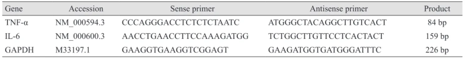
Table 1. Sequences of oligonucleotide primers used in real-time polymerase chain reactions
Gene Accession Sense primer Antisense primer Product
TNF-α NM_000594.3 CCCAGGGACCTCTCTCTAATC ATGGGCTACAGGCTTGTCACT 84 bp
IL-6 NM_000600.3 AACCTGAACCTTCCAAAGATGG TCTGGCTTGTTCCTCACTACT 159 bp
GAPDH M33197.1 GAAGGTGAAGGTCGGAGT GAAGATGGTGATGGGATTTC 226 bp
TNF-α = tumor necrosis factor-alpha; IL-6 = interleukin-6; GAPDH = glyceraldehyde-3-phosphate dehydrogenase.
Slides were mounted with ProLong Gold (ThermoFisher Scientific, Waltham, MA, USA), and images were captured by fluorescence microscopy (Axio Vision 4; Carl Zeiss, Jena, Germany). Fluorescence intensity was measured us- ing color histograms generated with ImageJ software.
Statistical analysis
Data were evaluated by one-way analysis of variance us- ing Tukey’s post-hoc test. Analyses were performed using GraphPad PRISM software ver. 5.02 (GraphPad PRISM Software Inc., La Jolla, CA, USA). The criterion for statis- tical significance was set at p < 0.05. All statistical tests were two-tailed.
Results
Effects of diquafosol ophthalmic solution on the viabil- ity of HCECs
To investigate the cytotoxicity of diquafosol ophthalmic solution, we measured cell viability using the CCK-8 assay kit. Diquafosol displayed a dose-dependent toxicity in HCECs under starved conditions. No significant toxicity was observed at diquafosol concentrations of up to 6 mg/
mL. However, cell viability notably decreased when HCECs were exposed to 30 mg/mL of diquafosol (Fig. 1A). We also measured the effects of diquafosol on apoptosis. Relative to control cells, the percentage of apoptotic HCECs increased with diquafosol in a dose-dependent manner, but the change was small and not significant (Fig. 1B).
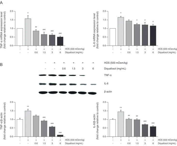
Effects of diquafosol on hyperosmotic stress-induced TNF-α and IL-6 expression
To evaluate the anti-inflammatory activity of diquafosol, we investigated the effects of diquafosol on hyperosmotic stress-induced inflammatory gene expression. Real-time PCR analysis showed an increase in hyperosmotic stress-in- duced TNF-α and IL-6 mRNA concentration (Fig. 2A).
However, pretreatment with diquafosol (1.5–6 mg/mL) resulted in the attenuation of the inflammatory response.
In addition, western blot results revealed that TNF-α and IL-6 protein levels were markedly decreased by diquafosol in hyperosmotic stress-induced inflammation (Fig. 2B).
C el l v ia bi lit y ( % o f c on tro l)
DQF (mg/mL)
015
10
5
- 0.6 1.5 3 6 30
A
A po pt ot ic c el ls ( % )
DQF (mg/mL)
015
9 6 12
3
- 0.6 1.5 3 6
Fig. 1. Effects of diquafosol (DQF) on the (A) viability and (B) apoptosis of human corneal epithelial cells. Cells were treated with different concentrations of DQF solution. After 20 hours, the apoptosis rate was assessed using the CCK-8 assay kit or the annexin V and dead cell assay kit. Results are expressed as the percentage of surviving cells over control cells. Data are expressed as the mean ± standard deviation from three separate experiments (p < 0.01 and p < 0.001, significantly different from the control).
DQF 0.6 (mg/mL) Control
DQF 3 (mg/mL)
DQF 1.5 (mg/mL)
DQF 6 (mg/mL)
B
Suppression of hyperosmotic stress-induced TNF-α and IL-6 expression by diquafosol
Non-hyperosmotic stress-stimulated cells exhibited weak staining, suggesting low basal levels of TNF-α and IL-6 (Fig. 3). After hyperosmotic stress stimulation, im- munocytochemistry analysis revealed that TNF-α and IL-6 expression increased significantly. Diquafosol appears
to block expression of TNF-α and IL-6 proteins in HCECs in response to hyperosmotic stress.
Effects of diquafosol on the NF-κB signaling pathway in hyperosmotic stress
The NF-κB signaling pathway contributes to the inflam- matory process of dry eye, so we investigated the effects A
TN F- α m R N A e xp re ss io n l eve l (fo ld c ha ng e r el at ive t o c on tro l)
-
- +
- +
0.6 +
1.5 +
3 +
6 0.0
0.5 1.5 1.0 2.0
HOS (500 mOsm/kg) Diquafosol (mg/mL)
*
##
### ###
###
IL -6 m R N A e xp re ss io n l eve l (fo ld c ha ng e r el at ive t o c on tro l)
-
- +
- +
0.6 +
1.5 +
3 +
6 0.0
0.5 1.5 1.0 2.0
HOS (500 mOsm/kg) Diquafosol (mg/mL)
*
# #
#
B
TN F- α/β -a ct in (fo ld c ha ng e r el at ive t o c on tro l)
-
- +
- +
0.6 +
1.5 +
3 +
6 0.0
0.5 1.5 1.0 2.0
HOS (500 mOsm/kg) Diquafosol (mg/mL)
**
###
###
###
IL -6 /β -a ct in (fo ld c ha ng e r el at ive t o c on tro l)
-
- +
- +
0.6 +
1.5 +
3 +
6 0.0
0.5 1.5 1.0 2.0
HOS (500 mOsm/kg) Diquafosol (mg/mL)
**
## ##
### ###
- -
+ -
+ 0.6
+ 1.5
+ 3
+ 6
HOS (500 mOsm/kg) Diquafosol (mg/mL) TNF-α
β-actin IL-6
Fig. 2. Diquafosol downregulates mRNA and protein expression of tumor necrosis factor-α (TNF-α) and interleukin-6 (IL-6) in human
corneal epithelial cells. (A) Total RNA was extracted from human corneal epithelial cells. RNA levels were measured by real-time poly-
merase chain reaction. The cells were exposed to hyperosmotic media (500 mOsm/kg DMEM/F12, serum-free) for 30 minutes, followed
by diquafosol (0.6–6 mg/mL) for 4 hours. The graph of multiple analyses shows the relative mRNA levels of TNF-α and IL-6. Glycer-
aldehyde-3-phosphate dehydrogenase was used as the reference gene. (B) Representative western blots of TNF-α and IL-6. Cells were
exposed to hyperosmotic media (500 mOsm/kg DMEM/F12, serum-free) for 30 minutes, followed by diquafosol (0.6–6 mg/mL) for 24
hours. Expression levels of TNF-α and IL-6 were determined using β-actin as a control. The densities of bands relative to those of β-actin
were measured using ImageJ software. Data are expressed as the mean ± standard deviation from three separate experiments (
*p < 0.01
and
**p < 0.01, significantly different from the control;
#p < 0.05,
##p < 0.01, and
###p < 0.001, significantly different from the hyperosmot-
ic group). HOS = hyperosmotic stress.
of diquafosol on hyperosmotic stress-induced NF-κB acti- vation. Phosphorylation of I-κB and nuclear translocation of NF-κB p65 were notably increased by hyperosmotic stress, whereas treatment with diquafosol decreased the hyperosmotic stress-induced levels of p-I-κB and nuclear translocation of NF-κB p65 in a dose-dependent manner (Fig. 4A). Hyperosmotic stress resulted in the translocation of p-NF-κB-p65 to the nucleus as seen by immunocyto- chemistry. Incubation with anti-NF-κB-p65 showed marked nuclear staining in HCECs under hyperosmotic stress conditions, whereas cells treated with diquafosol (6 mg/mL) showed only background staining (Fig. 4B).
The action of diquafosol through inhibiton of NF-κB ac- tivity is schematically summarized (Fig. 4C).
Discussion
Dry eye disease is the most common disorder of the eye, and its associated symptoms can cause corneal injury [16].
Dry eye disease is a condition caused by deficient tear pro- duction or an excessive loss of water from the tear film by evaporation. Increased tear osmolarity has been recog- nized as a hallmark of dry eye syndrome, and it appears to play an important role in the pathogenesis of ocular sur- face damage. Tear osmolarity has been reported to be the single best marker for dry eye disease [17,18]. Tear film hy- perosmolarity may cause pathological changes in the cor- neal epithelium, such as desquamation, fewer intercellular connections, loss of microplicae, cell membrane disrup- tions, and cellular swelling with decreased cytoplasmic Fig. 3. Fluorescence immunocytochemistry for tumor necrosis factor-α (TNF-α) and interleukin-6 (IL-6) in human corneal epithelial cells. Cells were exposed to hyperosmotic media (500 mOsm/kg DMEM/F12, serum-free) for 30 minutes, followed by diquafosol (DQF, 0.6–6 mg/mL) for 24 hours. Changes in the expression of cytoplasmic TNF-α and IL-6 were confirmed using fluorescence immunocyto- chemistry. Cells were counterstained with DAPI. The fluorescence intensity from green to blue was measured in ImageJ software using a color histogram. White scale bars: 100 μm (
***p < 0.001, significantly different from the control;
###p < 0.001, significantly different from the hyperosmotic group). HOS = hyperosmotic stress.
TN F- α p os iti ve c el l n um be r (% o f t ot al c el ls )
-
- +
- +
0.6 +
1.5 +
3 +
6 0
10 20 30
HOS (500 mOsm/kg) DQF (mg/mL)
***
### ###
### ###
IL -6 p os iti ve c el l n um be r (% o f t ot al c el ls )
-
- +
- +
0.6 +
1.5 +
3 +
6 0
10 20 30 40
HOS (500 mOsm/kg) DQF (mg/mL)
***
### ###
### ###
Control HOS (500 mOsm/kg) HOS+DQF 0.6 (mg/mL) HOS+DQF 1.5 (mg/mL) HOS+DQF 3 (mg/mL) HOS+DQF 6 (mg/mL)
density [19]. Other studies have reported that hyperosmotic stress not only stimulates the expression and production of inflammatory cytokines TNF-α, IL-8, and IL-1 in HCECs in vitro [8,9], but also stimulates the expression of these in-
flammatory mediators in an experimental dry eye mouse model in vivo [20]. Other studies have also shown that hy- perosmolarity induces the expression of inflammatory cy- tokines such as TNF-α, IL-6, and monocyte chemotactic Fig. 4. Effects of diquafosol on the nuclear factor-kappa B (NF-κB) signaling pathway in human corneal epithelial cells. (A) Expression levels of NF-κB, p-NF-κB, I-κB, and p-I-κB proteins in nuclear and cytosolic fractions were assessed using western blotting. The cells were exposed to hyperosmotic media (500 mOsm/kg DMEM/F12, serum-free) for 30 minutes, followed by diquafosol (1.5–6 mg/mL) for 30 minutes. Lamin B1 and ß-actin were used as standard proteins for quantitating the levels of the proteins of interest. (B) Expression of p-NF-κB in the nuclear fraction was confirmed using fluorescence immunocytochemistry. Cells were counterstained with DAPI. Fluores- cence intensity and the density of bands were measured using ImageJ software. (C) Schematic diagram showing the inhibitory effects of diquafosol on hyperosmotic stress-induced inflammatory cytokine production via the NF-κB signaling pathway in human corneal epithe- lial cells. Data are expressed as the mean ± standard deviation from three separate experiments (
***p < 0.001, significantly different from the control;
#p < 0.05,
##p < 0.01 and
###p < 0.001, significantly different from the hyperosmotic group). HOS = hyperosmotic stress.
A
pN F- κB /L am in B 1 r at io
Nuclear fraction
0.0 0.5 1.5 1.0 2.0
HOS (500 mOsm/kg) Diquafosol (mg/mL) -
- +
- +
1.5 +
3 +
6
***
######
###
pN F- κB /β -a ct in r at io
Cytosolic fraction
0.0 0.5 1.0 1.5
HOS (500 mOsm/kg) Diquafosol (mg/mL) -
- +
- +
1.5 +
3 +
6
***
## ## #
pI -κ B /β -a ct in r at io
Cytosolic fraction
0.0 0.5 1.5 1.0 2.0
HOS (500 mOsm/kg) Diquafosol (mg/mL) -
- +
- +
1.5 +
3 +
6
***
#### ###
- -
+ -
+ 1.5
+ 3
+ 6
HOS (500 mOsm/kg) Diquafosol (mg/mL) NF-κB
NF-κB
β-actin Lamin B1
I-κB p-I-κB p-NF-κB
p-NF-κB
C yt os ol ic f ra ct ion N uc le ar f ra ct io n
B
pN F- κB p os iti ve c el ls ( % o f t ot al c el l n um be r w ith D A P I)
0 10 30 20 40HOS (500 mOsm/kg) Diquafosol (mg/mL) -
- +
- +
6
***
###
Control HOS (500 mOsm/kg) HOS+diquafosol
Hyperosmotic stress
Corneal epithelial cells
IL-6 TNF-α NucleusCytoplasm NF-κB NF-κB
I-κBα I-κBα
NF-κB NF-κB