147
Inhibitory Effects of β-Glycyrrhetinic Acid on Tumor Necrosis Factor-α Production in RAW 264.7 Cells
Kyoung Sik Park*
Nutrition & Functional Food Research Team, Korea Food & Drug Administration, Seoul, Korea Received April 7, 2010; Accepted June 7, 2010
β-glycyrrhetinic acid (GA), the active principle of licorice (Glycyrrhiza glabra L.) has been reported to exhibit anti-inflammatory properties in different animal models. In this study, the effects of GA on the production of inflammatory mediators including tumor necrosis factor (TNF)-α, interleukin (IL)-6, nitric oxide (NO), and prostaglandin E (PGE)-2 were examined in RAW 264.7 cells in vitro.
Furthermore, to elucidate a possible mechanism for the inhibitory effect of GA on the production of TNF-α, it was investigated whether the treatment of GA affects the I-κBα degradation and subsequent nuclear translocation of NF-κB. Various inflammatory responses were induced in the culture system by treating with a lipopolysaccharide (LPS). GA showed anti-inflammatory activities in dose-dependant manner with IC50 of 5.4 µM by inhibiting the production of TNF-α in RAW 264.7 cells. In addition, the treatment of GA blocked both I-κBα degradation and the nuclear translocation of NF-κB from cytosol to nucleus. However, it did not affect the production of IL-6, NO, and PGE-2, implying the direct blocking of the production of TNF-α resulting from both the I-κBα degradation and the nuclear translocation of NF-κB. This finding might provide the underlying mechanism to explain the reported anti-inflammatory activities of GA in animal models.
Key words: β-glycyrrhetinic acid, anti-inflammatory herb, inflammatory mediators, triterpene
Root extracts of Glycyrrhiza glabra L. (licorice) and its main water-soluble constituent glycyrrhizin (GL), a pentacyclic triterpene derivative of the β-amyrin type (oleanane), have been widely used as a traditional medicine for the treatment of inflammatory diseases including abscesses, nervous disorders, asthma and peptic ulcers [Ohuchi et al., 1981; Kobayashi et al., 1993; Zhang et al., 1993; Kondo and Takano, 1994; Kimura and Watanabe, 1995]. After oral administration or intravenous injection, GL has been shown to be hydrolyzed by the glucuronidase in intestinal bacteria to its active principle aglycone, β-glycyrrhetinic acid (GA), which is then absorbed into the blood [Yamamura et al., 1992; Yim et al., 2004].
GA exhibits anti-inflammatory properties in different animal models [Capasso et al., 1983; Amagaya et al., 1984; Khaksa et al., 1996]. Previously, the mechanism of action was considered to be identical to that of glucocorticoids. This assumption was based on the structural resemblance between GA and corticosteroids. Recently, it was postulated that the anti- inflammatory activity of GA is probably due to the inhibition of the enzyme 11β-hydroxysteroid hydroxygenase [Walker and
Edwards, 1991]. Inhibition of this enzyme results in an accumulation of hydrocortisone, a natural steroid with anti- inflammatory properties. However, the overall anti-inflammatory mechanism of GA action has not been yet thoroughly understood.
A tumor necrosis factor, TNF-α was initially discovered in mammalian tumor tissues, but it is also recognized to be one of the most pleotropic cytokines acting as a host defense factor in immune and inflammatory responses. The TNF-α production during processes of inflammation and tumor necrosis is beneficial to the host. However, its excessive production arises various adverse responses e.g. the disseminated intravascular coagulation, the death in septic shock, and asthma and dermatitis, multiple sclerosis, inflammatory bowel disease, cystic fibrosis, rheumatoid arthritis and other immune diseases [Sekut and Connolly, 1996]. Therefore, either the suppression of over-production of TNF-α or inhibition of its functions is the major target for relieving those adverse reactions.
The present study was designed to examine whether the treatment of GA exhibits anti-inflammatory activities by affecting the production of TNF-α, the degradation I-κBα and subsequent nuclear translocation of NF-κB in RAW264.7 cells.
Maerials and Methods
Materials. 18β-glycyrrhetinic acid (GA), rolipram, quercetin,
*Corresponding author
Phone: +82-2-380-1665; Fax: +82-2-385-7081 E-mail: [email protected]
doi:10.3839/jabc.2010.027
MO). DMEM and RPMI1640 medium and other reagents for cell culture were obtained from Gibco BRL Life Biotechnologies (Gaithersburg, MD). Rabbit polyclonal Rel A (p65) and I-κBα antibody were from New England BioLabs Inc. (Beverly, MA).
Interferon (INF)-γ, anti-mouse TNF-α monoclonal antibodies and a biotinylated secondary antibody were purchased from Pharmingen International (San Diego, CA).
Cell Culture. RAW 264.7 cells, a murine macrophage cell line obtained from the American Type Culture Collection, were maintained at 37oC in DMEM medium (Gibco BRL) with containing 10% heat-inactivated fetal bovine serum (HyClone) under 5% CO2. Penicillin (100 U/mL) and streptomycin (100 µg/mL) were added to the culture medium. For stimulation of inflammatory activities, the cells were treated with 1µg/mL of LPS, and incubated for 1 hr.
MTT cell viability assay. The degree of mitochondrial respiration as an indicator of cell viability, was assayed by measuring the mitochondria-dependant reduction of 3-(4,5- dimethylthiazol-2-yl)-2,5-diphenyl-tetrazolium bromide (MTT) to formazan as follows; the cells (2.5×10⁵ cells/well) were seeded in a 96-well plate, and treated together with LPS and GA, and incubated for 4 hr at 37oC. Then the media was removed, and the cells were incubated with 0.1 mg/mL MTT for 4 hr at 37oC. After the medium was removed, DMSO (200µL) was added to dissolve the formazan crystals produced in the culture, followed by the addition of 25µL of 0.1 M glycine buffer containing 0.1 M NaCl (pH 10.5). Absorbance was measured at 450 and 650 nm using the Automatic ELISA microplate reader (EL808, Bio-Tek Instrument Inc.).
RT-PCR analysis. Cells (5×10⁶ cells/mL), treated with LPS in the presence or absence of GA, were incubated for 6 hr. Then total RNA was harvested from cultured cells by the manufacturer’s guidance using total RNA isolation kit (Promega). The harvested RNA (1.5µg) was reverse-transcribed into cDNA for 90 min at 37oC using cDNA synthesis kit (Amersham Pharmacia).
PCR was performed for 30 cycles, and the initial denaturation at 94oC was proceeded. An amplification profile of each cycle consisted of the denaturation for 30 sec at 94oC, annealing for 30 sec at 60oC, and elongation for 1 min at 72oC, followed by final extension for 7 min at 72oC. Each sample consisted of RNA (30 µL), KCl (50 mM), Tris-Cl buffer (10 mM), Triton X-100 (0.1%, v/v), MgCl2 (1.5 mM), dNTP (50µM), PCR primer (0.3 µM), Tag DNA polymerase (1 U) (Promega) and cDNA. The sense and antisense primers for TNF-α were 5'-GAT CTC AAA GAC AAC CAA CTA GTG-3' and 5'-CTC CAG CTG GAA GAC TCC TCC CAG-3', respectively [Ribeiro et al., 2000].
The sense (5'-ACC ACA GTC CAT GCC ATC AC-3') and
antisense primers (5'-TCC ACC ACC CTG TTG CTG TA-3') for GAPDH were used as a control for measuring total RNA content of each sample [Nabika et al., 1999]. Electrophoresis was performed for the products on a 1.5% agarose gel and visualized by staining with ethidium bromide.
Measurement of TNF-α and IL-6 production. The amount of TNF-α and IL-6 in the cell-culture supernatant was measured using an ELISA kit (eBioscience, San Diego, CA). RAW 264.7 cells were plated in a 12-well cell culture plate at a density of 5×
105 cells and incubated with a range of GA in 1µg/mL LPS for 24 hr. The cultured supernatant was collected and assayed, according to the manufacturer’s instructions, to determine the amount of TNF-α and IL-6 that had been released from the cell.
Measurement of nitrite and PGE-2 production. The Fig. 1. Inhibition of TNF-α mRNA production by GA in RAW 264.7 cells. (A) A representative RT-PCR analysis shows the effects of GA on TNF-α transcription in RAW 264.7 cells. Cells were treated with GA (10µM) for 1 hr and then stimulated with LPS (1 µg/mL) for 6 hr. Cells were lysed and total RNA was prepared for the RT-PCR analysis of gene expression. PCR amplification of the housekeeping gene, GAPDH, was performed for each sample. One of triplicate experiments is shown. (B) The relative TNF-α mRNA production level in RAW 264.7 cells. The ratio of RT-PCR products of TNF-α to those of GAPDH was calculated. All values were expressed as mean±SD of three separate experiments. *p<0.05, significantly different from LPS alone.
amount of nitrite and prostaglandin E (PGE)-2 produced by the mouse macrophage was indicated by the amount that was measured in the RAW 264.7 cell culture supernatant. RAW 264.7 cells were plated at a density of 5×10⁵ cells and in a 24- well cell culture plate with 500µL of culture medium and incubated for 12 hr. They were then treated with a range of GA in 1µg/mL LPS and incubated for another 18 hr. The amount of nitrite produced was measured using the Griess reagent system (Promega). The amount of PGE-2 produced was measured using an enzyme-linked immunosorbent assay (ELISA) kit (R&D, Minneapolis, MI) according to the manufacturer’s instructions.
Preparations of cytosolic and nuclear fractions. Cytosolic and nuclear fractions of cells were prepared as previously described [Hasko et al., 1998]. Briefly, cells were scraped and
suspended in 400µL of cold buffer A (HEPES 10 mM; pH 7.9;
KCl 10 mM; EDTA 0.1 mM; EGTA 0.1 mM; dithiothreitol 1 mM; phenylmethylsulfonyl fluoride 0.5 mM; pepstatin A 1µg/mL; leupeptin 10 µg/mL; aprotinin 10 µg/mL) in ice bath under the presence of 25µL 1% IGEPALCA-630 (Sigma).
Then, the above samples were vortexed and centrifuged for 1 min at 10,000×g. The supernatant layer and the precipitated cell pellet were used as the cytosolic fraction and the nuclear fraction for immunoblot assay, respectively. And the precipitated cell pellet was re-suspended with shaking in 100µL of buffer B (HEPES 20 mM; pH 7.9; NaCl 400 mM; EDTA 1 mM; EGTA 1 mM; dithiothreitol 1 mM; phenylmethylsulfonyl fluoride 0.5 mM; pepstatin A 1µg/mL; leupeptin 10 µg/mL; aprotinin 10 µg/mL), and centrifuged for 15 min at 10,000×g. The supernatant aliquots (70µL) were used for assay.
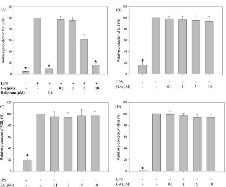
Fig. 2. Dose-dependent inhibition of TNF-α production by GA in RAW 264.7 cells. Cells were treated with various concentrations of GA (0.1- 10µM) and then stimulated with LPS (1 µg/mL). Using ELISA with anti-mouse TNF-α, IL-6, and PGE-2 monoclonal antibodies and a biotinylated secondary antibody measured the TNF-α (A), IL-6 (B), and PGE-2 (C) level, respectively. The absorbance was read using an ELISA reader at 405 nm. (D) The amount of nitrite was measured using the Griess reagent system. One of the quadruplicate experiments is shown. All values were expressed as mean±SD of four separate experiments. *p<0.05, significantly different from LPS alone.
fractionated protein bands were transferred to nitrocellulose paper in an electrophoresis, and they were immunoblotted with the specific antibody. A polyclonal anti-I-κBα antibody was used to assay the I-κBα protein in the cytosol. In addition, NF- κB protein in the cytosolic and nuclear fractions was assayed with a polyclonal anti-Rel A (p65) antibody. The secondary antibody was alkaline phosphatase-conjugated anti-rabbit antibody.
The nitrocellulose paper was developed using 5-bromo-4- chloro-3-indolylphosphate/4-nitroblue tetrazolium chloride.
Scanning densitometry. Scanning densitometry was performed with an imaging densitometer (Model GS-700, Biorad). The area of each lane was integrated using BIO-CAPT software (version 1.0), followed by background subtraction.
Statistics. All values expressed as mean±SD were obtained from three or four separated experiments. Statistical significance was tested by one-way analysis of variance followed by Dunnett’s test, and p<0.05 was considered statistically significant.
The values for the concentration required for 50% inhibition (IC50) were calculated by nonlinear regression analysis to
evaluate the anti-inflammatory potency of GA [Bowen and Jerman, 1995].
Fig. 3. No effect of various concentrations of GA on cell viability in RAW 264.7 cells. The cells (2.5×10⁵ cells/well) were seeded in a 96-well plate, and treated together with LPS and GA (1-50µM), and incubated for 4 hr at 37oC. Cell viability was measured by MTT reduction assay. All values were expressed as mean±SD of three separate experiments.
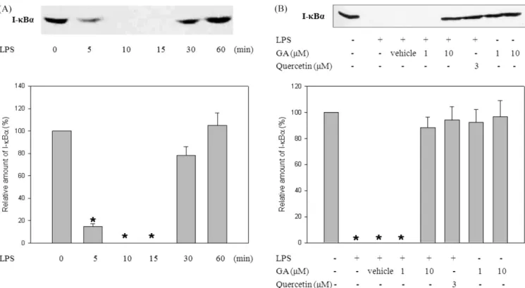
Fig. 4. The suppression of LPS-induced I-κBα degradation by GA in RAW 264.7 cells. (A) A representative immunoblot shows the time- dependent I-κBα degradation induced by LPS treatment. Cells were stimulated with LPS (1 µg/mL) for indicated times. Cellular extract were analyzed by Western blotting with an anti-I-κBα polyclonal antibody. One of the four representative experiments is shown. (B) A representative immunoblot shows the effect of GA on LPS-induced I-κBα degradation. Cells were treated with GA (10 µM) and then stimulated with LPS (1 µg/
mL) for 15 min. Cellular extract were analyzed by Western blotting with an anti-I-κBα polyclonal antibody. Quercetin (Sigma) was used as a positive control. One of the four representative experiments is shown. Using scanning densitometry assessed band intensities in the immunoblots.
All values were expressed as mean±SD of three separate experiments. *p<0.05, significantly different from no stimulation.
Results
Effects of GA on TNF-α mRNA synthesis and protein expression. The effects of GA on the production of TNF-α mRNA synthesis and subsequently expressed protein accumulation were examined in RAW 264.7 cells stimulated with LPS by using methods of RT-PCR and ELISA (Fig. 1 and 2A).
Rolipram (Sigma), employed as a positive control, reduced the production of TNF-α by 85 % even at a concentration of 1 µg/
mL. The 10µM of GA down-regulated TNF-α mRNA synthesis
by 72% in response to an LPS challenge (Fig. 1). The results with ELISA assay for TNF-α production indicated that GA suppressed TNF-α production in a dose-dependent manner with IC50 of 5.4µM (Fig. 2A).
It was also investigated whether GA has an influence on the production of other inflammatory mediators in RAW 264.7 cells. However, GA didn’t affect the production of IL-6, NO, and PEG-2 in RAW 264.7 cells stimulated with LPS (Fig. 2B, C, and D). These findings suggest that GA has a selective influence on the suppression of TNF-α production.
Cell viability was also measured by the MTT assay to determine whether the inhibitory effects of GA on TNF-α production were attributed to nonspecific cytotoxicity. Up to 50 µM of GA, a concentration at which TNF-α production was completely blocked, no significant cytotoxicity was observed (Fig. 3).
Effects of GA on degradation of I-κBα and subsequent nuclear translocation of NF-κB. A crucial step in the transcriptional activation of TNF gene is to need the mobilization of a nuclear factor (NF-κB) from cytoplasm into nucleus. To elucidate a possible mechanism of inhibitory effects of GA on the TNF-α production, influences on the I-κBα degradation and subsequent nuclear translocation of NF-κB were investigated.
The Western blot analysis was performed to measure the degree of the I-κBα degradation and subsequent nuclear translocation of NF-κB (Fig. 4 and 5). As a positive control, quercetin (1 µg/
mL) was used. Treatment with LPS led to the rapid disappearance of the immuno-reactive I-κBα band within 15 min; this band returned to basal levels within 60 min (Fig. 4A). In cells stimulated with LPS for 15 min, the treatment of GA prevented the degradation of I-κBα significantly (Fig. 4B). Furthermore, the treatment of GA (10µM) suppressed the translocation of subunit Rel A of NF-κB into the nucleus (Fig. 5B). It was also noted that the amount of Rel A in the cytosolic fraction increased significantly by the treatment of 10µM GA (Fig. 5A).
Disscussion
In the present study, the effects of GA on the production of inflammatory mediators in LPS-stimulated RAW 264.7 cells were examined. Pretreatment of RAW 264.7 cells with GA in vitro induced the significant decrease in TNF-α production in response to LPS stimulation, whereas GA didn’t affect the production of IL-6, NO, and PEG-2 in RAW 264.7 cells stimulated with LPS (Fig. 2). The suppressive activity of GA on TNF-α production is correlated with the degree of reduction on TNF-α mRNA synthesis (Fig. 1). A transcription factor, NF-κB, is a candidate for regulation of TNF-α gene. Activation of NF- κB is depending on the phosphorylation and degradation of I- κB, an endogenous inhibitor, which binds to NF-κB in the Fig. 5. The suppression of nuclear translocation of NF-κB (Rel A)
by GA in RAW 264.7 cells. A representative Western blot analysis indicates Rel A protein level in cytosolic (A) and nuclear (B) fractions.
Cells were treated with GA (10µM) and then stimulated with LPS (1 µg/mL). Cellular extract were analyzed by Western blotting with an anti-Rel A polyclonal antibody. The same antibody was used in both (A) and (B). Quercetin (Sigma) was used as a positive control. One of the three representative experiments is shown. (C) The relative Rel A protein level in cytosolic and nuclear fractions of RAW 264.7 cells.
Using scanning densitometry assessed band intensities in the immunoblots. All values were expressed as mean±SD of three separate experiments. *p<0.05, significantly different from LPS alone.
cytoplasm. Then the NF-κB eventually binds to the promoter region of its target genes of TNF-α in nucleus. As the data obtained from the Western blot analyses show, the treatment of GA almost abolished LPS-induced I-κBα degradation (Fig. 4).
In the consistency with this observation, the amount of subunit Rel A of NF-κB translocated into the nucleus was reduced by treatment with GA (10µM) (Fig. 5). These findings indicated that the selective inhibition of TNF-α production by GA might be attributed to the direct blocking of the production of TNF-α resulting from both the I-κBα degradation and the nuclear translocation of NF-κB.
NF-κB plays a crucial role in inflammatory responses through the regulation of genes encoding pro-inflammatory cytokines, adhesion molecules, chemokines, growth factors and inducible enzymes [Barnes and Karin, 1997]. However, in the present study, the nuclear translocation of NF-κB didn’t affect the production of IL-6, NO, and PEG-2 in RAW 264.7 cells stimulated with LPS (Fig. 2). A further study will be needed to explain the reason of these observations.
GA exhibits anti-inflammatory properties in different animal models [Capasso et al., 1983; Amagaya et al., 1984; Khaksa et al., 1996]. For example, Capasso et al. [1983] reported that oral administration of GA to rats resulted in inhibition of carrageenan-induced inflammation. So far, it was postulated that the anti-inflammatory activity of GA is probably due to the inhibition of the enzyme 11β-hydroxysteroid hydroxygenase [Walker and Edwards, 1991].
It has been also reported that GA exerted an anti- inflammatory action by inhibiting the generation of reactive oxygen species by neutrophils, the most potent inflammatory mediator at the site of inflammation [Akamatsu et al., 1991].
Furthermore, the derivatives of GA with a 3, 4-seco-structure or a lactone moiety (Fig. 6) exhibited potent inhibitory effect on superoxide anion generation in rat neutrophils and TNF-α formation in RAW 264.7 cells [Maitraie et al., 2009].
Compound 1, a 30-isoproylcarbamoyl seco-compound exhibited potent inhibitory effect on NO accumulation while compound 2 and 3 revealed suppressive effect on TNF-α formation in RAW Fig. 6. The chemical structures of β-glycyrrhetinic acid and its derivatives.
264.7 cells (Fig. 6).
Besides the well-known glucocorticoid-like activities, the present study demonstrated the anti-inflammatory potency of GA through the suppression of TNF-α production and its mechanism of action. Therefore, this report might provide the possible mechanism to explain the reported anti-inflammatory activities of GA in animal models.
Acknowledgments
I thank Dr. Miyoung Park for valuable comments on this manuscript.
References
Akamatsu H, Komura J, Asada Y, and Niwa Y (1991) Mechanism of anti-inflammatory action of glycyrrhizin: effect on neutrophil functions including reactive oxygen species generation. Planta Med 57, 119-121
Amagaya S, Sugishita E, Ogihara Y, Ogawa S, Okada K, and Aizawa T (1984) Comparative studies of the stereoisomers of glycyrrhetinic acid on anti-inflammatory activities. J Pharmacobiodyn 7, 923-8
Barnes PJ and Karin M (1997) NF-κB: a pivotal transcription factor in chronic inflammatory disease. N Engl J Med 336, 1066-1071
Bowen WP and Jerman JC (1995) Nonlinear-regression using spreadsheets. TIPS 16, 413-7
Capasso F, Mascolo N, Autore G, and Duraccio MR (1983) Glycyrrhetinic acid, leucocytes and prostaglandins. J Pharm Pharmacol 35, 332-5
Hasko G, Nemeth ZH, Szabo C, Zsilla G, Salzman AL, and Vizi ES (1998) Isoproterenol inhibits IL-10, TNF-α, and nitric oxide production in RAW 264.7 macrophages. Brain Res Bull 45, 183-7
Khaksa G, Zolfaghari ME, Dehpour AR, and Samadian T (1996) Anti-inflammatory and anti-nociceptive activity of disodium glycyrrhetinic acid hemiphthalate. Planta Med 62, 326-8 Kimura M, Watanabe H, and Abo T (1995) Selective activation of
extrathymic T cells in the liver by glycyrrhizin. Biotherapy 5, 167-76
Kobayashi M, Schmitt DA, Utsunomiya T, Pollard RB, and Suzuki
F (1993) Inhibition of burn-associated suppressor cell generation by glycyrrhizin through the induction of contrasuppressor T cells. Immunol Cell Biol 71, 181-9 Kondo Y and Takano F (1994) Nitric oxide production in mouse
peritoneal macrophages enhanced with glycyrrhizin. Bio Pharm Bull 17, 759-61
Laemmli UK (1970) Cleavage of structural proteins during assembly of the head of the bacteriophage T4. Nature (Lond ) 227, 680-5
Maitraie D, Hung CF, Tu HY, Liou YT, Wei BL, Yang SC, Wang JP, and Lin CN (2009) Synthesis, anti-inflammatory, and antioxidant activities of 18 beta-glycyrrhetinic acid derivatives as chemical mediators and xanthine oxidase inhibitors. Bioorg Med Chem 17, 2785-2792
Nabika T, Terashima M, Momose I, Hosokawa Y, Nagasue N, and Tanigawa Y (1999) Synergistic effect of ubiquitin on lipopolysaccharide-induced TNF-alpha production in murine macrophage cell line RAW 264.7 cells. Biochim Biophys Acta 1450, 25-34
Ohuchi K, Kamada Y, Levine L, and Tsurufuji S (1981) Glycyrrhizin inhibits prostaglandin E2 production by activated peritoneal macrophages from rats. Prostaglandins Med 7, 457- 63
Sekut L and Connolly KM (1996) Pathophysiology and regulation of TNF in inflammation. Drug News Perspectives 9, 261-9 Ribeiro RA, Vale ML, Ferreira SH, and Cunha FQ (2000)
Analgesic effect of thalidomide on inflammatory pain. Eur J Pharmacol 391, 97-103
Yamamura Y, Kawakami J, Santa T, Kotaki H, Uchino K, Sawada Y, Tanaka N, and Iga T (1992) Pharmacokinetic profile of glycyrrhizin in healthy volunteers by a new high-performance liquid chromatographic method. J Pharm Sci 81, 1042-6 Yim JS, Kim YS, Moon SK, Cho KH, Bae HS, Kim JJ, Park EK,
and Kim DH (2004) Metabolic activities of ginsenoside Rb1, baicalin, glycyrrhizin and geniposide to their bioactive compounds by human intestinal microflora. Biol Pharm Bull 27, 1580-3
Walker BR and Edwards CRW (1991) 11β-hydroxysteroid dehydrogenase and enzyme-mediated receptor protection: life after liquorice? Clin Endocrinol 35, 281-9
Zhang YH, Isobe K, Nagase F, Lwin T, Kato M, Hamaguchi M, Yokochi T, and Nakashima I (1993) Glycyrrhizin as a promoter of the late signal transduction for interleukin-2 production by splenic lymphocytes. Immunology 79, 528-34