Molecular Mechanisms Involved in Peptidoglycan-induced Expression of Tumor Necrosis Factor-α in Monocytic Cells
Ji-Young Jeong, Yonghae Son, Bo-Young Kim and Koanhoi Kim*
Department of Pharmacology, School of Medicine, Pusan National University, Yangsan, Gyeongnam 50612, Korea Received September 3, 2019 /Revised October 8, 2019 /Accepted November 8, 2019
Peptidoglycan (PG) is found in atheromatous lesions of arteries, where monocytes/macrophages ex- press inflammatory cytokines, including tumor necrosis factor-alpha (TNF-α). This study investigated the effects of PG on TNF-α expression and examined possible cellular factors involved in TNF-α upregulation. The overall aim was to identify the molecular mechanisms underlying inflammatory re- sponses to bacterial pathogen-associated molecular patterns in the artery. Exposure of human THP-1 monocytic cells to PG enhanced the secretion of TNF-α and induced its gene transcription. Inhibition of TLR-2/4 with OxPAPC significantly inhibited TNF-α gene expression, whereas inhibition of LPS by polymyxin B did not. The PG-induced expression of TNF-α was also significantly suppressed by pharmacological inhibitors that modulate activities of cellular signaling molecules; for example, U0126 (an ERK inhibitor), SB202190 (a p38 MAPK inhibitor), and SP6001250 (a JNK inhibitor) significantly attenuated PG-induced transcription of TNF-α and secretion of its gene product. TNF-α expression was also inhibited by rapamycin (an mTOR inhibitor), LY294002 (a PI3K inhibitor), and Akt inhibitor IV (an Akt inhibitor). ROS-regulating compounds, like NAC and DPI, also significantly attenuated TNFα expression induced by PG. These results suggest that PG induces TNF-α expression in mono- cytes/macrophages by multiple molecules, including TLR-2, PI3K, Akt, mTOR, MAPKs, and ROS.
Key words : Gene expression, monocytic cells, peptidoglycan, signaling molecules, tumor necrosis
factor-α
*Corresponding author
*Tel : +82-51-510-8064, Fax : +82-51-510-8068
*E-mail : [email protected]
This is an Open-Access article distributed under the terms of the Creative Commons Attribution Non-Commercial License (http://creativecommons.org/licenses/by-nc/3.0) which permits unrestricted non-commercial use, distribution, and reproduction in any medium, provided the original work is properly cited.
Journal of Life Science 2019 Vol. 29. No. 11. 1251~1257 DOI : https://doi.org/10.5352/JLS.2019.29.11.1251
서 론
Tumor necrosis factor-alpha (TNF-α)는 전신 염증과 관련 된 사이토카인이며 주로 homotrimer로 배열된 212개의 아미 노산의 type II 세포막 관통 단백질로 합성된다. 그리고 세포막 에 결합한(membrane-bound) 형태의 TNF-α가 metallopro- tease TNF-α converting enzyme (TACE)에 의해 가수분해되 어 soluble homotrimeric TNF (sTNF)로 분비된다[7]. TNF-α 는 두 가지의 수용체, 즉 TNF receptor type 1 (TNF-R1)과 type 2 (TNF-R2)와 결합, 작용함으로써 급성단계의 반응을 자 극한다. TNF-R1는 대부분의 조직에서 발현되고 수용성 삼량 체(soluble trimeric) 그리고 membrane-bound 형태의 TNF-α 에 의해 활성화되는 반면, TNF-R2는 면역계 세포에서만 발견 이 되고 TNF homotrimer의 memebrane-bound TNF-α에 반 응한다[23].
TNF-α와 그 수용체 TNF-R1과의 결합은 죽상경화 촉진효
과로 이어지는 신호전달체계를 활성화시킨다. TNF-α는 사이 토카인과 CXC3를 포함한 케모카인의 생산을 유도하고 T 림프 구의 활성화와 혈관평활근세포의 확장과 이동 그리고 내피세 포의 접착물질 발현을 증가시켜 단핵구/대식세포의 혈관벽 접착과 동맥의 intima로의 침윤을 초래한다[7, 21]. 사람의 죽 상경화 플락에서 TNF-α의 존재는 본 질환의 발병에 TNF-α가 관련되어 있음을 뒷받침한다. TNF-α는 모든 단계의 죽상경화 병변에서 발견된다[2, 18]. 또한, 동물 연구결과에 의하면 TNF- α 가 혈관질환과 연관되어 있다. ApoE 생쥐에서 TNF-α의 결핍 은 apoE-null (apoE
-/-) 생쥐보다 덜 진행된 죽상경화증을 보여 준다[4, 17]. 따라서 TNF-α 발현에 대한 연구 결과는 죽상경화 의 발병 기전을 이해하는데 중요하다.
펩티도글리칸(peptidoglycan)은 그람양성 세균의 세포벽을
구성하는 주요 물질이며 보통 사람의 소화관 및 다른 점막의
flora에 풍부하게 존재한다. 점막이 아닌 곳에서 선천면역계는
펩티도글리칸을 세균성 pathogen-associated molecular pat-
tern (PAMP)로 인지하고 Toll-like receptors (TLR)를 통하여
염증을 촉진한다[24, 26]. 또한 펩티도글리칸은 단핵구의 αMβ
2-integrin의 발현을 유도하고 β2-integrin-dependent 이동을
증가시킨다[16]. 펩티도글리칸은 내피세포에 의한 접착 물질
의 발현과[6] 단핵구/대식세포에 의한 염증성 사이토카인과
케모카인을 높일 수 있다[12, 25]. 펩티도글리칸이 사람의 죽상
경화증 병변, 특히 대식세포가 많은 부분에 존재하기 때문에
펩티도글리칸이 추가적인 염증성 요인으로 작용할 것이라 추 정한다[11]. 그리고 펩티도글리칸이 염증반응을 유도하는 신 호경로의 설명은 죽상경화에서 세균성 PAMPs의 역할을 규명 하는데 중요하다. 그러나 펩티도글리칸이 TNF-α 발현을 유도 하는 분자적 과정은 아직 밝혀지지 않았다.
본 연구에서는 펩티도글리칸이 단핵세포의 TNF-α 분비에 미치는 영향을 연구하였다. 실험적으로 펩티도글리칸이 단핵 세포주인 THP-1 세포의 TNF-α 분비를 증가시키고 그리고 전 사체를 양을 변화시키는지를 조사하였다. 그리고 펩티도글리 칸에 의한 TNF-α 발현에 TLR2, Akt, mammalian target of ra- pamycin (mTOR), mitogen-activated protein kinases (MAPKs), and reactive oxygen species (ROS)의 연관성을 규명하였다.
재료 및 방법
세포 및 시약
American Type Culture Collection (ATCC, Manassas, VA, USA)에서 인간 급성 monocytic leukemia THP-1 세포주를 구 입하였고 ATCC에서 제안한 방법에 따라 유지하였다. Staphy-
lococcus aureus에서 분리한 펩티도글리칸 그리고 polymyxinB와 oxidized 1-palmitoyl-2-arachidonosyl-sn-phosphatidylcho- line(OxPAPC)은 InvivoGen (San Diego, CA, USA)에서 구입 하였다. Endotoxin-free bovine serum albumin (BSA), LY294002, diphenyleneiodonium chloride (DPI), N-acetylcysteine (NAC), rapamycin, 그리고 SP600125는 Sigma-Aldrich Co. (St. Louis, MO, USA)에서 구입하였다. U0126, SB202190, 그리고 Akt in- hibitor IV (Akti IV)는 Cell Signaling Technology (Danvers, MA, USA)에서 구입하였다.
세포의 처치
Inhibition 실험을 위해서, THP-1 세포에 상기 화학 물질을 1시간 동안 전 처리하였다. 화학 물질이 존재하는 상태에서 펩티도글리칸을(1 μg/ml) 9시간 동안 처리하고 배지에 분비된 TNF-α의 양을 enzyme linked immunosorbent assay (ELISA) 로 측정하고 reverse transcription (RT)-polymerase chain re- action (PCR) 혹은 real-time PCR을 통하여 세포 내 TNF-α 유전자의 전사체를 조사하였다.
ELISA
ELISA kits (BD Biosciences, San Diego, CA, USA)를 이용 하여 분비된 TNF-α의 양을 분석하였다. THP-1 세포를 0.1%
BSA를 첨가한 RPMI medium 1640에 overnight incubation시 키고, 펩티도글리칸을 처리하고 일정시간 후 세포배양 배지를 모았다. 세포배양 배지와 TNF-α의 standard를 TNF-α에 대한 단일클론 항체로 미리 코팅되어 있는 microtiter plate에 첨가 하고, 2시간 배양 후에 plate를 세척하고 TNF-α에 대한 특정
enzyme-conjugated polyclonal 항체와 함께 배양하였다.
Plate를 여러 번 세척한 후 substrate를 첨가하고 색의 강도를 측정하였다. Standard curve를 만들고 이를 바탕으로 배지에 존재하는 TNF-α의 양을 정하였다.
Reverse transcription-polymerase chain reaction (RT- PCR)
TRIzol
TMReagent를 사용하여 THP-1 세포로부터 Total RNAs를 얻고 Moloney murine leukemia virus reverse tran- scriptase를 이용하여 역 전사하였고, PCR은 94℃ for 30 s, 55
℃ for 30 s, 72℃ for 30 s 조건에서 25cycles를 수행하였다.
TNF-α의 primer는 5- CAGCCTCTT CTCCTTCCTGA-3 (for- ward)와 5-GGAAGACCCCTCCCAGATAG-3 (reverse)이고, glyceraldehyde-3-phosphate dehydrogenase (GAPDH)의 pri- mer는 5-AAGCTCTGCGTGACTGTCCT-3 (forward)와 5-GC TTGC TTCTTTTGGTT TGG-3 (reverse)이었다. PCR 산물은 2% agarose gel에서 전기영동하여 분리하였고 ethidium bro- mide 염색을 이용하여 PCR 생산물을 확인하였다.
Quantitative Real-time PCR
Applied Biosystems Prism 7900 Sequence Detection Sys- tem (Applied Biosystems, Foster City, CA) 장비를 사용하여 real-time PCR을 수행하였다. 384-well plate의 각 well에 SYBR Green PCR Master Mix 와 TNF-α 및 GAPDH의 forward pri- mer and reverse primer (각각 10 pM)를 넣고 50 °C for 2 min, 95 °C for 10 min 반응시킨 다음 95 °C for 30 s, 60 °C for 30 s 조건에서 40 cycles 을 수행하였다. TNF-α의 primer는 5-ATGAGCACTGAAAGCATGATCC-3 (forward)와 5-GAG GGCTGATTAG AGAGAGGTC-3 (reverse)이고, GAPDH의 primer는 5-ATGGGGAAGGTGAAGGTCG-3 (forward) 와 5-GGGGTCAT TGATGGCAACAATA-3 이었다.
통계처리
통계학적 GraphPad PRISM, version 5.0 (GraphPad Soft- ware Inc., San Diego, CA, USA)를 이용하였고, p<0.05는 통계 학적으로 유의미한 것으로 처리하였다.
결 과
펩티도글리칸에 의해 TNF-α 발현의 증가
펩티도글리칸이 단핵세포의 TNF-α 발현에 미치는 영향을 알아보기 위해, TNF-α 유전자의 전사 정도를 조사하였다(Fig.
1A, Fig. 1B). THP-1 세포는 TNF-α 유전자의 전사체를 발현하
였고 펩티도글리칸에 의하여 TNF-α 전사체가 증가하였다. TNF-
α 전사의 유도는 펩티도글리칸 처리 후 3시간째부터 관찰되었
고 9시간까지 유지되었다. TNF-α 전사체의 발현은 펩티도글
A B
C
D
Fig. 1. Expression of TNF-α at the messenger and protein levels in response to peptidoglycan (PG). THP-1 cells (1×10⁶ cells/ml) were incubated for indicated time periods with 1 μg/ml PG (A) or for 9 hr with indicated amount of PG (B), and TNF-α gene transcripts were amplified by RT-PCR. Following stimulation of THP-1 cells (1×10⁶ cells/ml) for 9 hr with or without (control) the indicated concentration of PG, the amount of TNF-α released into the medium and levels of TNF-α transcripts within cells were assessed by ELISA (C) and real-time PCR (D), respectively. Data are expressed as mean ± SD (n=3 replicates/group). **p<0.01 vs. control; ***p<0.001 vs.
control.
A
B
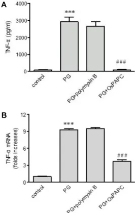
Fig. 2. Effects of OxPAPC and polymyxin B on peptidoglycan (PG)-mediated up-regulation of TNF-α. THP-1 cells were stimulated for 9 hr with or without PG (1 μg/ml) in the presence or absence of OxPAPC (30 μg/ml) and pol- ymyxin B (10 mg/ml). The amount of TNF-α released into the medium and TNF-α gene transcripts were as- sessed by ELISA (A) and real-time PCR (B), respectively.
Data are expressed as mean ± SD (n=3 replicates/group).
***p<0.001 vs. control; ###p<0.001 vs. PG.
리칸 10 ng/ml이 있을 때부터 유도되었고, 100과 1,000 ng/ml 일 때 더 분명히 유도되었다. 또한 펩티도글리칸이 TNF-α 단 백질의 분비에 어떤 영향을 미치는지 조사하였다(Fig. 1C).
THP-1 세포가 계속적으로 적은 양의 TNF-α를 분비하였고 펩 티도글리칸이 TNF-α 분비를 상당히 증가시켰다. 펩티도글리 칸이 없는 상태에서 배양된 대조군 세포와 비교하여 볼 때, 분비된 TNF-α의 양이 100 ng/ml일 때 2.8배, 1000 ng/ml일 때 15.6배 증가하였다. 또한 real-time PCR 결과에 의하면 펩티 도글리칸의 농도에 비례하여 TNF-α 전사체의 양이 증가하였 다(Fig. 1D).
펩티도글리칸에 의한 TNF-α 발현에 TLR2/4의 억제가 미 치는 영향
TLRs이 TNF-α 발현을 매개하는지 조사하기 위해 TLR2/4 억제제인 OxPAPC를 사용하였다. OxPAPC 존재 하에서 펩티 도글리칸에 의한 TNF-α의 분비는 대조군 세포만큼 거의 완벽 히 차단되고 또한 유도된 TNF-α 전사체의 발현 역시 OxPAPC 존재 하에 억제되었다. 펩티도글리칸을 준비할 때 염증성 사 이토카인과 케모카인의 분비를 증가시키는 LPS에 오염될 수 있다. 그러므로, LPS 억제제로 알려진 polymyxin B를 사용하 여 LPS가 TNF-α의 증가에 영향을 주는지 조사해보았다. Poly-
myxin B는 펩티도글리칸에 의한 TNF-α 분비와 전사체 발현 을 약화시키지 않았다(Fig. 2A, Fig. 2B).
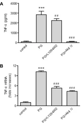
펩티도글리칸에 의한 TNF-α 발현에 mTOR과 Akt의 역할 펩티도글리칸은 인산화를 통하여 Akt pathway를 활성화 시킨다[13]. 그러므로 LY294002와 Akti IV, 두 가지 억제제를 사용하여 TNF-α 발현에 Akt가 관여하는지 조사하였다(Fig.
3A, B). LY294002는 phosphoinositide 3-kinases (PI3Ks)의 re- versible 억제제이고 Akti IV는 Akt protein kinase의 저해제이 다. Akti IV와 LY294002 둘 다 펩티도글리칸에 의한 TNF-α 유전자 발현을 약화시키고 TNF-α 분비를 저해하였다. 특히 Akti IV를 처리하였을 때 TNF-α의 분비가 완전히 차단되었다.
Akt는 mTOR의 활성화를 통해 효과를 나타낸다[8]. 그러므
로 mTOR의 저해제로 알려진 rapamycin을 이용하여 TNF-α
발현에 mTOR가 관여하는지 실험하였다(Fig. 4A, Fig. 4B). 펩
티도글리칸으로 인한 TNF-α 분비 및 유전자 전사는 rapamy-
cin이 존재할 때 현저히 감소하였다.
A
B
Fig. 3. Effects of LY294002 and Akti IV on peptidoglycan (PG)- mediated up-regulation of TNF-α. Following stimulation of THP-1 cells for 9 hr with PG (1 μg/ml) in the absence or presence of LY294002 and Akti IV (10 μM each), the amount of TNF-α released into the medium and TNF-α gene transcripts were assessed by ELISA (A) and re- al-time PCR (B), respectively. Data are expressed as mean
± SD (n=3 replicates/group). ***P<0.001 vs. control; ##p<
0.01 vs. PG; ###p<0.001 vs. PG.
A
B
Fig. 4. Effects of rapamycin on peptidoglycan (PG)-mediated up-regulation of TNF-α. After stimulation of THP-1 cells for 9 hr with PG (1 μg/ml) in the absence or presence of with rapamycin (100 nM), the amount of TNF-α re- leased into the medium and TNF-α gene transcripts were assessed by ELISA (A) and real-time PCR (B), respectively. Data are expressed as mean ± SD (n=3 rep- licates/group). ***p<0.001 vs. control; ##p<0.01 vs. PG;
###p<0.001 vs. PG.
펩티도글리칸에 의한 TNF-α 발현에 MAPKs의 역할 펩티도글리칸이 extracellular signal-regulated kinase (ERK), p38 MAPK, 그리고 c-jun N-terminal kinase (JNK)를 인산화 시키므로[13], MAPKs가 TNF-α 발현에 관여하는지 조사하기 위하여 다음과 같은 억제제들을 사용하였다: SB202190 (p38 MAPK 억제제), SP600125 (JNK 억제제), 그리고 U0126 (ERK 억제제) 세가지 억제제는 펩티도글리칸에 의한 TNF-α 분비를 현저히 줄였고, 또한 TNF-α 유전자 전사 역시 약화시켰다(Fig.
5A, Fig. 5B).
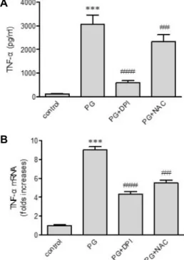
펩티도글리칸에 의한 TNF-α 발현에 ROS의 역할 ROS가 TNF-α 발현에 역할을 하는지 조사하기 위해, NAC 와 DPI를 이용하였다(Fig. 6A, Fig. 6B). DPI는 NADPH oxi- dase의 저해제로 펩티도글리칸에 의해 유도된 TNF-α 유전자 전사를 약화시킬 뿐만 아니라 펩티도글리칸으로 인한 TNF-α 의 분비를 상당히 저해시켰다. ROS의 direct scavenger NAC 도 역시 단백질 단계에서 TNF-α 발현에 영향을 주고, 펩티도 글리칸 인한 TNF-α 전사를 상당히 약화시켰다.
고 찰
본 연구에서는 죽상경화 병변에 존재하는 성분인 펩티도글 리칸이 사람의 THP-1 세포주 에서 TNF-α의 발현을 전사와 단백질 단계에서 증가시킨다는 것을 밝혔다. 이 연구 결과는 ribonuclease protection assay를 이용하여 인간 혈액 단핵구 에서 펩티도글리칸과 LPS이 염증성 사이토카인을 유도한다 고 보고한 연구와 일관되는 결과이다. LPS가 펩티도글리칸에 의한 TNF-α 발현에 관여하는지 조사하기 위해 이번 연구에서 는 LPS와 결합하여 LPS의 생물학적 효과를 저해한다고 알려 진 polymyxin B를 이용하였다[5]. Polymyxin B는 전사와 단 백질 수준 모두에서 펩티도글리칸에 의한 TNF-α 발현에 영향 을 끼치지 않는다는 것을 밝혔고 이는 이번 연구에서 관찰된 TNF-α의 증가는 펩티도글리칸에 의해 유도되었다는 것을 나 타낸다.
펩티도글리칸이 TNF-α 발현을 유도한다는 것은 사실이지 만, TNF-α 발현과 관련된 세포 인자는 아직 밝혀지지 않았다.
본 연구는 펩티도글리칸에 의한 TNF-α 발현에 역할을 하는
세포 요소를 밝히는 실험을 수행하였다. 펩티도글리칸은 염증
A
B
Fig. 5. Effects of inhibitors of MAPKs on peptidoglycan (PG)- mediated up-regulation of TNF-α. THP-1 cells were stimulated for 9 hr with or without PG (1 μg/ml) in the absence or presence of the indicated MAPKs inhibitors (10 μM each). The amount of TNF-α released into the medium and TNF-α gene transcripts were assessed by ELISA (A) and real-time PCR (B), respectively. Data are expressed as mean ± SD (n=3 replicates/group). ***p<0.001 vs. control; ##p<0.01 vs. PG; ###p<0.001 vs. PG.
A
B
Fig. 6. Effects of ROS quenchers on peptidoglycan (PG)-mediated up-regulation of TNF-α. THP-1 cells were stimulated for 9 hr with or without PG (1 μg/ml) in the absence or presence of DPI (10 μM) and NAC (5 mM), after which The amount of TNF-α released into the medium and TNF-α gene transcripts were assessed by ELISA (A) and real-time PCR (B), respectively. Data are expressed as mean ± SD (n=3 replicates/group). ***p<0.001 vs. control;
##p<0.01 vs. PG; ###p<0.001 vs. PG.
성 반응으로 이어지는 신호전달 경로의 활성화를 유도하는 TLR2에 의해 인지되는 세균성 PAMP이므로[1, 10, 24] TLR2/
4의 저해제인 OxPAPC를 이용하여 TNF-α 발현에 영향을 주 는지 조사하였다. OxPAPC는 TNF-α의 분비와 유전자의 전사 를 완벽하게 저해하였다. Polymyxin B에 의해서는 아니지만 OxPAPC에 의한 TNF-α 발현의 완벽한 저해는 TLR2가 펩티도 글리칸에 의해 유도된 TNF-α 발현에 책임이 있음을 의미한다.
펩티도글리칸은 Akt를 인산화시키는데[13], 이는 펩티도글 리칸에 의하여 kinase가 활성화됨을 의미한다. 그러므로 Akt 와 Akt의 activator인 PI3K가 펩티도글리칸에 의한 TNF-α 발 현에 연관이 있는지 조사하였다[14]. Akt inhibition은 TNF-α 분비를 완벽히 차단하였으며 유전자의 전사 역시 저해하였다.
PI3K inhibition 또한 TNF-α 발현의 약화를 초래하였다. 이러 한 결과들은 PI3K와 Akt 모두 펩티도글리칸에 의한 TNF-α 발현에 필요하다는 것을 의미한다.
Akt는 세포질에서 주로 발견되는 mTOR를 포함하여 pro- tein kinase targets를 통해 생물학적 효과를 가진다[8]. 본 연구 에서 mTOR가 펩티도글리칸에 의한 TNF-α 발현에 관련이 있
는지 조사하였고, mTOR의 저해제인 rapamycin은 TNF-α 유 전자 전사를 약화시킬 뿐만 아니라 TNF-α의 분비를 상당히 감소시키는 확인하였다. 이 결과는 rapamycin이 펩티도글리 칸에 의한 TNF-α 발현을 단백질 수준과 전사 수준에서 저해시 킴을 나타낼 뿐만 아니라 mTOR이 단백질 생성에 책임이 있다 는 사실과 부합한다[19]. 본 연구의 결과와 기존의 보고를 종합 해보면 PI3K 의존적인 Akt/mTOR 경로가 펩티도글리칸에 의 한 TNF-α 증가에 중요한 역할을 한다는 것을 의미한다.
MAPKs는 세포 외 자극들에 반응하고 다양한 세포 활성을 조절하는 serine/threonine-specific protein kinases로 펩티도 글리칸에 의해 활성화 된다[13]. MAPKs는 TLR-2, -4, -9의 활 성화에 대한 반응으로 케모카인을 생성하도록 한다[22]. 이러 한 사실은 TNF-α 발현에서 kinases의 관련을 시사한다. 따라 서 MAPKs가 TNF-α의 발현에 중요한 역할을 하는지 조사하 였고, ERK, p38 MAPK, JNK의 선택적 저해가 단백질 및 유전 자 수준에서 TNF-α 발현을 현저하게 약화시킴을 증명하였다.
이는 TNF-α 발현에 위 kinases가 적극적으로 관여함을 의미한
다. 이러한 사실들을 종합하면 펩티도글리칸에 의한 TNF-α
발현에 ERK, p38 MAPK, 그리고 JNK의 활성이 필수적임을
알 수 있다.
펩티도글리칸은 TLR2를 통해 백혈구에서 ROS 생산을 증 가시킨다[9]. 이것은 ROS가 TLR2 아래의 신호 전달 분자 중 하나라는 것을 나타낸다. 따라서 본 연구에서는 DPI와 NAC 를 이용하여 ROS가 TNF-α 발현에 연관이 있는지를 조사하였 다. DPI는 ROS를 생산하는 NADPH의 저해제로 ROS의 형성 을 억제한다[15]. 세포 내 glutathione의 충족을 위한 cysteine source로 작용하는 thiol 복합체인 NAC는 ROS의 direct scav- enger로 작용한다[20]. 본 연구결과에 의하면 DPI와 NAC는 펩티도글리칸에 의한 TNF-α의 증가를 상당히 약화시켰다. 이 러한 사실을 종합하면 ROS가 펩티도글리칸에 의한 TNF-α 증 가에 적극적으로 관여하고 있다는 것을 의미한다.
본 연구에서는 THP-1 세포가 펩티도글리칸에 노출되면 TNF-α 분비가 증가하고 TNF-α 유전자의 전사가 유도된다는 결과를 보여주었고, 그리고 이 과정에 TLR2, PI3K, Akt, mTOR, ERK, p38 MAPK, JNK, 그리고 ROS가 관여한다는 것을 밝혔 다. 그러나 이번 연구는 이러한 분자들이 TNF-α 발현에 독립 적으로 또는 협동적으로 역할을 하는지 정확하게 밝혀내지 못했다. 따라서 위에서 거론된 신호전달 물질들 간의 관련성 이나 가능한 crosstalk의 유형을 밝히는 연구가 필요하다.
감사의 글
이 과제는 부산대학교 기본연구지원사업(2년)에 의하여 연 구되었음.
References
1. Akira, S., Uematsu, S. and Takeuchi, O. 2006. Pathogen rec- ognition and innate immunity. Cell 124, 783-801.
2. Barath, P., Fishbein, M. C., Cao, J., Berenson, J., Helfant, R.
and Forrester, J. S. 1990. Detection and localization of tumor necrosis factor in human atheroma. Am. J. Cardiol. 65, 297- 302.
3. Black, R. A., Rauch, C. T., Kozlosky, C. J., Peschon, J. J., Slack, J. L. and Wolfson, M. F. 1997. A metalloproteinase disintegrin that releases tumour-necrosis factor-alpha from cells. Nature 385, 729-33.
4. Branen, L., Hovgaard, L., Nitulescu, M., Bengtsson, E., Nils- son, J. and Jovinge, S. 2004. Inhibition of tumor necrosis fac- tor-alpha reduces atherosclerosis in apolipoprotein E knock- out mice. Arterioscler. Thromb. Vasc. Biol. 24, 2137-2142.
5. Cardoso, L. S., Araujo, M. I., Goes, A. M., Pacifico, L. G., Oliveira, R. R. and Oliveira, S. C. 2007. Polymyxin B as in- hibitor of LPS contamination of Schistosoma mansoni re- combinant proteins in human cytokine analysis. Microb. Cell Fact. 6, 1.
6. Dobrina, A., Nardon, E., Vecile, E., Cinco, M. and Patriarca, P. 1995. Leptospira icterohemorrhagiae and leptospire pepti- dolgycans induce endothelial cell adhesiveness for poly- morphonuclear leukocytes. Infect Immu. 63, 2995-2999.
7. Getz, G. S. 2005. Thematic review series: the immune system
and atherogenesis. Immune function in atherogenesis. J.
Lipid Res. 46, 1-10.
8. Hahn-Windgassen, A., Nogueira, V., Chen, C. C., Skeen, J.
E., Sonenberg, N. and Hay, N. 2005. Akt activates the mam- malian target of rapamycin by regulating cellular ATP level and AMPK activity. J. Biol. Chem. 280, 32081-32089.
9. Kavoosi, G., Ardestani, S. K. and Kariminia, A. 2009. The involvement of TLR2 in cytokine and reactive oxygen spe- cies (ROS) production by PBMCs in response to Leishmania major phosphoglycans (PGs). Parasitology 136, 1193-1199.
10. Kawai, T. and Akira, S. 2006. TLR signaling. Cell Death Differ. 13, 816-825.
11. Laman, J. D., Schoneveld, A. H., Moll, F. L., van Meurs, M. and Pasterkamp, G. 2002. Significance of peptidoglycan, a proinflammatory bacterial antigen in atherosclerotic ar- teries and its association with vulnerable plaques. Am. J.
Cardiol. 90, 119-123.
12. Langer, M., Malykhin, A., Maeda, K., Chakrabarty, K., Wil- liamson, K. S. and Feasley, C. L. 2008. Bacillus anthracis pep- tidoglycan stimulates an inflammatory response in mono- cytes through the p38 mitogen-activated protein kinase pathway. PloS One 3, e3706.
13. Lee, S. A., Kim, S. M., Son, Y. H., Lee, C. W., Chung, S. W., Eo, S. K. and Kim, K. 2011. Peptidoglycan enhances secre- tion of monocyte chemoattractants via multiple signaling pathways. Biochem. Biophys. Res. Commun. 408, 132-138.
14. Manning, B. D. and Cantley, L. C. 2007. AKT/PKB signal- ing: navigating downstream. Cell 129, 1261-1274.
15. Miesel, R., Sanocka, D., Kurpisz, M. and Kroger, H. 1995.
Anti-inflammatory effects of NADPH oxidase inhibitors.
Inflammation 19, 347-362.
16. Nijhuis, M. M., Pasterkamp, G., Sluis, N. I., de Kleijn, D.
P., Laman, J. D. and Ulfman, L. H. 2007. Peptidoglycan in- creases firm adhesion of monocytes under flow conditions and primes monocyte chemotaxis. J. Vasc. Res. 44, 214-222.
17. Ohta, H., Wada, H., Niwa, T., Kirii, H., Iwamoto, N. and Fujii, H. 2005. Disruption of tumor necrosis factor-alpha gene diminishes the development of atherosclerosis in ApoE-deficient mice. Atherosclerosis 180, 11-17.
18. Rus, H. G., Niculescu, F. and Vlaicu, R. 1991. Tumor necrosis factor-alpha in human arterial wall with atherosclerosis.
Atherosclerosis 89, 247-254.
19. Shaw, R. J. and Cantley, L. C. 2006. Ras, PI(3)K and mTOR signalling controls tumour cell growth. Nature 441, 424-430.
20. Spagnuolo, G., D'Anto, V., Cosentino, C., Schmalz, G., Schweikl, H. and Rengo, S. 2006. Effect of N-acetyl-L-cys- teine on ROS production and cell death caused by HEMA in human primary gingival fibroblasts. Biomaterials 27, 1803-1809.
21. Tedgui, A. and Mallat, Z. 2006. Cytokines in atherosclerosis:
pathogenic and regulatory pathways. Physiol. Rev. 86, 515- 81.
22. Thobe, B. M., Frink, M., Hildebrand, F., Schwacha, M. G., Hubbard, W. J. and Choudhry, M. A. 2007. The role of MAPK in Kupffer cell toll-like receptor (TLR) 2-, TLR4-, and TLR9-mediated signaling following trauma-hemorrhage. J.
초록:펩티도글리칸에 의한 단핵세포의 Tumor necrosis factor-α 발현 기전 연구
정지영․손용해․김보영․김관회*
(부산대학교 의과대학 약리학교실)
본 연구에서는 펩티도글리칸이 단핵세포의 TNF-α 발현에 미치는 영향을 조사하였고, 또한 펩티도글리칸에 의 한 TNF-α 발현에 관련된 세포의 요소들을 연구하였다. 사람의 단핵세포주인 THP-1 세포를 펩티도글리칸에 노출 시키는 경우 TNF-α 분비 증가뿐만 아니라 TNF-α 유전자 전사를 유도하는 결과를 가져왔다. TLR-2/4의 억제제인 OxPAPC은 펩티도글리칸에 의한 TNF-α의 발현을 저해하였다. 그리고 U0126, SB202190, SP6001250, LY294002, Akti IV, rapamycin, NAC, DPI 같은 약리학적 저해제 또한 TNF-α 발현을 유전자/단백질 수준에서 상당히 약화 시켰다. 그러나 polymyxin B는 TNF-α 발현에 영향을 주지않았다. 따라서 펩티도글리칸이 TLR-2, PI3K, Akt, mTOR, MAPKs, ROS 등을 통하여 단핵세포의 TNF-α 발현을 증가시킴을 확인하였다.
Cell. Physiol. 210, 667-675.
23. Wajant, H., Pfizenmaier, K. and Scheurich, P. Tumor necrosis factor signaling. 2003. Cell Death Differ. 10, 45-65.
24. Wang, Q., Dziarski, R., Kirschning, C. J., Muzio, M. and Gupta, D. 2001. Micrococci and peptidoglycan activate TLR2--
>MyD88-->IRAK-->TRAF-->NIK-->IKK-->NF-kappaB signal transduction pathway that induces transcription of inter- leukin-8. Infect. Immu. 69, 2270-2276.
25. Wang, Z. M., Liu, C. and Dziarski, R. 2000. Chemokines
are the main proinflammatory mediators in human mono- cytes activated by Staphylococcus aureus, peptidoglycan, and endotoxin. J. Biol. Chem. 275, 20260-20267.
26. Yoshimura, A., Lien, E., Ingalls, R. R., Tuomanen, E., Dziar- ski, R. and Golenbock, D. 1999. Cutting edge: recognition of Gram-positive bacterial cell wall components by the in- nate immune system occurs via Toll-like receptor 2. J. Immu- nol. 163, 1-5.