대한외과학회지:제 73 권 제 5 호
□ 증 례 □
Vol. 73, No. 5, November, 2007
430
책임저자:박민호, 전남 화순군 화순읍 일심리 160
519-809, 화순전남대학교병원 외과 Tel: 061-379-7646, Fax: 061-379-7661 E-mail: [email protected]
접수일:2007년 5월 30일, 게재승인일:2007년 7월 24일
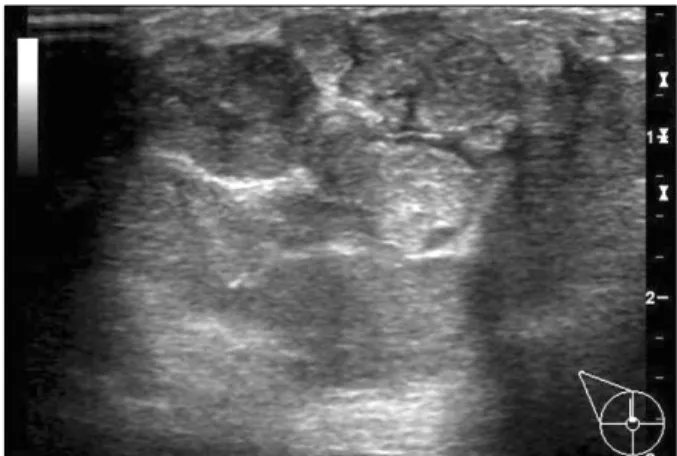
Fig. 1. Breast ultrasonogram showing a 4.5×4.5 cm sized, multi- lobulated, hypoechoic lesion in the right breast.
유방의 악성 선근상피종
전남대학교 의과대학 외과학교실 박 민 호
Malignant Adenomyoepithelioma of the Breast
Min Ho Park, M.D.
An adenomyoepithelioma of the breast is an uncommon tu- mor that is characterized by biphasic lesions, which consist of epithelial and myoepithelial cells. Most adenomyoepi- theliomas are benign, and a malignant transformation is rare.
It is generally limited to the epithelial component but in rare cases, the myoepithelial component may become malignant and give rise to a malignant myoepithelioma. We encoun- tered a case of a 29-year-old woman who presented with both an epithelial and myoepithelial carcinoma of the breast in an adenomyoepithelioma. The malignancy was evidenced by the presence of pleomorphism, a high mitotic rate and local invasion. Immunohistochemical staining demonstrated the expression of cytokeratin in epithelial cells and actin in myoepithelial cells. We report this case of a malignant ad- enomyoepithelioma of the breast with review of the relevant literature. (J Korean Surg Soc 2007;73:430-433)
Key Words: Breast, Malignant, Adenomyoepithelioma 중심 단어: 유방, 악성, 선근상피종
Department of Surgery, Chonnam National University Medical School, Gwangju, Korea
서 론
선근상피종은 1970년 Hamperl이 처음으로 보고하였으며 (1) 유방에 발생하는 경우는 드물어 전세계적으로 약 140예 가 보고되었다. 선근상피종은 대부분 양성으로 알려져 있 어 지금까지 선근상피종에서 발생한 악성종양은 약 20예에 서 문헌에 보고되었다.(2,3,4-9) 조직학적으로 악성 선근상
피종은 상피세포나 근상피세포 또는 두 세포 성분 모두를 침범할 수 있다. 그러나 상피세포와 근상피세포 성분 모두 에서 발생한 악성 선근상피종은 극히 드물어 지금까지 12 예 미만의 증례들이 보고되었을 뿐이다.(9) 저자들은 상피 세포와 근상피세포 모두 악성화한 유방의 선근상피종 1예 를 경험하였기에 문헌고찰과 함께 보고하는 바이다.
증 례
29세 여자 환자가 최근 갑자기 커지는 우측 유방의 종괴를 주소로 내원하였다. 과거력 및 가족력상 특이 소견은 없었 다. 신체검사에서 우측 유방의 12시 방향에 약 5×5 cm 크기 의 단단한 종괴가 만져졌으며 액와림프절은 만져지지 않았 다. 초음파 검사에서 우측 유방의 12시 방향에 4.5×4.5×2.7 cm 크기의 비교적 경계가 명확하고 다소엽상인(multilobu- lated) 저 음영의 종괴가 관찰되었다(Fig. 1). 초음파 유도하 핵생검 조직검사 결과 기질을 생산하는 병변은 보이나 암 종을 시사하는 소견은 보이지 않아 확진을 위해 절제생검 을 시행하였다. 수술 소견상 우측 유방의 12시 방향에 4.5×4.3×3.3 cm 크기의 경계가 명확하고 피막으로 잘 둘러 싸인 원형 종괴가 존재하였다(Fig. 2). 조직검사 결과 근상 피세포와 상피세포로 구성된 종양이고 다형성(pleomor- phism)과 10개 이상의 빈번한 유사분열이 관찰되며 종양이
박민호:유방의 악성 선근상피종 431
Fig. 3. Histologic findings. Small ducts are surrounded by my- oepithelial cells with clear cytoplasm. Myoepithelial cells show solid or trabecular arrangement. Several mitotic fig- ures are seen (H&E stain ×200).
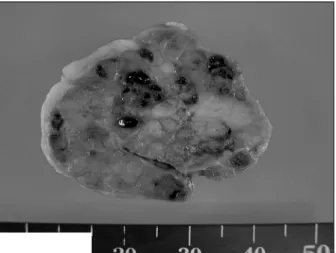
Fig. 2. Surgical specimen showing a 4.5×4.3 cm sized, well encap- sulated hard mass.
Fig. 4. Immunohistochemical stain for cytokeratin (CK). The glan- dular epithelial cells are stained with CK but the my- oepithelial cells are not immunoreactive.
Fig. 5. Immunohistochemical stain for actin. Myoepithelial cells are stained with actin but the glandular epithelial cells are not immunoreactive.
국소 침윤하는 부위가 있어 악성 선근상피종으로 진단하였 다(Fig. 3). 일부에서 근상피세포와 상피세포로 구성된 관상 구조가 남아있는 점으로 두 가지 성분 모두가 악성화한 경 우로 진단하였다. 면역조직화학 염색상 관상피세포에서 세 포각질(cytokeratin)에 양성소견을 보였고(Fig. 4), 근상피세 포에서 액틴(actin)에 양성소견을 보였다(Fig. 5). 악성 선근 상피종으로 전신마취하에 광범위 절제술을 시행하였고, 환 자는 수술 후 6일째 별다른 문제없이 퇴원하였다. 환자는 수술 후 1년 동안 국소재발이나 원격전이 소견 없이 외래 추적관찰 중이다.
고 찰
근상피세포는 정상 유방조직의 일부로 이것에서 기원하
는 종양은 매우 드물게 보고되고 있다. 1991년 Tavassoli(2) 는 근상피세포에서 기원하는 종양을 근상피증(myoepithe- liosis), 선근상피종(adenomyoepithelioma), 근상피암(myoepit- helial carcinoma)으로 분류할 것을 제시하였으며 양성의 선 근상피종은 방추상세포형, 관상형, 분엽형으로 다시 세분하 였다. 선근상피종의 대부분은 양성으로 보고되고 있으나 원발 종양을 절제한 후에도 국소 재발이 흔하게 발생하고 드물게 원격 전이를 하는 등 악성 변화를 보이는 경우도 보고되고 있다.(10) Rosen과 Oberman(11), Tavassoli(2)의 증 례를 합한 45명 중에서 13%인 6명에서 국소 재발이 발생하 였으며, Rasbridge와 Millis(7)는 7명의 증례 중 4명에서 국소 재발이 있었고 1명에서 뇌 전이가 발생하여 사망하였고 보 고하였다. 이러한 국소재발에 대한 보고의 차이는 선근상 피종의 정의에 있어서의 차이로 인한 것으로 생각하고 있
432 대한외과학회지:제 73 권 제 5 호 2007
다. 폐로의 원격전이는 Kihara 등(12)이 악성 선근상피종에 서 전이한 증례를 보고하였으며, Nadelman 등(13)은 양성 선근상피종에서 전이한 2예를 보고하였다.
임상적으로 선근상피종은 대부분 여자에서 관찰되었으 며, 남자에서 발생한 경우는 3예가 보고되었다.(14-16) 증상 은 촉지되는 단일 결절의 형태로 나타나는 경우가 흔하며 크기는 1 cm에서 7 cm에 이르기까지 다양하다. 일반적으로 종괴는 원형이나 가끔 소엽성인 경우도 있으며, 다소 불규 칙한 경계를 가지며 단단하거나 급속히 자라는 양상을 보 이기도 한다. 선근상피종의 영상학적 소견은 비 특이적이 고 상피세포암과 유사한 악성소견을 보일 수 있다. 유방촬 영술에서 양성 선근상피종은 양성 유방 병변의 소견을 나 타낸다고 보고되고 있고,(15) 초음파 소견은 낭종에서 고형 종괴 소견에 이르기까지 다양하다. 조직학적으로 악성 병 변은 2가지 군으로 분류할 수 있는데 한군은 명확한 악성 양상을 보여 쉽게 확인이 가능한 군이며, 다른 군은 본 증례 의 경우처럼 양성에 더 가까운 소견을 보이나 자세히 검사 하면 세포 이형성과 증가된 유사분열 활성을 보이는 군이 다.(2,7,17,18) 세포 이형성, 침윤성 경계, 결합조직증식증 (desmoplasia) 같은 반응성 기질반응, 괴사, 그리고 고 유사 분열 활성이 악성 선근상피종을 시사하는 소견으로 간주되 고 있다. 악성 선근상피종은 상피세포나 근상피세포 또는 두 세포 성분 모두를 침범할 수 있다. 일반적으로 상피세포 성분에 제한되어 침범하나, 드물게 근상피세포 성분을 침 범하는 경우 악성 근상피종으로 불린다.(19) 그러나 본 증 례의 경우처럼 상피세포와 근상피세포 성분 모두에서 발생 한 악성 선근상피종은 극히 드물어 지금까지 12예 미만의 증례들만이 보고되었다.(9) 선근상피종의 형태학적 양상은 관상피세포에 대한 근상피세포의 증식 비율, 종양세포의 형태, 그리고 섬유화의 정도에 따라 아주 다양하다. 형태학 적 변화의 정도가 너무 다양하기 때문에, 선근상피종은 선 증(adenosis), 대롱선종(tubular adenoma), 관선종(ductal ad- enoma), 섬유선종, 그리고 근상피종 등과 감별하여야 한다.
이런 감별진단은 절제된 조직에서 우세한 근상피세포의 증 식을 확인함으로써 판별할 수 있다. 따라서 적은 생검 조직 에서 선근상피종을 진단하는 것은 불가능할 수 있다. 결과 적으로 cytokeratin, actin, S-100 단백, desmin, vimentin 등에 대한 면역조직화학 염색과 전자현미경 등의 특수한 병리학 적 검사가 감별진단에 도움을 준다.(20,21) 선근상피종의 치 료는 대부분의 선근상피종이 양성이지만 국소재발의 위험 성이 있고, 드물지만 악성변화나 원격전이의 위험성이 있 어 음성 절제연을 확보한 광범위 절제술이 필요하다. 수술 적 치료방법 중 액와부 림프절 곽청술에 대한 정립된 견해 는 아직까지 보고되어있지 않으며 더 많은 증례를 통한 연 구가 필요한 실정이다. Ahmed와 Heller(9)는 악성 선근상피 종의 전이는 림프절을 통한 전이보다 혈행성 전이로 생각 되고, 종양의 크기가 2 cm 이상인 경우에만 보고되었다고
하였다. 그리고 Chen 등(4)은 지금까지 원격전이가 보고된 11예에서 액와부 림프절 전이를 보인 예는 1예뿐이었다고 하였다. 원격전이를 동반한 악성 선근상피종의 치료는 아 직 확립되어있지 않다.(22) 원발성 유방종양과 전이 병소를 수술적으로 절제하는 방법이 시행되어질 수 있지만, 불완 전한 절제로 인한 국소재발이 흔하다.(5,18) 일부 증례에서 항암요법이 시도되었지만 효과는 미흡하였고,(4,18,22) 호 르몬 치료 역시 시험적인 단계이다. 악성 선근상피종의 예 후는 질환의 희박함 때문에 잘 알려져 있지 않지만, 원격전 이를 동반한 경우는 대부분 몇 년 이내에 사망하여 예후가 좋지 않은 것으로 보고되고 있다.(12)
저자는 절제생검을 한 후에 진단된 유방의 악성 선근상 피종 1예를 경험하였기에 문헌고찰과 함께 보고하는 바이 다. 악성 선근상피종은 현재까지 효과적인 보조요법이 알 려져 있지 않기 때문에, 완전한 국소 절제술이 국소재발과 원격전이의 위험성을 줄이기 위한 유일한 방법으로 간주되 어지고 있다. 아울러 유방 병변을 진단할 때 임상의 들은 이와 같이 드문 질환의 가능성에 대해 주지하고 있어야 할 것으로 생각한다.
REFERENCES
1) Hamperl H. The myothelia (myoepithelial cells). Normal state;
regressive changes; hyperplasia; tumors. Curr Top Pathol 1970;53:161-220.
2) Tavassoli FA. Myoepithelial lesions of the breast. Myoepithe- liosis, adenomyoepithelioma, and myoepithelial carcinoma.
Am J Surg Pathol 1991;15:554-68.
3) Van Hoeven KH, Drudis T, Cranor ML, Erlandson RA, Rosen PP. Low-grade adenosquamous carcinoma of the breast. A clinicopathologic study of 32 cases with ultrastructural analy- sis. Am J Surg Pathol 1993;17:248-58.
4) Chen PC, Chen CK, Nicastri AD, Wait RB. Myoepithelial car- cinoma of the breast with distant metastasis and accompanied by adenomyoepitheliomas. Histopathology 1994;24:543-8.
5) Michal M, Baumruk L, Burger J, Manhalova M. Adenomyo- epithelioma of the breast with undifferentiated carcinoma com- ponent. Histopathology 1994;24:274-6.
6) Foschini MP, Pizzicannella G, Peterse JL, Eusebi V. Adeno- myoepithelioma of the breast associated with low-grade ad- enosquamous and sarcomatoid carcinomas. Virchows Arch 1995;427:243-50.
7) Rasbridge SA, Millis RR. Adenomyoepithelioma of the breast with malignant features. Virchows Arch 1998;432:123-30.
8) Van Dorpe J, De Pauw A, Moerman P. Adenoid cystic carci- noma arising in an adenomyoepithelioma of the breast. Vir- chows Arch 1998;432:119-22.
9) Ahmed AA, Heller DS. Malignant adenomyoepithelioma of the breast with malignant proliferation of epithelial and my- oepithelial elements: a case report and review of the literature.
박민호:유방의 악성 선근상피종 433
Arch Pathol Lab Med 2000;124:632-6.
10) Bult P, Verwiel JM, Wobbes T, Kooy-Smits MM, Biert J, Holland R. Malignant adenomyoepithelioma of the breast with metastasis in the thyroid gland 12 years after excision of the primary tumor. Case report and review of the literature. Vir- chows Arch 2000;436:158-66.
11) Rosen PP, Oberman HA. Tumors of mammary gland. AFIP 1993;7:91-6.
12) Kihara M, Yokomise H, Irie A, Kobayashi S, Kushida Y, Yamauchi A. Malignant adenomyoepithelioma of the breast with lung metastases: report of a case. Surg Today 2001;
31:899-903.
13) Nadelman CM, Leslie KO, Fishbein MC. “Benign,” metasta- sizing adenomyoepithelioma of the breast: a report of 2 cases.
Arch Pathol Lab Med 2006;130:1349-53.
14) Tamura G, Monma N, Suzuki Y, Satodate R, Abe H. Adeno- myoepithelioma (myoepithelioma) of the breast in a male.
Hum Pathol 1993;24:678-81.
15) Berna JD, Arcas I, Ballester A, Bas A. Adenomyoepithelioma of the breast in a male. Am J Roentgenol 1997;169:917-8.
16) Ryu KW, Kim AR, Kim CH, Lee JB, Lee ES, Bae JW, et al. Mammary adenomyoepithelioma in male patient. J Korean Breast Cancer Soc 2000;1:76-9.
17) Loose JH, Patchefsky AS, Hollander IJ, Lavin LS, Cooper HS, Katz SM. Adenomyoepithelioma of the breast: a spectrum of biologic behavior. Am J Surg Pathol 1992;16:868-76.
18) Trojani M, Guiu M, Trouette H, De Mascarel I, Cocquet M.
Malignant adenomyoepithelioma of the breast. An immunohis- tochemical, cytophotometric, and ultrastructural study of a case with lung metastases. Am J Clin Pathol 1992;98:598-602.
19) Van Dorpe J, De Weer F, Bekaert J, Lauweryns J, Moerman P. Malignant myoepithelioma of the breast: case report with immunohistochemical study. Arch Anat Cytol Pathol 1996;
44:193-8.
20) Schurch W, Potvin C, Seemayer TA. Malignant myoepithe- lioma (myoepithelial carcinoma) of the breast: an ultra- structural and immunohistochemical study. Ultrastruct Pathol 1985;8:1-11.
21) Cartagena N Jr, Cabello-Inchausti B, Willis I, Poppiti R Jr.
Clear cell myoepithelioma of the breast. Hum Pathol 1988;
19:1239-43.
22) Takahashi I, Tashiro H, Wakasugi K, Onohara T, Nishizaki T, Ishikawa T, et al. Malignant adenomyoepithelioma of the breast: a case with distant metastases. Breast Cancer 1999;
6:73-7.