102
책임저자:김용호, 서울시 동대문구 회기동 1번지 130-702, 경희의료원 외과
Tel: 02-958-8246, Fax: 02-966-9366 E-mail: [email protected]
접수일:2008년 4월 21일, 게재승인일:2008년 5월 15일
담낭암에서 Fascin 발현에 대한 연구
경희대학교 의과대학 외과학교실, 1병리학교실
나국영ㆍ김용호ㆍ김윤화1ㆍ홍성화
Expression of Fascin in Gallbladder Carcinoma
Kuk Young Na, M.D., Yong Ho Kim, M.D., Youn Wha Kim, M.D.1, Sung Wha Hong, M.D.
Departments of Surgery and 1Pathology, College of Medicine, Kyung Hee University, Seoul, Korea
Purpose: Gallbladder carcinoma is the most common malignancy of the biliary tract in Koreans. However the exact histopathological characteristics and its carcinogenesis are not well understood. Fascin is an actin bundling protein, and it induces membrane protrusions and increased cell motility in various transformed cells. The expression of fascin is known to be greatly increased in various human neoplasms, but its expression in gallbladder carcinoma is unknown.
Methods: A total 110 cases of gallbladder carcinoma, six cases of carcinoma in situ and 10 cases of chronic cholecystitis were immunohistochemically studied to evaluate the expression of fascin in the light of its relationship with various prognostic factors.
Results: Seventy eight gallbladder carcinomas (70.9%) showed positive staining for fascin, but none of the chronic cholecystitis and carcinoma in situ was positive. Fascin was strongly stained in the cytoplasm of the cancer cells.
The adjacent normal mucosa was negative for fascin staining. There was a significant correlation between lymph node metastasis (P=0.039) and the presence of residual tumor (P=0.016) but there was no significant correlation between age, gender, tumor invasion, histologic difference, neural invasion, lymphatic invasion, stage and recurrence. The 5-year overall survival rate of the fascin positive and negative groups were 48.5% and 53.8%, respectively (P=0.236). On the multivariate analysis, a fascin expression was not significant.
Conclusion: Our results suggest that a fascin expression is strongly associated with neoplastic progression in gallbladder carcinomas and fascin positive gallbladder carcinomas show more aggressive behavior. (J Korean Surg Soc 2008;75:102-108)
Key Words: Fascin, Gallbladder carcinoma, Neoplastic progression 중심 단어: Fascin, 담낭암, 종양 진행도
서 론
담낭의 원발성 악성종양은 비교적 드문 질환으로 국내에
서는 간외 담도암 중 세 번째로 흔하게 발생하며 위장관계 암 중 여덟 번째로 흔한 암종으로 알려져 있다. 담낭암은 임상적으로 암을 의심할 만한 특이적 증상이 없어서 상당 히 진행된 후에 진단되며, 결과적으로 근치적 절제가 불가 능한 경우가 많다. 또한 담낭암은 평균 생존기간이 약 10개 월 정도로 예후가 좋지 않은 종양 중 하나이다.(1) 담낭암은 다른 암종과 마찬가지로 기질 내로의 암세포의 침윤과 림 프절 전이가 생존율에 중요한 영향을 미치는 요소로 알려 져 있다. 이러한 암세포의 침윤과 전이는 세포와 세포 사이
의 부착과, 세포와 기질 사이의 부착을 끊어내는 암세포의 이동성 때문이며, 이러한 특성들은 암세포가 기저막을 침 투하게 만들고 주변의 조직으로 퍼져나가게 만든다.(2-4) 주변 기질 내로 침윤하는 암세포는 세포막이 돌출되어 있 고, 세포 사이의 유착이 손실된 특이적인 모습을 보이는데, 이런 것들은 액틴 교차결합 단백질의 활동에 의해 세포 골 격계의 미세섬유의 재배열에 기인한다고 알려져 있다.(5-7) 이런 액틴 교차결합 단백질 중에서 액틴 세사 다발을 만드 는데 관여하는 fascin은 신경세포, 아교세포, 모세혈관 내피 세포 및 항원제시 가지돌기세포의 세포질에서 높게 발현된 다.(8) Fascin은 정상 상피세포에서는 발현되지 않지만 최근 에 유방을 비롯한 폐, 식도, 췌장, 대장, 난소, 위, 담관계, 방광 및 피부 등 여러 상피기원 암에서 발현이 보고되고 있지만,(9-15) 아직까지 담낭암에서의 fascin 발현에 관한 연 구는 없다.
본 연구의 목적은 정상 담낭 상피, 담낭 상피내암, 그리고 담낭암에서의 fascin 발현을 면역조직화학적 방법으로 관찰 하고, fascin 발현이 다른 임상병리학적 인자들과의 관련이 있는 지 그리고 fascin 발현이 예후 인자로서의 의의가 있는 지를 알아보고자 하였다.
방 법
1) 대상
1983년 1월부터 2007년 1월까지 경희대학교 부속병원 외 과에서 담낭 절제술을 시행 받은 증례 중 담낭암으로 진단 된 110예, 담낭상피내암으로 진단된 6예, 그리고 정상 비교 군으로 만성담낭염으로 진단된 10예를 대상으로 하였다.
110예의 담낭암 환자들은 임상기록과 병리보고서 등을 재 검토하여 나이, 성별, 종양의 침윤정도, 림프절 전이 여부, 암종의 병리학적 분화도, 병기, 수술의 근치도 및 재발 여부 등을 조사하였고, 종양의 병기는 AJCC 분류에 의하여 나누 었다. 담낭암 환자의 성비는 남자가 53명, 여자가 56명이었 고, 1명의 환자는 임상기록의 미비로 성별을 알 수 없었다.
환자의 나이는 27세에서 86세까지 다양하게 분포하였으며, 평균 62세였다. 환자의 추적기간은 2개월에서 160개월로 평균 30개월이었다.
2) 면역조직화학적 염색
각 증례의 조직을 광학현미경으로 검색 후 병변과 주변 의 정상 상피가 포함된 가장 대표적인 부위의 파라핀 포매
조직을 1개씩 골라 5μm 두께로 박절한 후 poly-L-lysine으 로 처리한 슬라이드에 부착한 후 자동면역염색기(Vision Biosystems, Bond-Max, Australia)를 이용하여 제조사의 방식 에 따라 탈파라핀, 항원회복, 그리고 발색한 후 Mayor hem- atoxylin으로 대조 염색하여 봉입하여 광학현미경으로 관찰 하였다. Fascin 단백에 대한 면역염색은 미국의 DAKO 회사 의 anti-fascin (monoclonal, 55K-2, DAKO, Carpenteria, CA, USA)을 1:200으로 희석하여 사용하였다. Fascin 단백 염색 의 양성 대조군으로는 정상 편도선의 림프여포 안에 있는 수지상세포(dendritic cell)의 세포질에 강하게 염색이 되는 것을 사용하였다.
3) 면역조직화학적 염색 결과 판정
Fascin 단백에 대한 면역조직화학 염색의 판독은 한 명의 병리전문의가 환자의 임상적 정보를 모른 상태에서 광학현 미경으로 살펴보았을 때 양성 대조군으로 사용한 편도의 수지상세포와 비교하여 종양세포의 세포질 및 세포막에 비 슷하게 염색되었을 때 양성으로 판정하였고, 전체 종양세 포 중에서 양성으로 염색된 종양세포가 없거나 전체의 10%
이하인 경우를 0, 10∼30%에서 양성으로 염색된 경우 1+, 30∼60% 이상인 경우 2+, 그리고 60% 이상인 경우를 3+
로 판독하였다. 또한 종양세포에서 염색된 강도는 모든 예 에서 강한 발현을 보였기 때문에 따로 분류하지 않았다.
4) 통계방법
통계처리는 Window용 SPSS version 12.0 프로그램(SPSS, Chicago, USA)을 사용하였으며, fascin 발현과 임상병리학 적 인자들의 분석은 Pearson χ2 test를 이용하였고, 생존율 은 Kaplan-Meier 방법을 사용하였다. 예후인자들에 대한 다 변량 분석은 Cox regression을 사용하여 구했고, P값이 0.05 이하인 경우를 통계적으로 유의하다고 판정하였다.
결 과
1) Fascin의 발현 양상
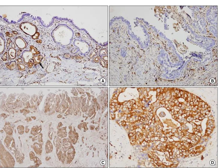
만성 담낭염 환자에서는 점막상피세포에서는 양성으로 염색된 것은 없었고 단지 유문화생(pyloric metaplasia)을 보 이는 부위에만 부분적으로 양성으로 염색되었다(Fig. 1A).
그리고 점막 아래에 존재하는 혈관내피세포와 섬유모세포 의 세포질에 강하게 염색되었다. 6예의 담낭상피내암에서 는 종양세포에서 모두 음성으로 염색되었다(Fig. 1B). 담낭
Fig. 1. (A) Immunohistochmical staining pattern for normal gallbladder mucosa and carcinoma. (A) In the normal gallbladder mucosa, fascin is not expressed in the epithelium, except areas of pyloric metaplasia (arrow). (B) Absent expression of fascin is observed in carcinoma in situ. (C, D) Expression of fascin is observed in Gallbldder carcinoma. Intense staining of fascin is identified at the invasive fronts or infiltrating borders.
Table 1. Fascin immunoreactivity in normal epithelium, carcino- ma in situ, and invasive cancer in gallbladder
Histologic diagnosis Negative Positive
Total
1+ 2+ 3+
Chronic cholecystitis Carcinoma in situ Invasive cancer
10 6 32
0 0 27
0 0 20
0 0 31
10 6 110 암에서 fascin 단백은 종양 세포의 세포질에 강하게 염색되
었는데 110예 중 78예(70.9%)에서 fascin 단백이 양성으로 발현하였고, 이 중 1+ 염색은 27예(24.6%), 2+ 염색은 20 예(18.2%), 3+염색은 31예(28.2%)였다. 염색양상은 종양의 부위에 따라 불균등하게 염색되었으며, 주로 종양 세포가 기저막을 뚫고 침윤하는 부위에서 좀 더 강하게 발현되었 다(Table 1, Fig. 1C, D).
2) Fascin 발현과 임상병리학적 인자들과의 상관관계
Fascin의 발현과 종양의 TNM 분류, 조직병리학적 분류, 병기 등 다양한 임상병리학적 인자들과의 관련은 Table 2에 정리하였다(Table 2). Fascin의 발현은 림프절 전이가 있던 군에서 림프절 전이가 없던 군보다(P=0.039) 그리고 근치적 절제를 하지 못한 군에서 근치적 절제를 한 군보다(P=
0.016) 통계학적으로 의의가 있게 더 높게 발현되었다. 또한 통계학적으로 의의는 없지만 분화가 좋은 암보다는 미분화 암에서 더 높게 발현되었다(P=0.084). 그러나 나이, 종양의 침윤도, 신경 침윤, 림프관 침윤 및 재발 등과 같은 인자들 은 fascin의 발현과 상관관계가 없었다.
Fig. 2. Overall and disease-free survival curves according to the fascin expression.
Table 2. Relationship between fascin overexpression and clinicopathologic characteristics of patients with gallbladder carcinoma
Status of fascin expression n¶ (%) Negative
n (%)
1+ & 2+
n (%)
3+
n (%) P
Age Median (range)
<62
≥62
Gender Male
Female
T stage T1-T2
T3-T4 Lymph node metastasis Absent (N0)
Present (N1)
Stage I-II
III-IV
Histology Differentiated*
Undifferentiated† Residual tumor Absent (R0‡)
Present (R1§, R2∥)
Recurrence Absent
Present
Neural invasion Absent
Present Lymphatic invasion Absent Present
62 (27∼80) 48 (54.1) 61 (55.9) 53 (58.6) 56 (51.4) 69 (62.7) 41 (37.3) 44 (69.8) 19 (30.2) 100 (90.9) 10 (9.1) 97 (88.2) 13 (11.8) 40 (49.4) 41 (50.6) 44 (51.8) 41 (48.2) 98 (89.1) 12 (10.9) 90 (81.8) 20 (18.2)
12 (25.0) 20 (32.8) 14 (26.4) 18 (32.1) 22 (31.9) 10 (24.4) 13 (29.5) 2 (10.5) 30 (30.0) 2 (20.0) 29 (29.9) 3 (23.1) 7 (17.5) 16 (39.0) 16 (36.7) 11 (26.8) 29 (29.6) 3 (25.0) 27 (30.0) 5 (25.0)
24 (50.0) 22 (36.1) 20 (37.7) 26 (46.4) 27 (39.1) 20 (48.8) 14 (31.8) 13 (68.4) 42 (42.2) 5 (50.0) 44 (45.4) 3 (23.1) 24 (60.0) 12 (29.3) 16 (36.7) 20 (48.8) 42 (42.9) 5 (41.7) 37 (41.1) 10 (50.0)
12 (25.0) 19 (31.1) 19 (35.8) 12 (21.4) 20 (29.0) 11 (26.8) 17 (38.6) 4 (21.1) 28 (28.0) 30 (30.0) 24 (24.7) 7 (53.8) 9 (22.5) 13 (31.7) 12 (27.3) 10 (24.4) 27 (27.6) 4 (33.3) 26 (28.9) 5 (25.0)
0.479
0.386
0.577
0.039
0.794
0.084
0.016
0.794
0.901
0.767
*Differentiated = papillary, well differentiated and moderately differentiated carcinoma; †Undifferentiated = poorly differentiated carcinoma;
‡R0 = no residual tumor; §R1 = microscopic residual tumor; ∥R2 = macroscopic residual tumor; ¶n = No. of case.
3) Fascin 발현과 예후와의 관계
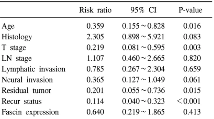
Fascin 발현이 없었던 군의 5년 생존율은 48.5%였고 발현 이 있었던 군의 생존율은 53.8%로 발현이 있는 군에서 예후 가 안 좋았지만 통계학적으로는 의의가 없었다(P=0.236) (Fig. 2). 다변량 분석 결과 연령(P=0.016), 종양의 침윤도(P=
0.003), 재발(P<0.001) 등이 독립적인 예후인자로 나왔고,
fascin 발현은 예후인자로는 의의가 없었다(P=0.413) (Table 3).
고 찰
Fascin은 가는 근육 미세섬유를 하나의 다발로 연결시켜 주는 55-kDa의 액틴 결합 단백(actin-bundling protein)으로, 미수정된 바다 성게 알의 추출물에서 발견되었고, 수정란
Table 3. Multivariate analysis between overall survival deter- mined by Cox progression model
Risk ratio 95% CI P-value Age
Histology T stage LN stage
Lymphatic invasion Neural invasion Residual tumor Recur status Fascin expression
0.359 2.305 0.219 1.107 0.785 0.365 0.201 0.114 0.640
0.155∼0.828 0.898∼5.921 0.081∼0.595 0.460∼2.665 0.267∼2.304 0.127∼1.049 0.055∼0.736 0.040∼0.323 0.219∼1.865
0.016 0.083 0.003 0.820 0.659 0.061 0.015
<0.001 0.413
의 표면에 있는 미세 융모의 섬유 다발과 filopodia에서 관찰 되었다.(16) Fascin은 포유동물에서는 세포막 주름과 세포의 운동성에 관련된 세포 섬유에서 높게 발현되고, 정상적으 로 가지돌기세포, 신경아교세포, 혈관내피세포에서 높게 발 현되며,(17) 정상 상피 세포에서는 발현하지 않거나 매우 미미하게 발현된다고 알려져 있다.(18,19) 최근 다양한 상피 기원 종양에서 fascin의 과발현이 보고되고 있는데 Hashimoto 등은 위암의 25.2%에서,(19) 식도암의 71.1%,(14) 그리고 대 장암의 26%에서(18) fascin의 과발현을 보였다고 보고하였 다. 그 밖에도 유방암, 폐암, 췌담도 종양, 난소암 등에서도 부분적으로 과발현을 보였다고 보고되었다.(15,20-23) Fascin의 발현을 보기 위해 면역조직화학염색을 시행한 본 연구에서 정상 담낭상피세포에서는 fascin이 발현되지 않았으며 단지 만성담낭염에서 유문화생을 보이는 부위에 만 부분적으로 양성으로 염색되었다. 또한 6예의 담낭 상피 내암에서도 모두 fascin 염색에 음성을 보였는데 이는 정상 상피세포에서는 fascin은 발현되지 않았고 단지 유문화생이 있는 부위에서 염색이 되었고, 담낭 상피내암의 42% (8/19) 에서 양성으로 염색되었다는 Sharon 등의 보고와는 차이를 보였다.(21) 이런 차이는 아마도 다른 위장관의 내암과 마 찬가지로 동서양간의 담낭상피내암의 진단 기준에 기인한 다고 할 수 있으며 앞으로 담낭의 상피내암에서는 더 많은 환자들을 대상으로 좀 더 연구가 필요할 것으로 생각된다.
본 연구에서는 담낭암 환자 110예를 대상으로 하였고, 이 중 78예(70.9%)에서 양성으로 발현하여, Sharon 등이 보고 한 담낭암에서의 발현율(67%)과 비슷하였지만, 같은 상피 세포에서 기원하는 췌장암(97%), 담도암(90%), 췌관 팽대부 암(76%) 등의 발현율보다는 약간 낮았다.(21,24) 또한 fascin 은 특히 종양의 침윤 가장자리에서 강하게 발현하였고, 위 암과 식도암에서도 같은 양상이 관찰되었다.(14,19,25) 이상
의 결과를 보면 담낭조직에서 fascin의 발현은 정상 상피세 포에서 발현되지 않으며, 담낭 상피내암에서도 발현이 되 지 않았지만 침윤성 종양의 경우에 높은 비율로 발현되어 fascin이 담낭암의 전단계가 아닌 말기에 담낭암의 발생과 관련이 있다는 것을 알 수 있었다.
본 연구에서 fascin은 림프절 전이가 있었던 군에서 림프 절 전이가 없던 군보다 그리고 근치적 절제가 가능하지 않 았던 군에서 근치적 절제를 한 군보다 더 높게 발현되었다.
이는 위암, 대장암, 식도암에서 림프절 전이가 있었던 군에 서 fascin의 발현이 더 높았다는 보고들과 일치하였 다.(14,18,19,26) Tsai 등(20)에 의하면 췌담도계암에서 fascin 은 조직학적 분화가 나쁠수록, 그리고 종양의 침윤이 심하 고, 높은 병기일 때 높게 발현된다고 보고하였고, 위암에서 도 조직학적 분화도가 나쁠 때 fascin 발현율이 높았다는 보 고가 있는데,(19,27) 본 연구에서도 통계학적 의의는 없었지 만 분화가 좋은 암 보다는 미분화암에서 발현이 더 높았다 (P=0.083). Fascin이 대부분 침윤성 종양에서 발현되고, 림프 절 전이와 근치적 절제가 불가능한 경우에 강하게 발현되 며, 종양의 침윤 가장자리에서 더 강하게 발현되는 점으로 볼 때 fascin의 발현은 침윤성 종양에서, 그 중에서도 병기 가 높은 진행성 암인 경우에 더 발현이 높을 것이라고 예측 할 수 있었다.
또한 본 연구에서는 생존율(P=0.236)과 무병 생존율(P=
0.396)이 낮은 환자에게서 fascin 단백이 강양성으로 발현되 는 양상이었으나 통계학적으로는 의미는 없었으며, 다변량 분석에서도 독립적 예후인자가 아니었다(P=0.413). 폐암, 대 장암, 위암, 식도암 등에서 fascin은 독립적 예후 인자로 의 의가 있다고 보고하였으나,(14,18,19,27,28) Hashimoto 등(19) 은 독립적 예후인자로서 의의가 없다는 상반된 보고가 있 어 추후 더 많은 연구가 필요하겠다.
정상 세포에서 발현하지 않는 fascin이 상피성 종양 세포 에서 과발현하는 기전에 대해선 아직 확실히 밝혀진 것은 없다. 종양과 연관되어 fascin의 발현기전을 밝히려는 연구 들이 활발히 진행되고 있으며, 그 중 fascin과 관련된 유전 자의 변화가 종양 세포의 이동성을 매우 높게 해주며, 침윤 성 종양으로 변화 시키는 가능성에 대한 연구들이 진행되 고 있다. Tao 등(23)은 대장암에서 Wnt 신호전달체계는 β- catenin의 안정화나 APC 유전자의 비활성화를 통해 종양 세 포에서 fascin의 과발현을 일으킨다고 하였고, Grothey 등 (11)은 유방암에서 c-erbB-2/HER-2의 과발현이나 증폭에 의 해 fascin이 과발현 한다고 하였다. 또한 암종에서 fascin 발
현의 증가가 세포 성장을 증가시키는지에 대해서도 활발하 게 연구 중이며, Hashimoto 등(19)은 fascin이 발현되는 부위 에서 Ki-67이 더 높이 발현되므로 결국 fascin도 세포 증식 에 관여한다고 발표하였고, Pelosi 등(23)은 편평상피세포암 의 경우는 관련성이 없으나, 샘암종의 경우에는 fascin 발현 이 높은 암에서 Ki-67의 발현이 낮았다고 발표하였다. 따라 서 아직까지는 이런 분자생물학적인 수준의 연구는 더 진 행되어야 하며, 담낭암에서 fascin이 임상적인 지표나 전향 적인 치료의 대상으로서 역할 등에 대하여 좀 더 많은 연구 가 필요할 것으로 생각된다.
결 론
Fascin은 110예의 담낭암 환자 중 78예(70.9%)에서 발현 되었으며, 상피내암과 정상 상피조직에서는 발현되지 않아 fascin이 담낭암의 말기에 암발생과 관계가 있다는 것을 알 수 있었고, fascin 발현과 림프절 전이와 근치적 절제 여부 가 통계적으로 의의가 있는 임상병리학적 인자들이며 또한 통계학적으로 의미가 없다고는 하나 생존율과 무병 생존율 이 낮은 환자에게서 fascin이 더 많이 발현되었다. 이상의 결과로 봤을 때 fascin의 발현은 병기가 높은 진행성 암인 경우에 더 발현이 높은 것을 알 수 있다. 비록 본 연구에서 는 fascin이 담낭암의 예후에 미치는 영향이 없다고 나왔으 나, 여러 암 종에서 예후 인자로서의 가치가 보고되고 있으 므로 앞으로 예후 인자로서의 역할에 대해서 더 많은 연구 가 필요할 것으로 사료된다.
REFERENCES
1) Grobmyer SR, Lieberman MD, Daly JM. Gallbladder cancer in the twentieth century: single institution's experience. World J Surg 2004;28:47-9.
2) Aznavoorian S, Murphy AN, Stetler-Stevenson WG, Liotta LA. Molecular aspects of tumor invasion and metastasis.
Cancer 1993:71:1368-83.
3) Partin AW, Schoeniger JS, Mohler JL, Coffey DS. Fourier analysis of cell motility: correlation of motility with metastatic potential. Proc Natl Acad Sci USA 1989;86:1254-8.
4) Liotta LA, Kohn EC. The microenvironment of the tumor-host interface. Nature 2001;411:375-9.
5) Otto JJ. Actin-bundling proteins. Curr Opin Cell Biol 1994;6:
105-9.
6) Tilney LG, Connelly PS, Vranich KA, Shaw MK, Guild GM.
Why are two different cross-linkers necessary for actin bundle
formation in vivo and what dose each cross-link contributes?
J Cell Biol 1998;143:121-33.
7) Matsudaira P. Actin crosslinking proteins at the leading edge.
Semin Cell Biol 1994;5:165-74.
8) Kureishy N, Sapountzi V, Prag S, Anilkumar N, Adams JC.
Fascins and their roles in cell structure and function. Bioessays 2002;24:350-61.
9) Hu W, McCrea PD, Deavers M, Kavanagh JJ, Kudelka AP, Verschraegen CF. Increased expression of fascin, motility as- sociated protein, in cell cultures derived from ovarian cancer and in borderline and carcinomatous ovarian tumors. Clin Exp Metastasis 2000;18:83-8.
10) Grothey A, Hashizume R, Sahin AA, McCrea PD. Fascin, an actin-bundling protein associated with cell motility, is up-regu- lated in hormone receptor negative breast cancer. Br J Cancer 2000;83:870-3.
11) Grothey A, Hashizume R, Ji H, Tubb BE, Datrick CW Jr, Yu D, et al. C-erbB-2/HER-2 up-regulates fascin, an actin-bun- dling protein associated with cell motility, in human breast cancer cell lines. Oncogene 2000;19:4864-75.
12) Guvakova MA, Boettiger D, Adams JC. Induction of fascin spikes in breast cancer cells by activation of the insulin-like growth factor-I receptor. Int J Biochem Cell Biol 2002;34:685- 98.
13) Maitra A, Iacobuzio-Donahue C, Rahman A, Sohn TA, Argan P, Meyer R, et al. Immunohistochemical validation of a novel epithelial and a novel stromal marker of pancreatic ductal ad- enocarcinoma identified by global expression microarrays: sea urchin fascin homolog and heat shock protein 47. Am J Clin Pathol 2002;118:52-9.
14) Hashimoto Y, Skacel M, Lavery IC, Mukherjee AL, Casey G, Adams JC. Prognostic significance of fascin expression in ad- vanced colorectal cancer: an immunohistochemical study of colorectal adenomas and adenocarcinomas. BMC Cancer 2006;6:241.
15) Yoder BJ, Tso E, Skacel M, Pettay J, Tarr S, Budd T, et al.
The expression of fascin, an actin-bundling motility protein, correlates with hormone receptor-negative breast cancer and a more aggressive clinical course. Clin Cancer Res 2005;11:186- 92.
16) Yamashiro-Matsumura S, Matsumura F. Intracellular local- ization of the 55-kD actin-bundling protein in cultured cell:
spatial relationships with actin, alpha-actin, tropomyosin, and fimbrin. J Cell Biol 1986;103:631-40.
17) Hashimoto Y, Ito T, Inoure H, Okumura T, Tanaka E, Tsunoda S, et al. Prognostic significance of fascin over- expression in human esophageal squamous cell carcinoma.
Clin Cancer Res 2005;11:2597-605.
18) Adam JC. Fascin protrusions in cell interactions. Trends Cardiovasc Med 2004;14:221-6.
19) Hashimoto Y, Shimada Y, Kawamura J, Yamasaki S, Imamura M. The prognostic relevance of fascin expression in human
gastric carcinoma. Oncology 2004;67:262-70.
20) Tsai WC, Chao YC, Sheu LF, Lin YF, Nieh S, Chen A, et al. EMMPRIN and fascin overexpression associated with clin- icopathologic parameters of pancreatobiliary adenocarcinoma in Chinese people. APMIS 2007;115:929-38.
21) Swierczynski SL, Maitra A, Abraham SC, Iacobuzio-Donanue CA, Ashfag R, Cameron JL, et al. Analysis of novel tumor markers in pancreatic and biliary carcinomas using tissue microassays. Hum Pathol 2004;35:357-66.
22) Cao D, Ji H, Ronnett BM. Expression of mesothelin, fascin, and preostate stem cell antigen in primary ovarian mucinous tumors and their utility in differentiating primary ovarian mu- cinous tumors from metastatic pancreatic mucinous carcinoma in ovary. Int J Gynecol Pathol 2005;24:67-72.
23) Tao YS, Edwards RA, Tubb B, Wang S, Bryan J, McCrea PD. β-catenin associates with the actin-bundling protein fas- cin in a non-cadherin complex. J Cell Biol 1996;134:1271-81.
24) van Heek NT, Maitra A, Koopmann J. Gene expression profil- ing identifies markers of ampullary. Cancer Biol Ther 2004;
3:651-6.
25) Xie JJ, Xu LY, Zhang HH, Cai WJ, Mai RQ, Xie YM. Role of fascin in the proliferation and invasiveness of esophageal carcinoma cells. Biochem Biophys Res Commun 2005;337:
335-62.
26) Pelosi G, Pastorino U, Pasini F, Maissoneuve P, Fraggetta F, Iannucci A, et al. Independent prognostic value of fascin im- munoreactivity in stage I nonsmall cell lung cancer. Br J Cancer 2003;88:537-47.
27) Goncharuk VN, Ross JS, Calson JA. Actin-binding protein fascin expression in skin neoplasia. J Cutan Pathol 2002;29:
430-8.
28) Chae SW, Sohn JH. Fascin-1 protein expression in gastric carcinoma. Korean J Pathol 2006;40:112-7.