635
제 2 형 당뇨 쥐 심장에서 고용량 라미프릴의 결합조직 성장인자 발현 및 섬유화 억제
가톨릭대학교 의과대학 응급의과학교실,1 순환기내과학교실2
최경호
1·백상홍
2·이원재
1·박규남
1·김범준
2·승기배
2·채장성
2·김재형
2·최규보
2High Dose Ramipril Inhibits Connective Tissue Growth Factor Expression and Fibrosis in Type 2 Diabetic Rat Heart
Kyoung Ho Choi, MD
1, Sang Hong Baek, MD
2, Won Jae Lee, MD
1, Kye Nam Park, MD
1, Pum Joon Kim, MD
2, Ki-Bae Seung, MD
2, Jang Seong Chae, MD
2, Jae-Hyung Kim, MD
2and Kyu-Bo Choi, MD
21Department of Emergency Medicine and 2Division of Cardiovascular Medicine, Department of Internal Medicine, The Catholic University of Korea, College of Medicine, Seoul, Korea
ABSTRACT
Background and Objectives:Connective tissue growth factor (CTGF) is a profibrotic cytokine, which may play an important role in the development of diabetic cardiovascular complications. ACE inhibition significantly prevents cardiovascular events in diabetics, although the mechanism remains obscure. The purpose of this study was to explore the effect of ACE inhibitors on the expression of CTGF and oxidative stress in the diabetic heart, and determine the effects of long term treatment with ACE inhibitors on diabetic cardiomyopathy. Materials and Methods:Thirty OLETF (Otsuka Long Evans Tokushima Fatty) diabetic and thirty LETO (Long Evans Tokushima Otsuka) nondiabetic control rats were randomized into four groups for 24 weeks of treatment with either ramipril (5 mg/kg/day, n=15, each groups) or saline (n=15, each groups). Results:The OLETF diabetic rats had prominent perivascular fibrosis, as shown by picrosirius red stains, compared to the LETO nondiabetic rats. ACE inhibition significantly prevented perivascular fibrosis in OLETF rats (p<0.01). Immunohistochemi- cal stains were used to detect proteins for the receptors of advanced glycation end products (RAGE), CTGF, collagen III and nitrotyrosine. Although there were no significant differences in the myocardiac collagen contents, as found by measuring the hydroxyproline concentration among the four groups, the OLFTF diabetic rats had significantly increased cardiac CTGF and collagen III protein expression compared with the nondiabetic rats.
The ACE inhibitor attenuated the increases in RAGE (-50.3%; p<0.01), CTGF (-37.5%; p<0.01) and collagen III (-52.3%; p<0.01) expression in the diabetic heart microvascular area. The OLFTF rats showed marked an increment in cardiac nitrotyrosine, a marker of protein oxidation. Ramipril also inhibited the expression of cardiac nitrotyrosine (-78.3%; p<0.01). Conclusion:The present study shows a possible role of RAGE/nitrotyrosine/
CTGF in the diabetic cardiomyopathy of OLETF rats. The long term treatment of high dose ACE inhibitors may have beneficial effects on the diabetic heart through both antioxidant and antifibrotic mechanisms. (Korean Circulation J 2006;36:635-643)
KEY WORDS:
Receptor of advanced glycation end products;Connective tissue growth factor;Fibrosis;
Ramipril.
논문접수일:2006년 6월 7일 심사완료일:2006년 9월 7일
교신저자:백상홍, 137-040 서울 서초구 반포동 505번지 가톨릭대학교 의과대학 순환기내과학교실 전화:(02) 590-2075·전송:(02) 591-1075·E-mail:[email protected]
서 론
당뇨병은 심혈관 질환의 주요 위험인자로, 동맥 경화성 관 상동맥질환, 심근증1)(diabetic cardiomyopathy), 및 미세 관 상동맥 질환 등의 합병증을 동반하는 것으로 알려져 있다.2) 임상적으로 심근 허혈의 임상적 증상은 있으나 혈관 병변 소견이 뚜렷하지 않은 당뇨병 환자의 부검 소견을 보면 심 장 소동맥 주위의 섬유화 소견이 흔히 관찰되며,3) 심부전과 는 무관하게 미세 관상동맥의 혈관주위 섬유화와 혈관벽의 비후가 관찰되었음을 보고하였다.4) 이러한 미세혈관의 변 화는 미세 혈관성 협심증(microvascular angina)은 물론, 당뇨병성 심근증의 발생기전에도 관여하는 것으로 알려져 있다.4)
당뇨 심장의 병태생리 기전 중, 특히, 콜라젠 이상에 관련 된 병리는 아직 규명되지 않았으나, 고혈당이 장기간 지속 될 경우, 종말당화산물(advanced glycosylation end products, 이하 AGEs로 약함)이 형성되어 심근증 발병에 영향을 미치 지는 것이 알려져 있지만, 그 기전은 밝혀져 있지 않다.5) 또 한, 고혈당이 장기간 지속되거나 AGEs가 형성되었을 경우 콜라젠 합성을 촉진하는 성장 인자들이 활성화되어 조직의 섬유화를 유발시키는 것이 알려져 있으며, 드물기는 하지만 profibrotic 인자인 결합조직 성장인자(Connective tissue growth factor, 이하 CTGF로 약함)도 심근증 발병에 중요 한 작용을 한다는 보고도 있다.6)
OLETF(Otsuka Long Evans Tokushima Fatty)쥐는 인 간의 제 2형 당뇨병과 임상경과가 유사한 새로운 동물 모 델로서, 20주령 이후에 당뇨가 발생하여 만성적인 경과를 보이며, 또한 경도의 비만과 고혈압을 동반하는 특징이 있 음을 보고하였다.7)
안지오텐신 전환효소(angiotensin converting enzyme, 이하 ACE로 약함)는 잘 알려진 혈압강하 효과 이외에도, 항산화, 항섬유화 효과 및 미세 관상동맥의 재형성 개선 효 과가 있음이 보고 되었다.8)
이 연구는 제 2형 당뇨병 쥐 모델에서 CTGF 발현 및 산 화 스트레스가 심장에 미치는 영향을 알아보고, 또한 당뇨 병이 발병한 후 ACE 억제제인 라미프릴을 투여하여 심장 의 섬유화에 미치는 영향을 알아보고자 시도하였다.
재료 및 방법
실험 동물
실험 동물은 제 2형 당뇨병 쥐 모델로 OLETF 쥐, 대조군 은 LETO 쥐(Long Evans Tokushima Otsuka rat)를 사용 하였고 일본 Otsuka 연구소에서 공여 받았다. 모든 쥐는 5 주령 상태로 제공받아, 의과학 연구원 동물실에서 19주간 사육 한 후 24주령이 된 체중 400~450 g의 수컷만을 사용 하였다. OLETF 쥐와 LETO 쥐 각각 30마리를 대상으로 각
군을 다시 고용량 라미프릴(5 mg/kg/day) 투여군과 식염 수 투여군으로 세분하여, 네 군(즉, OL-RMP군; 라미프릴 을 투여한 OLEFT 쥐 15마리, OL-CON군; 생리식염수를 투여한 OLEFT 쥐 15마리, LT-RMP군: 라미프릴을 투여한 LETO 쥐 15마리, 그리고 LT-CON군; 생리 식염수를 투여 한 LETO 쥐 15마리)으로 구분하였다. 실험동물의 관리 및 처치는 가톨릭대학교 의과학연구원 실험동물 연구실 규정 에 따랐다.
라미프릴(Ramipril)의 투여
24주령 된 OLETF 쥐와 LETO 쥐의 라미프릴 투여군은 라미프릴 정제(Aventis Pharmaceutical, Germany)를 혈압 강하 용량보다 많은 고용량인 5 mg/kg을 매일 음용수에 녹 여 24주간 경구 투여하였고, 대조군은 동일 용량의 생리 식 염수를 투여하였다. 매주 8시간 금식 후 쥐의 체중을 측 정하여 증량된 체중을 기준으로 라미프릴 용량을 다시 계산 하여 투여하였다.
혈압, 체중 측정 및 경구 당부하 검사
실험에 앞서 약물 투여 직전인 24주령에 모든 대상 쥐를 12시간 금식 후 안정상태에서, 오전에 꼬리에서 tail-cuff plethysmography(BP-2000, Visitech system, Apex, NC) 를 이용하여 혈압을 측정한 후 곧 이어서 체중을 측정하였 다. 모든 실험군과 대조군의 쥐에서 약물 투여 24주에 다 시 같은 방 법으로 혈압 및 체중을 측정 하였다. 경구 당 부하 검사는 금식 후 오전 8시에 25% 포도당용액(총량 2 g/kg)을 도관을 이용하여 경구로 투여하였다. 당부하 전과 당부하 후 각각 30분, 60분, 90분, 120분에 꼬리정맥에서 채혈하여 글루코미터를 이용하여 혈당을 측정하였다.
조직학적 검사
라미프릴 투여를 종료된 후 모든 실험 쥐에게 ketamine (75 mg/kg)과 xylazine(5 mg/kg)을 복강 내 주사하여 마 취한 후 희생시켰다. 좌심실에 도관을 설치하여 PBS(phos- phate-buffered saline)에 녹인 10% formalin 용액을 100 mmHg의 압력으로 관류시켜 고정하였다. 심장은 한꺼번에 빨리 적출한 후 총 무게를 잰 뒤 두 부분으로 나누어, 한 부분은 다시 10% 포르말린(phosphate-buffered formalin) 용액에 24시간 고정하였다가 파라핀에 포매하였고, 나머지 다른 부분은 냉동 보관하여 심장내 hydroxyproline의 양을 측정에 사용하였다. 파라핀 포매 조직은 4 μM의 박편으로 picrosirius red 염색을 시행한 뒤,9) 심근 간질성 콜라젠 및 혈관주위 섬유화의 정도를 평가하였다. 모든 조직 슬라 이드는 광학 현미경 하에서 200배의 배율로 관찰 하였고 컴 퓨터 영상분석 프로그램(OPTIMAS 6.51)을 이용하여 분석 하였다. 미세 혈관은 혈관의 직경이 300 μm 이하인 미세 관상동맥을 대상으로, 한 개의 심장에서 각각 5개 이상을 검
사하였다. 혈관 주위 섬유화의 정도는 혈관주위 섬유화 면적 분율(perivascular fibrosis area fraction)을 다음과 같은 방법으로 정하여 구하였고,8) 두 명의 실험자가 독립적으로 조직 검사를 분석하였다.
Perivascular fibrosis area fraction(%)=(perivascular fibrosis area/vessel wall area+perivascular fibrosis area)
×100.
심장 조직의 Hydroxyproline과 콜라겐의 정량
조직을 건조시킨 후 6 N 염산액에 24시간 동안 담가두어 가수분해 시켰다. 분해 된 산물을 다시 건조 시킨 후 3 mL 의 증류수에 담은 후 에탄올과 chloramines T 용액과 섞어 20분간 실온에서 산화시켰다. 산화물을 에탄올과 H2SO4용 액에 녹인 p-dimethylaminobenzaldehyde와 60℃에서 15 분간 반응시켜 나온 발색단(chromo phore)을 분광 광도계 를 사용하여 572 nm에서 hydroxyproline을 정량 측정하였 다. Hydroxyproline은 콜라겐에만 결합되어 있고 콜라겐은 14%의 hydroxyproline을 함유하고 있으므로, 심장의 건조 중량당 총 콜라겐 양을 측정할 수 있었다.
면역 조직화학 검사
심장의 4 μM의 연속적인 파라핀 조직으로 Receptor for AGE(receptor for advanced glycosylation end products, RAGE), collagen III, CTGF, nitrotyrosine 검사를 시행하 였다. 일차 항체로는 polyclonal goat anti-human type III collagen 항체(Southern Biotech, Birmingham, AL; 희석 배수 1:20), polyclonal rabbit anti-mouse CTGF 항체 (Abcam, Cambridgeshire, UK; 희석 배수 1:500), poly- clonal goat anti-human receptor for AGE(Abcam, Cam- bridgeshire, UK; 희석 배수 1:300)를 사용하였고, 단백 질산화의표지인 nitrotyrosin은 rabbit antinitrotyrosine (Upstate Biotechnology; Lake Placid, NY 희석 배수 1:
1,000)을 사용하였다. 세척 후 biotinylated 이차항체를 첨 가한 후 3,3-diaminobenzidine tetrahydrochloride(DAB;
Sigma, St. Louis, MO)로 발색하였다. 면역 조직 검사 결과 는 컴퓨터 프로그램(OPTIMAS ver. 6.51, Media Cybernet-
ics, USA)을 이용하여 optical density를 측정한 후 분석하 였다.
통계적 분석
통계적 분석은 응용 통계 프로그램인 SAS(version 8.1, SAS institute, Cary, North Carolina)를 이용하였다. 측정 수치는 mean±SD로 표시하였으며 각 군간의 비교는 two- way ANOVA를 이용하였다. P<0.05인 경우를 통계적 의의 가 있는 것으로 정의 하였다.
결 과
혈압 및 심장 무게의 변화
라미프릴 투여 전 생후 24주령의 OL-CON군 혈압은 수 축기; 135.4±10.9 mmHg, 이완기; 109.3±9.8 mmHg이었 고, LT-CON군 혈압은 수축기; 109.6±11.2 mmHg, 이완 기; 95.4±8.8 mmHg로 관찰되었다. 그러나 OL-RMP군 과 LT-RMP군은 OL-CON군과 LT-CON군과 비교하여 수 축기 및 이완기 혈압이 모두 약제 투여 후 유의하게 감소 하였다(p<0.05)(Table 1).
라미프릴 투여 전 생후 24주령 된 OL-CON군의 체중은 435.5±38.9 g이었고 LT-CON군의 체중은 380.4±27.9 g이었으나, 약제 투여 24주 후에는 OL-RMP군과 LT-RMP 군은 OL-CON군과 LT-CON군과 각각 비교하여 체중의 증 가 정도가 둔화되었다(p<0.05)(Table 1).
심장의 무게는 OLETF 쥐가 LETO 쥐보다 유의하게 증가 하였으나(p<0.005), OL-RMP군과 LT-RMP군은 OL-CON 군과 LT-CON군과 비교하여 유의한 감소를 보였다(p<0.05) (Table 1).
OL-RMP군과 LT-RMP군은 수축기와 이완기 혈압도 OL- CON군과 LT-CON군에 비교하여 유의한 감소를 보였다(p<
0.05)(Table 1).
심장 조직의 섬유화
심근의 hydroxyproline 값은 심장의 건 중량 당 콜라겐 양의 비(collagen content/dry heart weight ratio)로, OL- RMP군은 22.09±7.2 μg/mg이고 OL-CON군은 23.25±
Table 1. Characteristics of the animals at the 48 weeks of the study
Parameter OL-RMP (n=15) OL-CON (n=15) LT-RMP (n=15) LT-CON (n=15)
BW (g) 612.6±41.9∗ 0668.8±37.1 479.4±23.5∗0 548.1±21.9
HW (mg) 931.1±39.9∗ 1099.3±35.4 792.6±32.9∗0 934.9±15.6
HW/BW (mg/g) 01.58±0.01∗ 001.76±0.01 1.74±0.03∗ 01.84±0.01
SBP (mmHg) 111.4±11.9∗ 0143.5±25.6 100.8±12.11∗ 0127.4±10.21
DBP (mmHg) 0089±48.5∗ 000113±42.7 72.2±14.2∗ 098.5±12.8
Glucose (mg/dl) 0213±48.5 000211±42.7 117.2±14.20 124.5±12.8
*: p<0.05 vs. conrol groups. BW: indicates body weight, HW: heart weight, SBP: systolic blood pressure, DBP: diastolic blood pressure, Glucose: blood glucose level during oral glucose tolerance test. OL-RMP indicates ramipril-treated OLETF rats, OL-CON: OLETF control rats, LT-RMP: ramipril-treated LETO rats, LT-CON: LETO control rats. Data are expressed as mean±SD
4.38 μg/mg이었다. LT-RMP군은 21.76±3.5 μg/mg이 고 LT-CON군은 21.79±3.7 μg/mg로 각각 hydroxyproline 농도는 통계학적으로 유의한 차는 없었다.
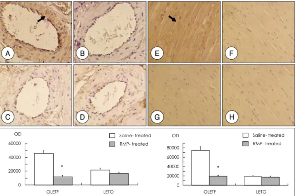
그러나 심근의 콜라젠 농도에는 차이가 없었으나, picrosi- rius red 염색을 이용한 간질성 콜라젠의 분포에서는 네군 가운데 OL-CON군이 다른 군들에 비하여 관상동맥 주위에 섬유화의 정도가 심하였다(Fig. 1A). LT-RMP군에서는 라 미프릴의 투여가 미세 동맥 주위 섬유화의 정도를 감소시 키지 못하였고(Fig. 1D), OL-RMP군은 혈관 주위 섬유화 가 OL-CON군에 비하여 27.3% 감소하여 당뇨가 발생하지
않은 LETO 쥐와 비슷한 정도를 보였다(OL-RMP군 및 OL- CON군: 0.44±0.03 및 0.32±0.01; p<0.01)(Fig. 1B, C).
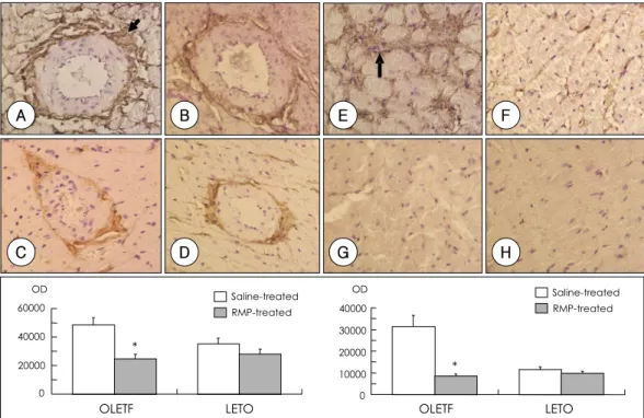
Collagen III의 면역 조직 화학검사 소견에서 OL-CON 군의 미세 관상동맥 주위와 심근 간질조직에서 Collagen III의 발현이 현저히 증가되었으나, OL-RMP군의 발현은 미 세 관상동맥 주위에서 52.3%, 심근에서 71.3%가 감소 되 었다(p<0.01)(Fig. 2).
그러나, LT-RMP군은 LT-CON군에 비하여 미세 관상동 맥 주위와 심근 간질조직에서 발현이 미세하게 감소하였으 나 통계적 의미는 없었다(p<0.01)(Fig. 2).
Fig. 1. Upper panel shows photomicrographs of picrosirius red stained (arrow) control OLETF rats (OL-CON) (A), ramipril-treated OLETF rats (OL-RMP)(B), control LETO rats (LT-CON)(C), ramipril-treated LETO rats (LT-RMP)(D). Ramipril attenuated the periarterial fibrosis. Lower panel shows perivascular fibrosis area fraction. Magnifications, ×200. *: p<0.01 vs. control, OLETF: otsuka long evans tokushima fatty, LETO: long evans tokushima otsuka.
A B
C D
*
0 0.1 0.2 0.3 0.4 0.5 0.6
OLETF LETO
Perivascular fibrosis area fraction (%)
Saline-treated RMP-treated
Fig. 2. Immunohistochemistry for collagen III in the hearts of control OLETF rats (OL-CON)(A and E), ramipril-treated OLETF rats (OL- RMP)(B and F), control LETO rats (LT-CON)(C and G), ramipril-treated LETO rats (LT-RMP)(D and H). Positive staining is shown as brown color (arrow). Lower left panel shows the image quantitaion of stain intensity of periarterial area. Lower right panel shows image quantitaion of stain intensity of myocardial area. OD indicates optical density. Magnifications, ×400. *: p<0.01 vs. control, OLETF: otsuka long evans tokushima fatty, LETO: long evans tokushima otsuka.
C D G H
A B E F
*
OLETF LETO 0
10000 20000 30000 40000 OD
*
OLETF LETO 0
20000 40000 60000
OD Saline-treated
RMP-treated
Saline-treated RMP-treated
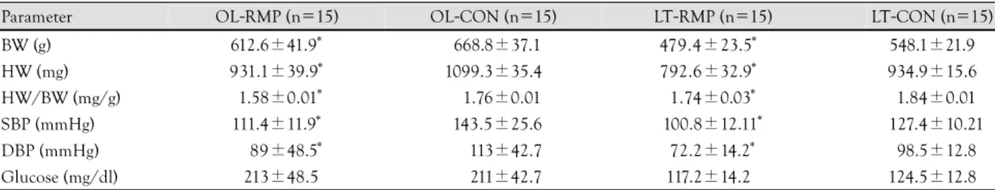
Fig. 3. Immunohistochemistry for CTGF in the hearts of control OLETF rats (OL-CON)(A and E), ramipril-treated OLETF rats (OL-RMP)(B and F), control LETO rats (LT-CON)(C and G), ramipril-treated LETO rats (LT-RMP)(D and H). Positive staining is shown as brown color (arrow). Lower left panel shows the image quantitaion of stain intensity of periarterial area. Lower right panel shows image quantitaion of stain intensity of myocardial area. OD indicates optical density. Magnifications, ×400. *: p<0.01 vs. control, OLETF: otsuka long evans tokushima fatty, LETO: long evans tokushima otsuka.
C D G H
A B E F
*
*
OLETF LETO OLETF LETO
0 4000 8000 12000 16000
Saline-treated RMP-treated OD
0 4000 8000 12000 16000
OD Saline-treated
RMP-treated
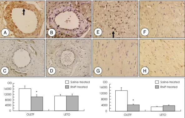
Fig. 4. Immunohistochemistry for RAGE in the hearts of control OLETF rats (OL-CON)(A and E), ramipril-treated OLETF rats (OL-RMP)(B and F), control LETO rats (LT-RMP)(C and G), ramipril-treated LETO rats (LT-RMP)(D and H). Positive staining is shown as brown color (arrow). Lower left panel shows the image quantitaion of stain intensity of periarterial area. Lower right panel shows image quantitaion of stain intensity of myocardial area. OD indicates optical density. Magnifications, ×400. *: p<0.01 vs. control, OLETF: otsuka long evans tokushima fatty, LETO: long evans tokushima otsuka.
C D G H
A B E F
OD
*
OLETF LETO 0
4000 8000 12000 16000 OD
*
OLETF LETO 0
20000 40000 60000
80000 Saline-treated
RMP-treated
Saline-treated RMP-treated
심장 CTGF의 발현
OL-CON군의 미세 관상동맥과 심장 조직에서 CTGF 단 백질의 발현이 LT-CON군에 비하여 증가하였으며, 특히, 혈관 내 평활근세포 및 내피세포에서 뚜렷하였다. 심장 내 CTGF 단백질의 발현 위치는 주로 심근 간질세포였으며, OL-RMP군의 발현은 미세 관상동맥에서 37.5%, 심근에서 는 70.1%가 억제되었다(p<0.01)(Fig. 3). 그러나, LT-RMP 군은 LT-CON군과 비교하여 발현에 전혀 차이가 없었으며 통계적 의미도 없었다(p<0.01)(Fig. 3).
심장 RAGE의 발현
OL-CON군의 미세 관상동맥의 벽과 심장 간질성 조직에 서 RAGE 단백질의 발현이 LT-CON군에 비하여 현저하게 증가되었다. OL-RMP군의 발현은 미세관상 동맥에서 50.3%
가, 그리고 심근에서는 80.1%가 감소되었다(p<0.01)(Fig. 4).
그러나, LT-RMP군은 LT-CON군에 비하여 미세 관상동 맥 주위와 심근 간질 조직에서 발현이 미세하게 감소하였 으나 통계적 의미는 없었다(p<0.01)(Fig. 4).
심장 Nitrotyrosine의 발현
단백질 산화 표지인 nitrotyrosine은 reactive nitrogen species와 유관한 조직 손상지수로, 이 물질의 발현은 OL- CON군의 미세 관상 동맥 및 심근에서 LT-CON군에 비하 여 증가하였고, 이들은 대개 세포질에 국한되어 존재 하였
다. OL-RMP에서 nitrotyrosine 발현은 미세 관상동맥 주위 에서 78.3%, 심근에서 73.5%가 감소되었다(p<0.01)(Fig. 5).
그러나, LT-RMP군은 LT-CON군에 비하여 미세 관상동 맥 주위와 심근 간질조직에서 발현이 미세하게 감소하였으 나 통계적 의미는 없었다(p<0.01)(Fig. 5).
고 찰
ACE 억제제를 장기간 투여하면 당뇨병의 합병증인 심혈관 질환 발생과 사망률을 현저히 감소시킨다는 HOPE 연구10)와, 동맥 경화의 진행을 억제하는 긍정적 효과가 있다는 SECURE 연구11)에 의하면, ACE 억제제는 혈압 강압 효과 이외에도 다 양한 작용이 있을 것으로 생각된다.
ACE 억제제의 주요 작용기전은 안지오텐신 II형성 억제뿐 아니라 bradykinin생성과 nitric oxide(NO) 또는 prostacy- cline의 분비를 촉진하여 혈관을 확장에 의한 혈압 강하작용 이 있다.8)
안지오텐신 II는 강력한 혈관 수축 작용뿐 아니라, 혈관 평 활근 세포의 MCP-1, VCAM-1의 발현을 증진시켜 혈관벽 에 염증반응을 유발시키고 평활근 세포에 mitogen으로 작 용하여 profibrotic cytokines에 직접 또는 간접적으로 작용 하여 세포 외 기질의 축적을 증가시킨다.12)
혈관벽의 산화적 스트레스에 관여하는 NAD(P)H oxidase 및 superoxide 음이온은 동맥경화의 진행에 관여하며, 안지
Fig. 5. Immunohistochemistry for nitrotyrosine in the hearts of control OLETF rats (OL-CON)(A and E), ramipril-treated OLETF rats (OL- RMP)(B and F), control LETO rats (LT-CON)(C and G), ramipril-treated LETO rats (LT-RMP)(D and H). Positive staining is shown as brown color (arrow). Lower left panel shows the image quantitaion of stain intensity of periarterial area. Lower right panel shows image quantitaion of stain intensity of myocardial area. OD indicates optical density. Magnifications, ×400. *: p<0.01 vs. control, OLETF: otsuka long evans tokushima fatty, LETO: long evans tokushima otsuka.
C A
D B
G H
E F
Saline- treated RMP- treated
*
OLETF LETO 0
20000 40000 60000 OD
OLETF LETO
* 0
20000 40000 60000 80000
OD Saline- treated
RMP- treated
오텐신 II에 의하여 활성화된다. Superoxides는 일산화질소 (NO)와 반응하면 강력한 반응물질인 peroxynitrite(OONO-) 를 생성하여,13) NO의 생물활성을 감소시키며, 단백질에 결 합된 tyrosine 잔유기를 질산화하여 단백질의 구조 및 기 능을 변화시키고 지질의 산화도 촉진한다.
안지오텐신 II에 의한 고혈압과 평활근 세포의 비후에도 NAD(P)H oxidase 활성화가 관여하며,14) Receptor of advanc- ed glycosylation end products(RAGE)의 촉 진은 NAD(P)H oxidase 의존하여 유착 물질의 발현과 tissue factor 생성을 유도한다.15) 결과적으로 혈관 세포에서의 NAD(P)H oxidase 활성은 혈관세포의 증식과 염증반응을 유발하여 혈관 질병 을 진행시킨다. ACE 억제제는 NO의 생물학적 활성도를 개 선시켜 혈관 superoxide생성과 NAD(P)H oxidase 활성을 근 원적으로 차단할 수 있는 우수한 항산화물로 생각할 수 있다.
만성 고혈당 상태의 조직에서 종말당화산물(AGEs)의 형 성 및 축적이 증가하고, 축적된 AGEs는 공유 결합으로 상호 결합하여 단백질의 생화학적 구조를 변형시키는데, 단백질 특 히 교원섬유의 기능에 영향을 미친다.16) Collagen-associ- ated AGEs가 증가하면 당뇨 심장의 경화와 섬유화를 촉진 하므로, AGEs는 당뇨 심장의 병태 생리에 중요하며,5) 특히 수용체 의존성 기전에 의하여 cytokines과 성장인자를 유도 하여 당뇨성 미세혈관 합병증을 유발한다.17) 만성 고혈당이 지속되면 직접적으로 세포내에 당이 증가하여, 산화스트레 스 및 비효소적 glycation의 증가하여 심장 구조가 병적으 로 재형성(remodeling)된다.18) 이처럼 고혈당에서 혈청이나 조직에 AGEs가 증가하고 콜라젠 등의 세포외 기질에 구조 적 변화가 발생하며,19)20) 당뇨쥐에서 심근 콜라젠에 AGEs가 축적되면 비록 섬유화가 발생하지 않더라도 심근의 탄성이 감소할 수도 있다.5)
AGEs 수용체(Receptor for AGEs, RAGE)는 AGEs와 함 께 병적 상태에서 유발되는 염증성지표이며, 산화적 스트레 스의 증가에 영향을 미치고, profibrotic cytokine 및 성장 인자들을 유도하고, 전염증성(pro-inflammatory) 경로에 주 요 역할을 한다. RAGE는 세포 표면 물질의 Ig superfamily 의 구성원으로서 CML-modified adducts와 같은 AGEs의 신호 전달에 주요 역할을 한다. AGE 수용체는 RAGE, AGE- R1, AGE-R2, galectin-3(AGE-R3), macrophage scaven- ger receptor 등으로 구성되어 있다.21) 이들 단백질들은 평 활근 세포, 대식 세포, 혈관 내피 세포, 족세포(podocyte) 등 의 세포에서 발현된다. 당뇨에서 AGE의 증가는 AGE 수용 체를 활성화 시키며, 당뇨병의 혈관 및 신장에서 RAGE 발 현이 증가한다는 연구 결과가 보고 되었다.21)
Candido 등22)은 쥐 심장에서 증가한 AGE 수용체의 유전 자와 단백질 발현을 최초로 보고 하였다. 당뇨 쥐 심장에서 의 RAGE 단백질 발현의 증가는 AGE-유인성 심근 구조 변 형에 역할을 하는 수용체임을 의미한다. 이 수용체는 AGE 리간드에 의존하지 않고 세포 부착 및 성장 조절에 의하여
심근 재형성에 직접적으로 작용한다.23)
제 2 형 당뇨 심장병에는 경화성 성장 인자인 transforming growth factor(TGF)-β1, basic fibroblast growth factor (bFGF)와 혈관 성장 인자인 vascular endothelial growth factor(VEGF)가 관련이 있다.24) 특히 최근에 관심을 끄는 CTGF는 세포 외 기질 및 혈관 생성에 관여하며, 당뇨성 신 증에서 이들 CTGF의 유전자 및 단백질 발현이 증가한다는 보고가 있다.25)26) 최근에 apoE-결핍 당뇨 쥐의 대동맥에서 CTGF 발현의 증가는 당뇨성 동맥경화증과의 관련성을 시 사한다.22)
CTGF는 TGF-β에 의하여 유도되는 profibrotic agent 로서 세포 외 간질생성25) 및 혈관 형성27)에 관여하지만, 최 근에는 당뇨성 심혈관 합병증에서 TGF-β 활성화와는 별 도로 독립적으로 심장의 섬유화 및 심기능 저하에 관여한 다고 알려지고 있다.22)
AGE와 CTGF 관련성은 배양된 사람 진피섬유 아세포와 신장의 간질세포에서 AGE에 의하여 CTGF 발현이 증가한 다는 것과, 더구나 AGE에 의하여 세포 외 간질 단백질인 fibronectin의 유도가 증진하는것에 기인한 것으로, 이들의 특별한 관계는 당뇨성 심혈관 질환에 주요 역할을 한다고 생 각된다.25)
본 연구에서도 당뇨 심장조직에서 당뇨 합병증과 관련된 RAGE 발현이 증가하였고, 섬유화와 관련된 collagen III 및 CTGF 단백질의 발현이 증가하였으나, 장기간의 ACE 억제 제 투여로 이들의 발현이 감소되었다.
산화 스트레스가 당뇨병 합병증의 주요 인자이지만,27) 고 혈당과 합병증 간의 초기 연결고리 인지 또는 일차 병태 기 전의 결과인지는 아직 규명 되지않았다.29)
AGE는 산화 스트레스의 증가와 관련이 있고, 이들 반응 성 산소기(reactive oxygen species, 이하 ROS로 약함)의 억제는 고혈당에 의한 세포 손상을 차단 한다.15) ACE 억제 제와 NO 생성과의 관계에서 라미프릴(1 mg/kg/일)을 투여 한 고혈압 쥐의 대동맥에서 endothelial nitric oxide syn- thase가 증가한다는 보고29)와는 달리, 최근에 ACE 억제제 인 quinapril(3 mg/kg/일)이 실험적 당뇨신증에서 endothe- lial nitric oxide synthase를 억제한다는 보고30)를 비교하 면, 현 단계로는 NO 생성에 미치는 ACE 억제제의 관계를 단정하기는 어렵다.
본 결과에서도 당뇨 심장에서 nitrotyrosine 발현이 증가 하였고, ACE 억제제인 라 프릴 투여로 nitrotyrosine 발현 이 현저히 감소하였다. 이는 엔지오텐신 II에 의한 유리 산 소기의 파생을 라미프릴이 차단하는 것을 의미하지만, 라미 프릴에 의한 NO의 생산 감소가 심장조직에서 nitrotyrosine 발현을 저하시키는지는 분명하지 않다.
이 연구는 제 2 형 당뇨쥐에서 당뇨병이 발병한 이후부터 24주간 장기적으로 ACE 억제제인 라미프릴을 고용량 투여 한 후 심장 조직에서 RAGE와 CTGF의 발현이 감소하고,
이는 단백질 산화의 지표인 nitrotyrosine 감소와 관련되었 음으로, 당뇨 심장의 산화적 스트레스와 섬유화에 RAGE, nitrotyrosine, CTGF의 역할이 중요함을 제시하였다. 그리 고 고용량의 라미프릴을 장기간 투여하면 당뇨병이 이미 진행된 당뇨 심장에서 강압 효과뿐 아니라 및 섬유화에 관 련된 단백질의 발현을 방지하여 심장 보호 효과가 있을 것 으로 생각되었다.
연구의 제한점
당뇨 심장에서 기술상의 제한으로 AGEs 대신 RAGE를 측정 비교한 것과, 고용량의 라미프릴을 장기간 투여전, 후 당뇨심장 조직에서 NAD(P)H oxidase 등 발생기산소의 발 현을 직접 비교 관찰하지 못한 것은 본 연구의 제한점이라 하겠다.
요 약
배경 및 목적:
결합조직 성장인자(Connective tissue growth factor, 이 하 CTGF로 약함)는 profibrotic cytokines의 일종으로, 당 뇨성 심혈관계 합병증인 섬유 화의 발생에 중요한 역할을 하 는 것으로 알려져 있다. 이러한 섬유화 합병증을 예방하기 위하여 안지오텐신 전환효소(Angiotensin converting en- zyme, 이하 ACE로 약함) 억제제가 유효하다는 보고가 많이 있으나, 정확한 기전은 아직 규명되지 않았다.
이 연구는 ACE 억제제의 한 종류인 라미프릴(ramipril) 고용량을 장기간 투여하여 당뇨 심장에서 CTGF의 발현 및 산화 스트레스에 대한 영향을 분석하고자 시행 하였다.
방 법:
OLETF(Otsuka Long Evans Tokushima Fatty) 쥐와 LE- TO(Long Evans Tokushima Otsuka) 쥐 각각 30마리를 대상으로 각 군을 다시 고용량 라미프릴(5 mg/kg/day) 투 여군과 식염수 투여군으로 세분하여, 네 군(즉, OL-RMP 군; 라미프 릴을 투여한 OLEFT 쥐 15마리, OL-CON군;
생리 식염수를 투여한 OLEFT 쥐 15마리, LT-RMP군: 라 미프릴을 투여한 LETO 쥐 15마리, 그리고 LT-CON군; 생 리식염수를 투여한 LETO 쥐 15마리)으로 구분하였다. 이들 네 군의 쥐에게는 라미프릴과 생리 식염수를 24주간 경구 투여하였다.
심장 조직에서 당뇨 합병증을 유발하는 receptor of ad- vanced glycation end products(RAGE)와 단백질 산화 지표 인 nitrotyrosine, 그리고 섬유화에 관여하는 CTGF 및 콜라 젠(collagen)III에 대한 면역 조직화학 검사를 시행하였다.
결 과:
OL-CON군에서 picrosirius red 염색에 의한 동맥 주위 의 섬유화는 LT-CON군에 비하여 현저하였다. 이들 네 군 에서 hydroxyproline 농도 측정에 의한 심근 콜라젠 농도의
차이는 없었지만, OL-CON군에서 CTGF 및 콜라젠 III의 발현은 LT-CON군에 비하여 증가하였다. 그러나, 고용량 라 미프릴을 투여한 OL-RMP군에서 미세 관상 동맥 주위의 RAGE, CTGF 및 콜라젠 III의 발현은 각각 50.3%, 37.5%
및 52.3%가 감소되었다. OL-CON군에서 nitrotyrosine 발 현은 타 군에 비하여 현저히 증가하였으나, 고용량 라미프 릴을 투여한 OL-RMP군에서는 78.3%가 감소되었다.
결 론:
고용량의 라미프릴을 24주간 장기 투여한 당뇨 쥐는 혈 압이 강하될 뿐 아니라, 심장 조직에서 산화 스트레스 및 섬 유화를 약화시켜 심장에 유익한 효과를 나타낼 것으로 생각 된다.
중심 단어:CTGF;RAGE;섬유화;Ramipril.
■ 감사문
실험 기간 동안 여러모로 도와주신 가톨릭대학교 의과학 연구원 순 환기내과학 연구실 이지은씨에게 감사를 드립니다.
REFERENCES
1) Haffner SM, Lehto S, Ronnemaa T, Pyorala K, Laakso M.
Mortality from coronary heart disease in subjects with type 2 diabetes and in nondiabetic subjects with and without prior myocardial infarction. N Engl J Med 1998;339:229-34.
2) Akasaka T, Yoshida K, Hozumi T, et al. Retinopathy identifies marked restriction of coronary flow reserve in patients with diabetes mellitus. J Am Coll Cardiol 1997;30:935-41.
3) Murakami H, Urabe K, Nishimura M. Inappropriate microvas- cular constriction produced transient ST-segment elevation in patients with syndrome X. J Am Coll Cardiol 1998;32:1287-94.
4) Ihm SH, Park IS, Youn HJ, Kim JH, Hong SJ, Choi KB.
Impairment of right ventricular diastolic function in early type 2 diabetes. Korean Circ J 2003;33:499-506.
5) Ihm SH, Youn HJ, Kim SR, et al. Relation between serum car- boxy-terminal propeptide of type 1 procollagen (PIP), a marker of myocardial fibrosis, and left ventricular diastolic function in patients with early type 2 diabetes mellitus. Korean Circ J 2005;
35:500-6.
6) Candido R, Forbes JM, Thomas MC, et al. A breaker of ad- vanced glycation end products attenuates diabetes-induced myo- cardial structural changes. Circ Res 2003;92:785-92.
7) Kawano K, Hirashima T, Mori S, Saitoh Y, Kurosumi M, Natori T. Spontaneous long-term hyperglycemic rat with diabetic com- plications: Otsuka Long-Evans Tokushima Fatty (OLETF) strain.
Diabetes 1992;41:1422-8.
8) Chang KY, Baek SH, Ko SH, et al. Angiotensin-converting enzyme inhibitor ramipril improves coronary microvascular remodeling in type II diabetic rats. Korean Circ J 2002;32:680-8.
9) Junqueira LC, Bignolas G, Brentani RR. Picrosirius staining plus polarization microscopy, a specific method for collagen detec- tion in tissue sections. Histochem J 1979;11:447-55.
10) Yusuf S, Sleight P, Pogue J, Bosch J, Davies R, Dagenais G.
Effects of an angiotensin-converting enzyme inhibitor, ramipril, on cardiovascular events in high-risk patients. N Engl J Med 2000;342:145-53.
11) Lonn E, Yusuf S, Dzavik V, et al. Effects of ramipril and vitamin E on atherosclerosis: the study to evaluate carotid ultrasound changes in patients treated with ramipril and vitamin E (SECU-
RE). Circulation 2001;103:919-25.
12) Omura T, Kim S, Takeuchi K, Iwao H, Takeda T. Transforming growth factor beta 1 and extracellular matrix gene expression in isoprenaline induced cardiac hypertrophy: effects of inhibition of the rennin-angiotensin system. Cardiovasc Res 1994;28:1835-42.
13) Beckman JS, Koppenol WH. Nitric oxide, superoxide, and per- oxynitrite: the good, the bad, and the ugly. Am J Physiol 1996;
271:C1424-37.
14) Laursen JB, Rajagopalan S, Galis Z, Tarpey M, Freeman BA, Harrison DG. Role of superoxide in angiotensin II-induced but not catecholamine-induced hypertension. Circulation 1997;95:
588-93.
15) Wautier MP, Chappey O, Corda S, Stern DM, Schmidt AM, Wautier JL. Activation of NADPH oxidase by AGE links oxidant stress to altered gene expression via RAGE. Am J Physiol Endo- crinol Metab 2001;280:E685-94.
16) Brownlee M, Cerami A, Vlassara H. Advanced glycosylation end products in tissue and the biochemical basis of diabetic com- plications. N Engl J Med 1988;318:1315-21.
17) Cooper ME. Interaction of metabolic and haemodynamic factors in mediating experimental diabetic nephropathy. Diabetologia 2001;44:1957-72.
18) Young ME, McNulty P, Taegtmeyer H. Adaptation and malad- aptation of the heart in diabetes: part II. potential mechanisms.
Circulation 2002;105:1861-70.
19) Sell DR, Lapolla A, Odetti P, Fogarty J, Monnier VM. Pentosi- dine formation in skin correlates with severity of complications in individuals with long-standing IDDM. Diabetes 1992;41:1286- 92.
20) Beisswenger PJ, Makita Z, Curphey TJ, et al. Formation of immunochemical advanced glycosylation end products precedes and correlates with early manifestations of renal and retinal disease in diabetes. Diabetes 1995;44:824-9.
21) Schmidt AM, Hori O, Brett J, Yan SD, Wautier JL, Stern D.
Cellular receptors for advanced glycation end products: impli- cations for induction of oxidant stress and cellular dysfunction in
the pathogenesis of vascular lesions. Arterioscler Thromb 1994;
14:1521-8.
22) Candido R, Jandeleit-Dahm KA, Cao Z, et al. Prevention of ac- celerated atherosclerosis by angiotensin-converting enzyme in- hibition in diabetic apolipoprotein E-deficient mice. Circulation 2002;106:246-53.
23) Pugliese G, Pricci F, Leto G, et al. The diabetic milieu modulates the advanced glycation end product-receptor complex in the mesangium by inducing or upregulating galectin-3 expression.
Diabetes 2000;49:1249-57.
24) Jesmin S, Sakuma I, Hattori Y, Fujii S, Kitabatake A. Long- acting calcium channel blocker benidipine suppresses expression of angiogenic growth factors and prevents cardiac remodelling in a Type II diabetic rat model. Diabetologia 2002;45:402-15.
25) Twigg SM, Cao Z, MCLennan SV, et al. Renal connective tissue growth factor induction in experimental diabetes is prevented by aminoguanidine. Endocrinology 2002;143:4907-15.
26) Ko SH, Kwon HS, Kim SR, et al. Ramipril treatment suppresses islet fibrosis in Otsuka Long-Evans Tokushima fatty rats. Biochem Biophys Res Commun 2004;316:114-22.
27) Babic AM, Chen CC, Lau LF. Fisp12/mouse connective tissue growth factor mediates endothelial cell adhesion and migration through integrin v3, promotes endothelial cell survival, and induces angiogenesis in vivo. Mol Cell Biol 1999;19:2958-66.
28) Yan SD, Schmidt AM, Anderson GM, et al. Enhanced cellular oxidant stress by the interaction of advanced glycation end products with their receptors/binding proteins. J Biol Chem 1994;
269:9889-97.
29) Wiemer G, Linz W, Hatrik S, Scholkens BA, Malinski T. An- giotensin-converting enzyme inhibition alters nitric oxide and superoxide release in normotensive and hypertensive rats. Hy- pertension 1997;30:1183-90.
30) Onozato ML, Tojo A, Goto A, Fujita T, Wilcox CS. Oxidative stress and nitric oxide synthase in rat diabetic nephropathy:
effects of ACEI and ARB. Kidney Int 2002;61:186-94.