387
및 분자생물학적 변화
Functional, Morphologic, and Molecular Biological Changes in the Bladder of OLETF Diabetic Rats according to Duration of Diabetes Mellitus
Jun Ha Lee, Hong Jin Suh, Hyuk Jin Cho, Yong Seok Lee,
Hyun Woo Kim, Sang Hoon Kim, Sae Woong Kim, Tae Kon Hwang, Sun-Ju Lee1, Ji Youl Lee
From the Department of Urology, The Catholic University of Korea College of Medicine, 1School of Medicine, Kyung Hee University, Seoul, Korea
Purpose: Our study was undertaken to investigate changes in the bladder according to duration of diabetes mellitus in the Otsuka Long Evans Tokushima Fatty (OLETF) rat model, which is similar to type 2 diabetes.
Materials and Methods: OLETF rats (n=14) and Long Evans Tokushima Otsuka (LETO, n=14) rats were used. LETO is a normal control of OLETF.
The animals were assigned to 4 groups: L-40 group, LETO rats 40 weeks after birth (n=7); O-40 group, OLETF rats 40 weeks after birth (n=7); L-60 group, LETO rats 60 weeks after birth (n=7); and O-60 group, OLETF rats 60 weeks after birth (n=7). At 40 weeks or 60 weeks after birth, blood glucose, cystometry, bladder weight, detrusor contractility, and mRNA expression of nerve growth factor (NGF) were assessed.
Results: Cystometry showed that diabetic bladders had increased compliance compared with the control groups at 40 and 60 weeks, and the O-60 group had greater compliance than the O-40 group. Contractile responses to electrical stimulation, bethanecol (250μM), and ATP (10 mM) were decreased in the experimental groups compared with the control groups at 40 and 60 weeks, and the O-60 group had a lower contractile response than the O-40 group. The mRNA expression of NGF was decreased in the experimental groups compared with the control groups, and the O-60 group had lower expression than the O-40 group. Changes in NGF were identified through immunohistochemical staining.
Conclusions: The degree of diabetic cystopathy in OLETF rats was changed by duration of type 2 diabetes mellitus. Our results showed that the changes in the bladder in type 2 diabetes mellitus can be identified through a new rat model. (Korean J Urol 2009;50:387-395)
Key Words: Type 2 diabetes mellitus, Neurogenic bladder, Nerve growth factor
Korean Journal of Urology Vol. 50 No. 4: 387-395, April 2009 DOI: 10.4111/kju.2009.50.4.387
가툴릭대학교, 1경희대학교
의과대학 비뇨기과학교실 이준하ㆍ서홍진ㆍ조혁진ㆍ이용석 김현우ㆍ김상훈ㆍ김세웅
황태곤ㆍ이선주1ㆍ이지열
Received:November 21, 2008 Accepted:December 24, 2008 Correspondence to:Ji Youl Lee
Department of Urology, Seoul St.
Mary's Hospital, College of Medicine, The Catholic University of Korea, 505, Banpo-dong, Seocho-gu, Seoul 137-040, Korea TEL: 02-2258-6227
FAX: 02-2258-6227 E-mail: [email protected]
Ⓒ The Korean Urological Association, 2009
서 론
당뇨병은 당뇨병의 기간 및 조절유무에 따라 다양한 합 병증을 동반하는 전신질환으로 여러 가지 합병증 중에 비 뇨기과적으로 중요한 합병증은 당뇨병성방광병증이다. 당
뇨병에 의해 방광의 지각신경 및 자율신경 침범으로 유발 되는 당뇨병성방광병증의 유병률은 성 (sex)과 나이에 관련 이 없으며, 당뇨병의 유병기간과 관련이 있다.1 또한 당뇨병 성방광병증의 경우 요폐, 잔뇨감, 복압배뇨, 빈뇨, 요실금 등 다양한 배뇨장애를 보인다. 당뇨병에 따른 이러한 방광 기능의 장애에 대한 기전은 아직 확실하지는 않으나 방광
근의 변화, 지각신경 및 자율신경의 변화 및 요로상피의 변 화로 발생한다고 생각되고 있다.2
당뇨병이 발생 시 방광 내에는 여러 형태학적, 생화학적 그리고 기능적인 변화가 일어나며 이러한 변화를 알아보고 자 많은 연구들이 이루어졌다. 또한 최근 들어 당뇨병으로 인하여 발생하는 방광병변의 원인을 세포학적인 또는 분자 생물학적인 수준에서 알아보고자 하는 노력들이 이루어지 고 있다. Koo 등3은 streptozotocin (STZ)에 의해 유도된 제1 형 당뇨쥐에서 당뇨병 초기의 방광 내 유전자 변화를 연구 하였으며, Steinbacher와 Nadelhaft4는 제1형 당뇨쥐에서 방광과 배근신경절 (dorsal root ganglion)에서 신경성장인자 (nerve growth factor; NGF)의 변화를 관찰하였다. 이와 같이 현재 까지의 연구는 주로 STZ를 투여하는 제1형 당뇨쥐에 대한 연구가 이루어졌으며 또한 분자생물학적인 연구에서도 당 뇨병 유도 후 초기변화에 대한 연구만 시행되었다. 그러나 실질적으로 대다수를 차지하고 있는 제2형 당뇨병은 증상 및 그 합병증에 있어서 제1형 당뇨병에 비하여 경한 것으로 되어 있고, 따라서 방광 내에 나타나는 기능변화 및 기전이 제1형 당뇨병과는 다르게 나타날 수 있을 것으로 생각한다.
이에 저자들은 STZ으로 유발된 제1형 당뇨병과는 다른 자 연발생적인 제2형 당뇨쥐인 Otsuka Long Evans Tokushima Fatty (OLETF)쥐에서 당뇨시기에 따른 방광의 기능적, 형태 학적 및 분자생물학적 변화를 알아보고자 하였다.
재료 및 방법 1. 실험동물
대조군으로는 Long Evans Tokushima Otsuka (LETO, n=14) 쥐, 실험군으로는 OLETF (n=14) 당뇨쥐를 사용하였다.
L-40군 (생후 40주 LETO쥐), O-40군 (생후 40주 OLETF쥐), L-60군 (생후 60주 LETO쥐) 및 O-60군 (생후 60주 OLETF 쥐)으로 나누어 각각 7마리씩 실험에 사용하였다.
2. 당뇨쥐의 각 시기별 혈당 측정
실험군과 대조군의 쥐를 각 시기별로 꼬리의 정맥에서 공복 시 혈액을 채취하여 혈당치를 측정하여 당뇨가 형성 되었음을 확인하였다.
3. 방광 기능 검사
정상 대조군과 실험군에서 모두 흡입 마취 후 복부를 종 절개하고 25 G needle을 방광 내로 유치시킨 후 6시간의 안 정기를 거쳐 harvard syringe pump를 이용하여 생리 식염수 를 0.04 ml/min의 속도로 방광 내로 주입하였다. 수축력, 수 축간 간격 등은 변환기를 통해 Polygraph (Grass 7D, Grass
Inst. Co., Quincy, USA)에 기록하였다.
4. 방광근 절취 및 처치
각 군의 실험동물은 방광기능 검사가 끝난 후 방광 및 요 도를 주위조직으로부터 박리하여, 방광 삼각부 직상방에서 방광을 제거하였다. 방광의 무게를 측정한 후 종절개를 가 하여 넓게 펴고 1/2은 조직절편 및 신경성장인자 (NGF)에 대한 분석을 위해 −70oC의 액화질소 탱크에 보관하고, 나 머지 1/2은 방광근 수축력 측정을 위해 3x10 mm 크기로 2 개의 근육절편을 만들었다.
5. 방광근 수축력 측정
각 군의 근육절편을 30 ml의 Tyrode's solution (Nacl 124.9 mM, KCl 2.6 mM, NaHCO3 23.8 mM, MgCl2ㆍ6H2O 0.5 mM, NaH2PO4ㆍH2O 0.4 mM, dextrose 5.5 mM and CaCl2 1.8 mM) 이 담긴 수조에 종으로 길게 매달았다. 수조를 37oC로 유지 하고 95% O2와 5% CO2를 통기시키면서 1 g의 초기 장력을 가하여 60분간 안정시킨다. 그 후 3분 간격으로 2, 8, 16, 32 Hz의 전기자극 (80 V, 1 msec duration)을 30초간 가하여 최 대 수축력을 측정하고, 약물반응에 대한 검사로서 bethane- chol (250μM) 및 ATP (10 mM)에 대한 방광근의 수축력을 측정하며, 자극에 대한 반응은 force transducer를 통해 polygraph (Grass 7D, Grass Inst. Co., Quincy, USA)에 기록하 였다.
6. 방광근 내 신경성장인자 (NGF)에 대해 RT-PCR을 이용한 mRNA의 변화 측정
1) Total RNA 분리와 cDNA 합성: 보관된 조직에서의 total RNA 분리는 RNeasy minikit (Qiagen Inc., Hilden, Ger- many)를 이용하여 분리하였으며, 자외선 흡광기 (Ultrospec 3,000, Pharmacia Biotech Inc., Cambridge, England)를 이용하 여 260 nm/280 nm 흡광도의 비를 구하여 RNA의 순도를 검 정하고 전기영동으로 RNA의 파괴 유무를 확인하였다.
분리된 총 RNA로부터 cDNA 합성은 First Strand cDNA Synthesis Kit (Life Technologies Inc., Grand Island, USA)를 이용하였다. 분리된 RNA 1μg과 10x reaction buffer (100 mM Tris, 500 mM KCl, pH 8.3) 2μl, 25 mM MgCl2 4μl, dNTP (10 mM mixture of dATP, dCTP, dGTP and dTTP) 2μl, oligo (dT)15 primer 2μl, RNase inhibitor 1μl, AMV reverse transcriptase 0.8μl를 넣고 DEPC-처리 증류수를 혼합한 20 μl의 용액을 25oC에서 10분간 반응시켰다. 그 후 42oC에서 60분간 반응시키고 역전사 효소의 불활성화를 위해 99oC에 서 5분간 방치시킨 후 −20oC에 보관하였다.
2) 중합효소 연쇄반응 (polymerase chain reaction; PCR):
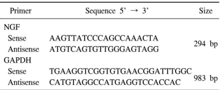
Table 1. Primer sequences used for polymerase chain reaction ana- lysis
Primer Sequence 5' → 3' Size
NGF
Sense AAGTTATCCCAGCCAAACTA
294 bp Antisense ATGTCAGTGTTGGGAGTAGG
GAPDH
Sense TGAAGGTCGGTGTGAACGGATTTGGC 983 bp Antisense CATGTAGGCCATGAGGTCCACCAC
NGF: nerve growth factor, GAPDH: glyceraldehyde 3-phosphate dehydrogenase
중합효소 연쇄반응에는 신경성장인자와 internal control 유 전자인 glyceraldehyde 3-phosphate dehydrogenase (GAPDH)의 sense primer와 antisense primer에 대한 염기서열은 Table 1과 같다.
신경성장인자의 경우 PCR tube에 0.5μg의 cDNA, 2μl의 10x PCR buffer (15 mM MgCl2, 100 mM tris-HCl, 500 mM KCl), 0.4μl의 dNTP (10 mM mixture of dATP, dCTP, dGTP and dTTP), 20 pM씩의 sense primer와 antisense primer, 0.2μg 의 Taq DNA polymerase (Life Technologies Inc.)를 혼합하고 DEPC-처리 증류수를 넣은 20μl의 용액으로 중합효소 연쇄 반응을 시행하였다. 중합효소 연쇄반응은 thermal cycler (Gene cyclerTM, Bio-Rad Laboratories, Hercules, USA)를 이 용하여 94oC 30초, 58oC 30초 그리고 72oC에서 45초로 30주 기 동안 반응시키고 4oC에서 반응을 정지시켰다. GAPDH의 경우 sense primer와 antisense primer를 각각 20 pM씩을 혼합 하고 94oC 1분, 72oC 2분으로 30주기 동안 반응시키고 4oC 에서 반응을 정지시켰다. 5μl씩의 PCR 생성물을 100-bp ladder (Bioneer Corporation, 대전, 한국)와 함께 1.5% agarose gel에 전기영동을 시행한 다음 ethidium bromide (Sigma, Saint Louis, USA)로 염색하여 자외선하에서 형광띠를 확인 하고 Gel-doc (Bio-Rad, USA)으로 촬영하였다. 관찰하고자 하는 mRNA 발현정도는 densitometer (ImageMaster VDS, Pharmacia Biotech Inc., San Francisco, USA)를 이용하여 GAPDH의 발현 정도에 대한 상대치를 구하였다.
7. 신경성장인자 (NGF)에 대한 면역조직화학염색 액체질소에 냉동 보관된 조직을 4% paraformaladehyde로 4oC에서 4시간 고정하고, 20% sucrose 용액에 넣어 4oC에서 12시간 방치한 후, Polyethylene glycol인 optimal cutting temperature (OCT) 용액을 이용하여 포매시켰다. 10μm 조 직 절편을 만들어 slide에 부착시킨 후 phosphate buffered saline (PBS) 용액 (pH 7.4)을 이용하여 5분씩 3번 세척하였
다. 내인성 peroxidase의 활성을 저지 (blocking)하기 위하여 3% 과산화 수소로 처리하고, 비특이적 염색반응을 없애기 위해서 10% normal goat serum solution으로 상온에서 1시간 동안 반응시켰다. 1차 항체로 rabbit anti-mouse NGF anti- serum (1:1000; Chemicon, Temecula, USA)을 사용하여 상온 에서 2시간 동안 반응시킨 후 PBS 용액으로 10분씩 3번 세 척하고, 2차 항체로 Alexa FluorⓇ 568 goat anti-rabbit IgG (1:500; Molecular Probe, Eugene, USA)를 이용하여 상온 암 실에서 1시간 동안 반응시키고 PBS 용액으로 3분간 3번 세 척하였다. Anti-fading solution (Vectashield, Vector Labora- tories Inc., Burlingame, USA)으로 mounting하여 형광 현미경 으로 관찰하였다.
8. 통계분석방법
모든 실험 성적은 평균±표준오차 (mean±SEM)로 표시하 였고, 통계학적 분석은 Student's t-test와 분산분석법 (ANOVA) 을 이용하여 분석하였으며 각 군 간의 비교는 Neuman- Keuls multiple comparison test를 시행하여 p값이 0.05 미만일 때 의의 있는 것으로 판정하였다.
결 과 1. 공복 시 혈당 수치의 변화
L-40, O-40, L-60 및 O-60군의 혈당수치는 106±3.4, 265±
14.0, 112.4±3.1 및 312±14.2 mg/dl로 LETO쥐에 비하여 OLETF 당뇨쥐에서 유의하게 높은 혈당 수치를 보였다 (p<0.05) (Fig. 1).
2. 방광 기능의 변화
방광기능검사에서 각 군의 수축간 간격은 139.6±5.7, 171.4±7.6, 122.4±6.9 및 412.7±13.3초 (sec)이고, 각 군의 방 광용적은 106.4±3.4, 265.7±14.0, 112.4±3.1 및 312.7±14.2 ml 이며, 각 군의 방광 유순도 (ΔVolume/ΔPressure)는 0.178±
0.011, 0.253±0.019, 0.162±0.015 및 0.312±0.023 ml/cmH2O로 40주, 60주 모두 대조군에 비해 실험군에서 유의하게 증가 하였다 (p<0.05). 60주 OLETF 당뇨쥐에서는 40주 OLETF 당뇨쥐에 비하여 수축간격, 방광용적 및 유순도가 더욱 증 가하였다 (p<0.05) (Fig. 2).
3. 방광무게의 변화
40주 LETO쥐의 방광 무게는 113.95±2.80 mg이었으며 40 주 OLETF 당뇨쥐는 135.53±5.63 mg이었고, 60주 LETO쥐 의 방광 무게는 127.38±3.14 mg이었으며 60주 OLETF 당뇨 쥐는 157.83±5.37 mg으로, 40주에는 대조군과 실험군 사이
Fig. 1. Comparison of blood glucose level in Long Evans Tokushima Otsuka (LETO) and Otsuka Long Evans Tokushima Fatty (OLETF) rats. Blood glucose levels of the experimental groups were increased compared with the control groups at 40 and 60 weeks, and the O-60 group had a higher level than the O-40 group. The data are expressed as mean±SEM. L-40 group: LETO rats 40 weeks after birth (n=7), O-40 group: OLETF rats 40 weeks after birth (n=7), L-60 group: LETO rats 60 weeks after birth (n=7), O-60 group: OLETF rats 60 weeks after birth (n=7). a: significantly different from the control groups (p<0.05), b: significantly different from the O-40 group (p<0.05).
Fig. 2. Comparison of bladder compliance in Long Evans Tokushima Otsuka (LETO) and Otsuka Long Evans Tokushima Fatty (OLETF) rats. The diabetic bladders had increased com- pliance compared with the control groups at 40 and 60 weeks, and the O-60 group had greater compliance than the O-40 group. The data are expressed as mean±SEM. L-40 group: LETO rats 40 weeks after birth (n=7), O-40 group: OLETF rats 40 weeks after birth (n=7), L-60 group: LETO rats 60 weeks after birth (n=7), O-60 group: OLETF rats 60 weeks after birth (n=7). a: significantly different from the control groups (p<0.05), b: significantly different from the O-40 group (p<0.05).
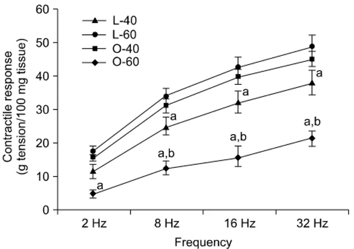
Fig. 3. Contractile response of the bladder muscle strip to 2, 8, 16, and 32 Hz electrical stimulation in Long Evans Tokushima Otsuka (LETO) and Otsuka Long Evans Tokushima Fatty (OLETF) rats. Contractile response to electrical stimulation was decreased in the experimental groups compared with the control groups at 40 and 60 weeks, and the O-60 group had a lower contractile response than the O-40 group. The data are expressed as mean±
SEM. L-40 group: LETO rats 40 weeks after birth (n=7), O-40 group: OLETF rats 40 weeks after birth (n=7), L-60 group: LETO rats 60 weeks after birth (n=7), O-60 group: OLETF rats 60 weeks after birth (n=7). a: significantly different from the control groups (p<0.05), b: significantly different from the O-40 group (p<0.05).
에 유의한 차이는 없었다. 그러나 60주에는 대조군에 비해 실험군에서 의미 있는 증가를 보였다 (p<0.05).
4. 방광근 수축력의 변화
모든 수축력은 방광조직 100 mg에 대한 수축력으로 교정 하여 g tension으로 표시하였다. 전기자극에 대한 방광근의 수축력은 모든 군에서 주파수가 증가할수록 방광근의 수축 력은 점차 증가되었고 실험군의 방광근 수축력이 대조군에 비하여 유의하게 감소하였으며, 특히 고주파로 갈수록 더 욱 많이 감소하였다 (p<0.05). 60주 OLETF 당뇨쥐의 방광 근 수축력은 40주 OLETF 당뇨쥐에 비하여 현저하게 감소 하였다 (p<0.05) (Fig. 3).
각 군에서 bethanechol (250μM)에 대한 방광근의 수축력 은 45.9±3.5, 35.9±5.7, 42.9±3.3 및 24.1±2.5로 실험군에서 대 조군에 비하여 유의하게 감소하였으며, 60주 OLETF 당뇨 쥐의 경우 40주 OLETF 당뇨쥐에 비하여 더욱 많이 감소하 였다 (p<0.05) (Fig. 4).
각 군에서 ATP (10 mM)에 대한 방광근의 수축력은 15.4±0.9, 11.2±1.7, 13.9±0.7 및 8.2±0.7로 실험군에서 대조군에 비하여 유 의하게 감소하였으며, 60주 OLETF 당뇨쥐의 경우 40주 OLETF 당뇨쥐에 비하여 더욱 많이 감소하였다 (p<0.05) (Fig. 5).
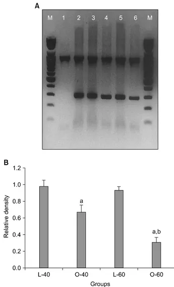
Fig. 6. (A) Reverse transcript-polymerase chain reaction (RT-PCR) measurement of nerve growth factor (NGF) mRNA expression.
Glyceraldehyde 3-phosphate dehydrogenase (GAPDH) was used for the internal control. M: 100 bp marker; lane 1: GAPDH (983 bp); lane 2: GAPDH+NGF (294 bp); lane 3: L-40 group; lane 4: O-40 group; lane 5: L-60 group; lane 6: O-60 group. (B) Results of densitometric scanning of NGF mRNA expression. The mRNA expression of NGF was decreased in the O-40 and O-60 groups compared with the control groups. The mRNA expression of NGF was lower in the O-60 group than in the O-40 group. The data are expressed as mean±SEM. L-40 group: Long Evans Tokushima Otsuka (LETO) rats 40 weeks after birth (n=7), O-40 group:
Otsuka Long Evans Tokushima Fatty (OLETF) rats 40 weeks after birth (n=7), L-60 group: LETO rats 60 weeks after birth (n=7), O-60 group: OLETF rats 60 weeks after birth (n=7). a: sig- nificantly different from the control groups (p<0.05), b: signi- ficantly different from the O-40 group (p<0.05).
Fig. 5. Contractile response of the bladder muscle strip to ATP (10 mM) in Long Evans Tokushima Otsuka (LETO) and Otsuka Long Evans Tokushima Fatty (OLETF) rats. The contractile response to ATP (10 mM) was decreased in the experimental groups compared with the control groups at 40 and 60 weeks, and the O-60 group had a lower contractile response than the L-40 group. The data are expressed as mean±SEM. L-40 group: LETO rats 40 weeks after birth (n=7), O-40 group: OLETF rats 40 weeks after birth (n=7), L-60 group: LETO rats 60 weeks after birth (n=7), O-60 group: OLETF rats 60 weeks after birth (n=7). a: significantly different from the control groups (p<0.05), b: significantly different from the O-40 group (p<0.05).
Fig. 4. Contractile response of bladder muscle strip to bethanechol (250μM) in Long Evans Tokushima Otsuka (LETO) and Otsuka Long Evans Tokushima Fatty (OLETF) rats. The contractile re- sponse to bethanecol (250μM) was decreased in the experimental groups compared with the control groups at 40 and 60 weeks, and the O-60 group had a lower contractile response than the O-40 group. The data are expressed as mean±SEM. L-40 group: LETO rats 40 weeks after birth (n=7), O-40 group: OLETF rats 40 weeks after birth (n=7), L-60 group: LETO rats 60 weeks after birth (n=7), O-60 group: OLETF rats 60 weeks after birth (n=7). a: significantly different from the control groups (p<0.05), b: significantly different from the O-40 group (p<0.05).
5. 방광근 내 NGF mRNA의 발현도
각 군의 NGF mRNA에 대한 RT-PCR을 시행하였으며 모 든 군에서 NGF mRNA의 발현이 확인되었다. NGF mRNA
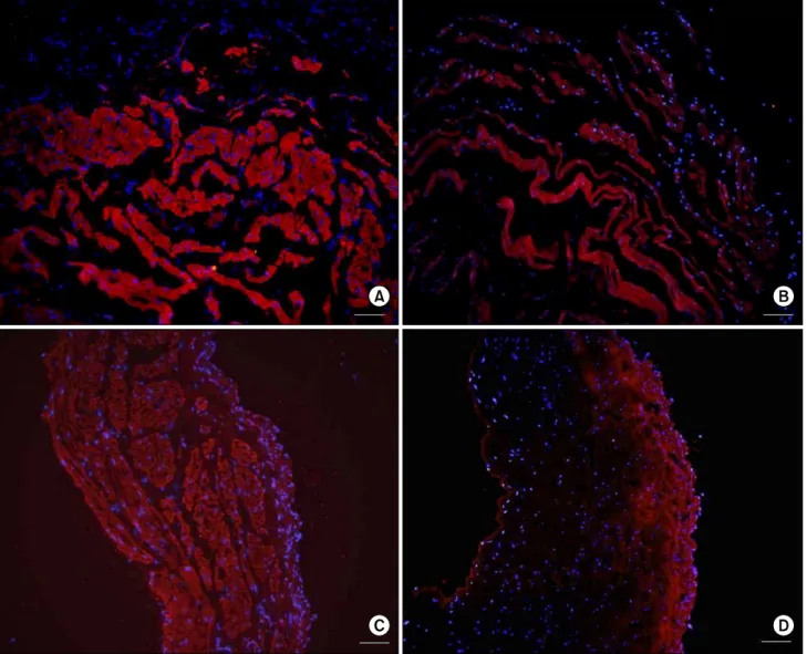
Fig. 7. Nerve growth factor (NGF) immunohistochemical staining of the bladder. NGF expression was decreased in the O-40 and O-60 groups compared with the control groups. NGF expression of the O-40 group was lower than in the O-60 group. (A) L-40 group (scale bar=25μm), (B) O-40 group (scale bar=25μm), (C) L-60 group (scale bar=25μm), (D) O-60 group (scale bar=25μm). L-40 group:
Long Evans Tokushima Otsuka (LETO) rats 40 weeks after birth (n=7), O-40 group: Otsuka Long Evans Tokushima Fatty (OLETF) rats 40 weeks after birth (n=7), L-60 group: LETO rats 60 weeks after birth (n=7), O-60 group: OLETF rats 60 weeks after birth (n=7).
의 발현은 실험군에서 그 발현이 대조군에 비하여 모두 감 소하였으며 (p<0.05), 60주 OLETF 당뇨쥐에서는 40주 OLETF 당뇨쥐에 비하여 더욱 감소하였다 (p<0.05) (Fig. 6).
6. 신경성장인자 (NGF)에 대한 면역조직화학염색 각 군의 조직에 대하여 신경성장인자의 분포 및 염색정 도를 관찰하였다. 실험군에서는 대조군에 비하여 방광근 내 형광반응이 감소되어 있었고, 60주 OLETF 당뇨쥐에서 는 40주 OLETF 당뇨쥐에 비하여 현저하게 감소되었으나, 요로상피에서의 신경성장인자의 형광반응은 실험군과 대 조군 사이에 차이가 없었다 (Fig. 7).
고 찰
당뇨병은 췌장의 인슐린 분비장애와 인슐린 작용기능의 결함으로 발생하는 대사질환으로써 여러 장기에 영향을 미 쳐 많은 합병증을 초래하게 되며, 그 분류는 크게 제1형 당 뇨병, 제2형 당뇨병으로 나눌 수 있으며, 그 외에 이차성 당 뇨병과 임신성당뇨병이 있다. 최근 우리나라는 식생활의 서구화로 인해 비만과 함께 제2형 당뇨병이 증가하고 있는 추세이다.5 이러한 제2형 당뇨병과 유사한 특성을 가지는 당뇨쥐인 OLETF쥐는 당뇨를 유도하는 여러 가지 열성유전
자를 가지고 있으며, 경도의 비만을 가지고 만성질환의 과 정을 거치게 된다. 생후 18주가 지나서 고혈당이 나타나게 되고, 췌장의 변화가 서서히 일어나서 40주가 지나면 췌장 의 위축이 발생하게 된다. OLETF쥐의 이러한 특징은 인간 의 제2형 당뇨병과 유사하다.6
당뇨병은 여러 장기에 영향을 미칠 뿐 아니라 평활근의 기능에도 영향을 미쳐 많은 합병증을 초래하게 되며, 특히 비뇨기과적으로는 당뇨병에 따른 방광기능의 장애가 가장 중요한 합병증이 되고 있다. 당뇨병이 발병 시 방광에는 형 태학적인 변화를 비롯하여 많은 기능적인 변화와 분자생물 학적 변화가 일어나게 된다.7
당뇨병에 따른 형태학적인 변화로써 여러 구조적인 변화 가 발생하는데 Andersson 등8은 STZ로 유도된 당뇨쥐에서 방광내압검사를 시행한 결과 방광의 용적이 증가되었다고 보고하였으며 Longhurst와 Belis9는 방광무게와 용적이 당뇨 쥐에서 증가된다고 하였다. 또한 평활근비대와 함께 방광 근 내에 콜라겐 (collagen)의 침착이 증가된다고 알려져 있 다.10 이러한 변화들은 화학적으로 유도된 당뇨병 초기에서 도 관찰되어질 수 있다고 하였으며11 이러한 것이 방광기능 의 장애와 관련이 될 수 있을 것으로 보고되었다.12,13 그러 나, 저자들의 경우 40주 OLETF 당뇨쥐에서 방광의 무게와 40주 LETO쥐에서 방광의 무게사이에 약간의 증가는 있었 지만 통계학적인 의의는 없었으나, 60주 LETO와 OLETF쥐 사이에서는 의의 있는 증가소견을 보였다. 성인쥐에서 STZ 를 투여하여 유도된 당뇨병은 췌장의 β 세포를 급속히 괴 사시켜 제1형 당뇨병을 형성시키며 일반적으로 매우 빠른 급성 변화를 일으킴으로써 단기간에 방광무게와 용적을 증 가시킨다. 그러나 제2형 당뇨쥐인 OLETF쥐는 여러 가지 열 성유전자에 의해 당뇨병이 발생하여, 서서히 진행하게 되 며 출생 약 18주가 지나야 고혈당이 나타나게 되고 40주가 되어야 췌장의 변화가 생기기 시작한다. 따라서 저자들의 경우에 STZ로 유도된 제1형 당뇨쥐와는 다르게 당뇨병 발 생 초기에 방광무게의 변화가 심하지 않았던 원인은 이와 같은 발생과정의 차이에 기인한 것으로 생각한다.
당뇨병이 존재할 때 비뇨기계의 여러 장기에서도 합병증 이 발생한다.14,15 방광에도 여러 가지 기능적인 문제가 발생 하게 된다. 이러한 기능적인 문제는 자율 신경장애에 따른 것으로 알려지고 있다. 당뇨가 진행함에 따라 방광의 지각 신경에 손상이 발생하면서 방광에 대한 감각의 저하가 일 어나고 결과적으로 방광의 과팽창이 발생하게 된다. 그 후 이러한 과정이 지속적으로 반복되면서 배뇨근대상부전 (decompensation)상태로 진행하여 방광근 기능에 문제가 발 생하고, 결국 방광근이 무력해지면서 방광근 수축력에 장 애가 발생하게 된다. 현재까지 이러한 당뇨병에 따른 기능
적인 변화에 대한 많은 연구가 이루어져 왔다. Andersson 등8은 STZ로 유도된 제1형 당뇨쥐에서 방광기능검사를 한 결과, 배뇨 시작을 유도하는 요량이 증가하고 방광의 유순 도가 증가한다고 하였다. 그러나 Starer와 Libow16는 이러한 전형적인 배뇨장애 형태보다 불수의적 배뇨근수축의 중요 성을 강조하는 의견을 제시하였다. Kaplan 등17도 조사대상 환자의 55%에서 불수의적 배뇨근수축이 있었다고 보고하 였다. 저자들의 경우 40주 OLETF 당뇨쥐와 60주 OLETF 당 뇨쥐에서 각각의 대조군에 비해 방광 유순도가 모두 증가 했으며, 60주 OLETF 당뇨쥐는 40주 OLETF 당뇨쥐에 비해 유순도가 더욱 증가하였다. 이는 당뇨가 진행될수록 반복 적인 과팽창과 배뇨근의 대상부전으로 인한 방광근 수축력 의 장애가 더 심해진 것으로 생각할 수 있으며 일부 개체에 서 발생한 불수의적 배뇨근 수축에 대한 연구도 필요할 것 으로 생각한다. Longhurst와 Belis9는 2개월된 당뇨쥐에서 전기 자극, acetylcholine 및 KCL에 대한 방광체부의 배뇨근 수축력이 모두 감소한다고 하였다. 반면에 STZ로 유도된 당뇨쥐에서 무스카린작용제를 투여했을 때 그 반응성이 증 가한다는 보고도 있으나 작용기전에 대해서는 명확히 밝혀 지지 않았다.18 40주 OLETF 당뇨쥐의 경우 전기 자극, bethanechol 및 ATP에 대한 방광근의 수축력이 대조군에 비 해 감소하였으나 그 감소 폭이 작았으며, 60주 OLETF 당뇨 쥐의 경우에는 전기 자극, bethanechol 및 ATP에 대한 방광 근의 수축력이 의미 있게 감소하는 현상을 나타냈다. 따라 서 방광근의 수축력의 감소도 당뇨가 진행될수록 더욱 더 심해진다는 것을 알 수 있었으며, 이와 같은 것을 확인하기 위해서 조직염색을 통한 방광근의 상태 및 수용체에 대한 연구가 병행되어야 할 것으로 생각한다.
최근 당뇨병성 방광병증의 원인을 세포학적인 또는 분자 생물학적인 수준에서 알아보고자 하는 노력이 이루어지고 있다. 또한 당뇨병성 방광병증은 당뇨의 흔한 합병증인 말 초신경병증의 하나로 알려져 있으며 이러한 말초 신경병증 에 NGF가 중요한 역할을 하는 것으로 알려져 있다. Koo 등3 은 STZ에 의해 유도된 인슐린 의존성 당뇨쥐에서 NGF가 4주까지 증가한 후 다시 감소하는 현상을 나타냈다고 하였 다. 이러한 당뇨병에 따른 초기 현상은 세포비대, 성장 및 재형성 (remodeling)에 따른 현상이라고 보았으며 NGF의 증 가는 당뇨병에서 계속적인 신경의 분포와 공급 및 혈관재 생에 따른 보상작용의 결과라고 하였다. Steinbacher와 Nadelhaft4는 제1형 당뇨쥐에서 방광과 배근신경절에서 NGF가 증가하였다고 하였으며, 실험 첫 주에 NGF가 증가 한 후 천천히 감소하는 현상을 관찰하였다. 이와 같이 당뇨 병에 따른 방광의 기능장애에 있어서 NGF의 중요성이 강 조되고 있으며 최근 이에 대한 연구가 많이 진행되고 있다.
NGF는 말초 교감신경과 신경릉에서 기원한 감각신경계가 분포된 조직에서 생산된다. 생산된 NGF는 그 조직에 분포 된 신경말단의 수용체에 의해 결합되며, 역행성으로 운반 되어 그 기능을 나타내게 된다. 따라서 모든 병리적인 현상 은 이러한 NGF 기능결핍이나 직, 간접적인 역행성 축삭운 반 (retrograde axonal transport)의 장애에 따라 나타날 수 있 다. 이와 같이 당뇨병이 존재 시 여러 조직에서의 NGF 단백 질 및 NGF mRNA의 감소가 보고되었고19-21 여러 신경에서 의 NGF의 감소 및 NGF의 역행성 축삭운반의 감소22-24가 일 어난다고 알려졌다. 반면에 Hellweg와 Hartung25은 당뇨병 을 유도한 후 수 주 뒤 대부분의 말초 신경 지배조직에서 최대 145-300%까지 NGF의 양의 증가를 관찰하였다. 그리 고 그 이유를 NGF에 민감한 신경의 퇴화에 의해 NGF의 생 산 증가가 일어나거나 또는 퇴화된 신경에 의한 NGF 제거 감소에 따른 것이라고 보고하였다. 이와 같이 NGF 변화에 차이가 나는 것은 조직의 종류나 당뇨발생 후 그 측정시기 에 따라 그 양이 달라질 수 있기 때문이며, 당뇨병성 방광병 증의 경우 방광 내 NGF 생산의 감소와 역행성 축삭운반의 장애가 발생한다고 알려져 왔다.26,27 저자들의 경우에도 OLETF 당뇨쥐에서 방광근 내에 NGF mRNA의 발현 감소 를 보였는데 이는 NGF의 합성 감소, 수용체와 결합 장애로 인한 NGF mRNA의 발현 감소가 일어났다고 생각되며, 이 를 확인하기 위해서는 NGF와 수용체의 결합 장애와 역행 성 축삭운반 장애의 문제에 대해 좀 더 연구해 볼 필요성이 있다고 생각한다.
결 론
제2형 당뇨병을 가진 OLETF 당뇨쥐의 당뇨병성방광은 그 시기에 따라 형태학적, 기능적 그리고 분자생물학적 변 화에 차이가 있다. 당뇨병 초기 (40주 OLETF 당뇨쥐)에는 정상 대조군에 비해 변화가 크지 않았지만, 시간이 지날수 록 당뇨가 진행되어 60주 OLETF 당뇨쥐에서는 정상 대조 군과 큰 차이를 나타냈다. 따라서 당뇨는 오랜 시간에 걸쳐 미세혈관 및 말초신경의 장애를 일으켜 방광의 변화를 유 발시켰으며, 그 변화를 관찰함으로써 시기에 따른 당뇨병 성 방광의 형태적, 기능적 변화와 RT-PCR을 이용한 NGF mRNA의 변화를 알 수 있었다. 이러한 실험을 통해서 자연 발생적인 제2형 당뇨병의 시기에 따른 방광병변을 이해하 는데 많은 도움을 줄 수 있을 것이며, 추후 제2형 당뇨병성 방광의 기초적인 실험 자료로 사용될 수 있을 것이다.
REFERENCES
1. Frimodt-Moller C. Diabetic cystopathy: epidemiology and re- lated disorders. Ann Intern Med 1980;92:318-21
2. Yoshimura N, Chancellor MB, Andersson KE, Christ GJ. Re- cent advances in understanding the biology of diabetes-associated bladder complications and novel therapy. BJU Int 2005;95:
733-8
3. Koo HP, Santarosa RP, Buttyan R, Shabsigh R, Olsson CA, Kaplan SA. Early molecular changes associated with streptozo- tocin-induced diabetic bladder hypertrophy in the rat. Urol Res 1993;21:375-81
4. Steinbacher BC Jr, Nadelhaft I. Increased levels of nerve growth factor in the urinary bladder and hypertrophy of dorsal root ganglion neurons in the diabetic rat. Brain Res 1998;782:
255-60
5. Kim NS, Moon OR, Kang JH, Lee SY, Jeong BG, Lee SJ, et al. Increasing prevalence of obesity related disease for Koreans associated with overweight and obesity. Korean J Prev Med 2001;34:309-15
6. Kawano K, Hirashima T, Mori S, Natori T. OLETF (Otsuka Long-Evans Tokushima Fatty) rat: a new NIDDM rat strain.
Diabetes Res Clin Pract 1994;24(Suppl):S317-20
7. Kim JC, Seo SI, Park YH, Hwang TG. Changes of detrusor contractility and growth factors in streptozotosin-induced NIDDM rat. Korean J Urol 2000;41:615-21
8. Andersson PO, Malmgren A, Uvelius B. Cystometrical and in vivo evaluation of urinary bladder function in rats with strepto- zotocin-induced diabetes. J Urol 1988;139:1359-62
9. Longhurst PA, Belis JA. Abnormalities of rat bladder contra- ctility in streptozotocin-induced diabetes mellitus. J Pharmacol Exp Ther 1986;238:773-7
10. Eika B, Levin RM, Longhurst PA. Collagen and bladder func- tion in streptozotocin-diabetic rats: effects of insulin and ami- noguanidine. J Urol 1992;148:167-72
11. Lincoln J, Crockett M, Haven AJ, Burnstock G. Rat bladder in the early stages of streptozotocin-induced diabetes: adrene- rgic and cholinergic innervation. Diabetologia 1984;26:81-7 12. Steers WD, Mackway AM, Ciambotti J, de Groat WC. Effects
of streptozotocin-induced diabetes on bladder function in the rat. J Urol 1990;143:1032-6
13. Longhurst PA, Kauer J, Levin RM. The ability of insulin treat- ment to reverse or prevent the changes in urinary bladder func- tion caused by streptozotocin-induced diabetes mellitus. Gen Pharmacol 1991;22:305-11
14. Chung YG, Yoo HG, Kwon YH, Park CS, Lim WS, Ryu JK, et al. The significance of periurethral fibrosis and the change of nitric oxide synthase containing nerves in the urethra of diabetic rats. Korean J Urol 2007;48:1050-7
15. Kang DI, Kim SH, Lee SD, Kwak HS, Choi SH, Kim DR,
et al. Effects of Alpha-lipoic acid on nitric oxide synthase expression and ultrastructural changes in the bladder of rats with streptozotocin-induced diabetes. Korean J Urol 2007;48:
212-8
16. Starer P, Libow L. Cystometric evaluation of bladder dysfunc- tion in elderly diabetic patients. Arch Intern Med 1990;150:
810-3
17. Kaplan SA, Te AE, Blaivas JG. Urodynamic findings in pa- tients with diabetic cystopathy. J Urol 1995;153:342-4 18. Kanda M, Eto K, Tanabe N, Sugiyama A, Hashimoto K, Ueno
A. Effects of ONO-2235, an aldose reductase inhibitor, on muscarinic receptors and contractile response of the urinary bladder in rats with streptozotocin-induced diabetes. Jpn J Pharmacol 1997;73:221-8
19. Ordonez G, Fernandez A, Perez A, Sotelo J. Low contents of nerve growth factor in serum and submaxillary gland of dia- betic mice. A possible etiological element of diabetic neuro- pathy. J Neurol Sci 1994;121:163-6
20. Fernyhough P, Diemel LT, Brewster WJ, Tomlinson DR. De- ficits in sciatic nerve neuropeptide content coincide with a reduction in target tissue nerve growth factor messenger RNA in streptozotocin-diabetic rats: effects of insulin treatment.
Neuroscience 1994;62:337-44
21. Fernyhough P, Diemel LT, Brewster WJ, Tomlinson DR. Al-
tered neurotrophin mRNA levels in peripheral nerve and skeletal muscle of experimentally diabetic rats. J Neurochem 1995;64:1231-7
22. Jakobsen J, Brimijoin S, Skau K, Sidenius P, Wells D. Re- trograde axonal transport of transmitter enzymes, fucose-labeled protein, and nerve growth factor in streptozotocin-diabetic rats.
Diabetes 1981;30:797-803
23. Schmidt RE, Modert CW, Yip HK, Johnson EM Jr. Retrograde axonal transport of intravenously administered 125I-nerve growth factor in rats with streptozotocin-induced diabates.
Diabetes 1993;32:654-63
24. Hellweg R, Raivich G, Hartung HD, Hock C, Keutzberg GW.
Axonal transport of endogenous nerve growth factor (NGF) and NGF receptor in experimental diabetic neuropathy. Exp Neurol 1994;130:24-30
25. Hellweg R, Hartung HD. Endogenous levels of nerve growth factor (NGF) are altered in experimental diabetes mellitus: a possible role for NGF in the pathogenesis of diabetic neuro- pathy. J Neurosci Res 1990;26:258-67
26. Sasaki K, Yoshimura N, Chancellor MB. Implications of dia- betes mellitus in urology. Urol Clin North Am 2003;30:1-12 27. Pittenger G, Vinik A. Nerve growth factor and diabetic neuro-
pathy. Exp Diabesity Res 2003;4:271-85