24
제 2 형 당뇨쥐와 비당뇨쥐에서 클라미디아 폐렴균에 의한 죽상동맥경화증의 발생
- 혈소판 유도 성장인자의 역할 -
가톨릭대학교 의과대학 내과학교실
신우승·장기육·전희경·김범준·김동빈 임상현·승기배·김재형·홍순조·최규보
Chlamydia Pneumoniae Infection Initiates the Development of Atherosclerosis
in OLETF and LETO Rats- The Role of Platelet-derived Growth Factor-B -
Woo Seung Shin, MD, Kiyuk Chang MD, Hui Kyung Jeon, MD, Pum Joon Kim, MD, Dong Bin Kim, MD, Sang Hyun Ihm, MD, Ki Bae Seung, MD,
Jae Hyung Kim, MD, Soon Jo Hong, MD and Kyu Bo Choi, MD
Department of Internal Medicine, College of Medicine, The Catholic University of Korea, Uijeongbu, Korea
ABSTRACT
Background and Objectives:Chlamydia pneumoniae (CP) has been linked with atherosclerosis. While several studies have shown that CP contributes to the acceleration of atherosclerotic lesions, any studies on the initiation of atherosclerosis are sparse. The present study investigated whether CP infection could initiate atherosclerotic lesions in rats that are known to be resistant to atherosclerosis; further, we investigated if these lesions do form, then how does the CP participate in this and develop of atherosclerosis in these rats. Materials and Methods:
Thirty 11-week-old Otsuka Long-Evans Tokushima Fatty (OLETF) rats, thirty type 2 diabetic rats and thirty age- matched Long-Evans Tokushima Fatty (LETO) rats that were maintained on a high-cholesterol diet were either mock-inoculated or inoculated intranasally 3 times at 11, 13 and 15 weeks of age. The serum levels of the lipid profiles, plasminogen activator inhibitor-1 (PAI-1), monocyte chemoattractant protein-1 (MCP-1) and C-reactive protein (CRP) were measured by performing ELISA at 24 weeks and 40 weeks of age. The atherosclerotic lesion areas were analyzed, and immunohistochemical staining using chlamydia genus-specific monoclonal antibody and PDGF-B was performed in the ascending aorta at 40 weeks of age. Results:Immunohistochemical staining with using specific monoclonal antibody demonstrated CP infection in the vessel walls. The serum PAI-1 level of the OLETF rats was higher than that of the LETO rats (p<0.05) regardless of the state of the CP infection, but there were no differences in the serum MCP-1 and CRP levels between the OLETF rats and the LETO rats. While no atherosclerotic lesion was observed in the mock-infected LETO rats, early-to-advanced atherosclerotic lesions were found in the other rat groups. CP-infected OLETF rats showed more advanced atherosclerotic lesions and greater mean lesion areas than the other rat groups (LT-N, 0 mm2; LETO-CP, 3.29±1.23 mm2; OT-N, 4.91±
2.11 mm2; OT-CP, 9.20±4.62 mm2)(p<0.05). The characteristics of the atherosclerotic lesions in the rats were intimal thickening that was mainly composed of smooth muscle cells. The atherosclerotic lesion area positively correlated with the presence and the extent of PDGF-B staining in the aortic wall (p<0.01). Conclusion:Chronic infection of CP in the vessel walls initiated the development of atherosclerosis in the LETO rats and it accelerated the atherosclerosis in the OLETF rats. CP-induced smooth muscle proliferation and the resultant intimal thickening may be mediated by PDGF-B in these atherosclerotic lesions. (Korean Circulation J 2006;36:24-31)
KEY WORDS:Atherosclerosis;Aorta;Chlamydia pneumoniae;Rats, inbred OLETF.
논문접수일:2005년 18월 26일 심사완료일:2005년 11월 09일
교신저자:장기육, 480-821 경기도 의정부시 금오동 65-1 가톨릭대학교 의과대학 내과학교실 전화:(031) 820-3000·전송:(031) 821-4471·E-mail:[email protected]
서 론
죽상동맥경화증의 새로운 위험인자로 관심을 받고 있는 감 염은 당뇨와 고혈압등의 전통적인 위험인자의 개념에서 벗 어난 새로운 인자로서 죽상동맥경화증의 발생기전인 염증성 질환이론에도 잘 부합되고 있다.1) 죽상동맥경화증과 관련된 여러 감염인자 중 클라미다아 폐렴균은 가장 주목받고 있 는 균으로,2) 혈청학적연구에서 Danesh 등3)은 심혈관질환이 있는 환자군의 클라미디아 폐렴균 항체 양성 빈도가 건강 한 군에 비해 2배 이상 높다고 하였으며, 한국인을 대상으 로 한 이 등4)의 연구에서도 관상동맥질환자와 클라미디아 폐 렴균 항체 고역가 양성군 간의 연관성이 있음을 보고하였다.
죽상동맥경화병변 조직을 이용한 병리학적 연구에서는 죽상 동맥경화병변의 절반이상에서 클라미디아 폐렴균이 검출되 었으며,5)in vitro 연구에서도 클라미디아 폐렴균은 죽상동 맥경화증의 병태생리와 밀접한 관계가 있는 세포인 혈관내 피세포, 혈관평활근세포, 그리고 대식구등을 모두 감염시킬 수 있으며, 감염된 세포들은 혈관 내에서 염증반응을 일으 켜 죽상동맥경화증의 병태생리에 관여하는 것으로 알려져 있다.6-10)
다양한 동물모델에서 클라미디아 폐렴균 감염시 죽상동맥 경화병변이 발생하거나 악화된다면 죽상동맥경화증의 발생 과 진행에 있어 클라미디아 폐렴균의 역할을 규명하는데 큰 도움을 줄 것이다. 실제로 apolipoprotein E 결핍 마우스와 New Zealand산 흰토끼를 이용한 동물실험을 살펴보면 클 라미디아 폐렴균의 감염은 전신으로 혈행성 파종(hematoge- nous dissemination)을 하여 혈관벽에 만성감염을 일으켜 죽상동맥경화증의 발생과 진행에 관여하고 있다.11-14) 그러 나 클라미디아 폐렴균에 의한 죽상동맥경화증 동물모델의 제 한점은 동물모델이 토끼와 마우스에 한정되어 있으며, 마우 스 동물모델에서는 클라미디아 폐렴균과 죽상동맥경화증 간 에 연관성이 없다고 한 보고도 있으며,15)16) 또한 대부분의 보고가 죽상동맥경화증의 악화와 진행에 관한 것이며 클라 미디아 폐렴균이 직접적으로 죽상동맥경화증의 발생에 관여 한다는 보고는 많지 않다.
쥐(rat)는 일반적으로 보통의 콜레스테롤 식이로는 죽상동 맥경화증이 발생하지 않는 동물로 알려져 있으며,17) 극히 일 부의 연구에서만 쥐에서 고콜레스테롤 식이후 죽상동맥경화 증의 발생을 보고하였는데, 다른 동물모델과 달리 쥐에서는 거품세포나 지질의 침착은 거의 없으며 혈관평활근세포의 증 식과 이주에 의한 내막의 비후(intimal thickening)로 특징 지워지는 죽상동맥경화병변이 관찰되었다.18) 인간의 제 2 형 당뇨병과 경과가 유사한 동물모델인 Otsuka Long-Evans Tokushima Fatty(OLETF) 쥐에서도 죽상동맥경화증에 대 한 조직학적 보고는 Saito 등19)이 62주의 OLETF 쥐에서 대 동맥의 내막과 중막이 Long-Evans Tokushima(LETO) 쥐 에 비하여 경도로 두꺼워져 있음을 보고한 것이 유일하다.
혈소판 유도 성장인자(PDGF)는 PDGF-A와 PDGF-B의 두 가지 종류의 peptide chain의 동형이량체(homodimer)나 이 형이량체(heterodimer)로 존재하며, 혈관 평활근세포에 대 한 강력한 유사분열 촉진인자(mitogen)일뿐 아니라 화학주 성물질(chemoattractant)로 작용하여 죽상동맥경화시 신내 막의 비후형성과정에 주요하게 작용한다.20)21) 실제로 인간 의 죽상동맥경화병변에서 PDGF-B의 mRNA와 단백질이 검 출되었으며,22) 백서 경동맥손상모델에서 PDGF-B의 주입은 혈관평할근세포의 증식과 이주에 의한 현저한 신내막과증 식을 발생하게 하였다.23) 따라서 쥐의 죽상동맥경화증 병변 의 특징을 살펴 보면 쥐에서는 다른 어떤 인자보다도 죽상 동맥경화증의 발생과 진행에 있어 PDGF가 중요한 역할을 할 것으로 생각된다.
본 연구에서는 고콜레스테롤 식이를 한 OLETF 쥐에서 클라미디아 폐렴균이 죽상동맥경화증의 발생과 진행에 관여 하는지를 우선적으로 보고자 하였고 비당뇨쥐인 LETO 쥐 에서도 클라미디아 폐렴균이 죽상동맥경화증을 발생시킬 수 있는지 보고자 하였다. 또한 OLETF와 LETO 쥐에서도 상 기의 특징적인 죽상동맥경화병변이 관찰되는지 보고자 하였 으며 죽상동맥경화증 발생에 있어 PDGF와 다른 인자들의 역할을 알아보고자 하였다.
재료 및 방법
실험동물
실험동물로는 당뇨쥐로 OLETF(Otsuka Long-Evans To- kushima Fatty)쥐, 비당뇨쥐로 LETO(Long-Evans Toku- shima Otsuka)쥐를 일본 Otsuka 연구소에서 공여받아 이 용하였다. 모든 쥐는 5주령 상태로 제공받아 본 연구소 동 물실에서 사육하였으며 11주령부터 OLETF쥐와 LETO쥐에 서 모두 1.25% cholesterol, 0.5% cholic acid가 포함된 고 지방식이요법을 실시하였다. 당뇨쥐와 비당뇨쥐군을 2개의 소군으로 나누어 각각 클라미디아 폐렴균 감염군과 감염하 지 않은 군으로 배정하여 OLETF쥐에서 클라미디아 폐렴균 으로 감염한 군(OL-CP, n=20), OLETF쥐에서 클라미디아 폐렴균을 감염하지 않은 군(OL-N, n=10), LETO쥐에서 클 라미디아 폐렴균으로 감염한 군(LT-CP, n=20)과 LETO쥐 에서 클라미디아 폐렴균을 감염하지 않은 군(LT-N, n=10) 으로 하여 총 4군, 60마리를 비교 분석하였다(Table 1).
클라미디아 폐렴균의 배양과 증폭
균주는 클라미디아 폐렴균 AR-39(American Type Cell Culture, Manassas, VA)를 이용하였다. SPG(sucrose phos- phate glutamate) 수송배지에 넣어져 -70℃ 냉동고에 보 관된 AR-39 균주를 vortex mixer에 1분간 충분히 진탕한 후에 200×g에 10분간 원심분리하여 상청액을 0.2 mL씩 McCoy cell(American Type Cell Culture, Manassas, VA)
의 단층 세포로 배양된 shell vial에 접종하였다. AR-39 균 주가 접종된 shell vial을 35℃, 1500×g에 1시간 원심분리 한 후 상층액을 버리고 cycloheximide(Sigma, 1 μg/mL)와 10% 우태아혈청이 첨가된 RPMI1640 배지에서 48시간을 배 양하였다. 배양된 shell vial에 trypsin을 처리한 후 30,000×
g에서 30분간 원심 분리하여 CP(Chlamydia pneumoniae)를 농축시켰다. 그 후 CP 농축액을 phosphate buffered sa- line(PBS)과 혼합하였다. 클라미디아 폐렴균의 titer는 inclu- sion-forming units(IFU) per mL로 표현하였다.
실험 동물에 클라미디아 폐렴균의 접종
각각 20마리의 OLETF쥐와 LETO쥐에 11주령, 13주령, 그 리고 15주령에 0.1 mL의 클라미디아 폐렴균 농축액(1×10⁷ inclusion-forming unit)을 카테터를 이용하여 비강인두내 로 접종하였고, 나머지 20마리는 클라미디아 폐렴균이 포함 되지 않은 PBS를 접종하였다.
대동맥 조직 적출과 조직학적 평가
실험동물에 ketamine 75 mg/kg(유한양행, 서울, 한국)과 xylazine 5 mg/kg(바이엘코리아, 서울, 한국) 를 복강 내로 주사하여 마취한 후 희생시켰다. 25주령에 클라미디아 폐 렴균을 감염시킨 12마리와 감염되지 않은 6마리, 그리고 40 주령에 나머지 쥐들을 모두 희생시켜 상행 대동맥을 적출하 여 동맥경화정도를 관찰하였으며, 희생 시킬 때 좌심실의 심 첨부에서 혈액을 채취하였다.
동맥경화병변을 분석하기 위해 적출된 쥐의 상행 대동맥 을 파라핀에 고정한 뒤 1 mm 간격, 5 μm 두께로 연속절편 을 만들어 hematoxylin-eosin 염색을 하였다. 모든 슬라이 드는 컴퓨터 영상분석 프로그램(OPTIMAS 6.51)을 이용하여 상행 대동맥의 내막면적(내탄력층으로 둘러싸인 면적-내강 면적)을 구하였다. 각 혈관당 5개 절편의 면적을 측정하여 평균치를 구하였다.
면역 조직화학적 염색
Paraffin에 고정된 조직을 xylene에 5분씩 3회, 100% eth- anol에 5분 1회, 95% ethanol에 5분 1회, 70% ethanol에
5분 1회 세척을 하고, 마지막으로 5분씩 3회 증류수로 세척 하여 paraffin을 제거하고 다시 수화시켰다. 조직내의 pero- xidase에 의한 반응을 억제하기 위해서 3% hydrogen per- oxide로 5분간 반응시킨 다음 1×PBS로 세척하였다. 비특 이적인 배경염색을 억제하기 위하여 goat serum으로 30분 간 반응시킨 후 일차 항체를 반응시켰다. 대동맥조직에서 클 라미디아 폐렴균의 감염을 확인하기 위하여는 anti-Chal- mydia pneumoniae 단일클론 일차항체인 CF-2(Washington Research Foundation, Seattle, WA)를 사용하였고, 대동맥 내 형성된 죽상동맥경화병변의 특징을 알아보기 위하여 hu- man anti-α smooth muscle actin 단일클론항체(R&D sys- tems, minneapolis, MN)와 mouse anti-macrophage 단 일클론항체(ABcam Limited, Cambridge, UK)를 사용하였 고, PDGF-BB의 활성도에 대해서는 rabbit anti-PDGF B 다클론항체(ABcam Limited, Cambridge, UK)를 사용하였 다. PBS로 수세한후 2차항체인 biotinylated 항체에 30분 간 반응시킨 후 streptavidin-biotin kit(Dakocytomation, Copenhagen, Denmark)를 이용하였으며 3,3 ’-diaminob- enzidine tetrahydrochloride(DAB)에 100 uL의 3.0% H2O2
를 첨가하여 5분 동안 발색시키고 광학현미경으로 관찰하 였다.
혈청 지질검사와 ELISA 검사
실험 24주와 40주에 혈액을 채취하여 총 혈장 콜레스테 롤과 중성지방, 그리고 HDL 콜레스테롤의 농도를 측정하 였고, 혈청 Enzyme-Linked ImmunoSorbent Assay(ELI- SA)(R&D systems, minneapolis, MN)을 시행하여 Plasm- inogen Activator Inhibitor-1(PAI-1), Monocyte Chemo- attractant Protein-1(MCP-1), 그리고 C-Reactive Protein (CRP)를 측정하였다.
통 계
One-way analysis of variance(ANOVA)를 이용하여 군 전체의 차이를 분석하고, 사후 검정(Bonferroni ’s multiple comparison t-test)을 통하여 군 사이의 분석을 하였다.
p<0.05를 통계적으로 유의하다고 간주하였다. 통계적 검정 은 SPSS(SPSS 11.5, SPSS Inc, Chicago, IL) 프로그램을 이용하였다.
결 과
혈중 지질 농도와 당검사
24주령에서 실험동물 네 군의 총콜레스테롤, 중성지방, LDL 콜레스테롤, HDL 콜레스테롤 수치는 통계학적으로 유 의한 차이는 없었다. 실험 40주가 경과한 시점에 총콜레스 테롤, 중성지방, LDL-C, HDL-C 그리고 공복시 혈당은 클 라미다아 폐렴균 감염과 관계없이 OLETF군이 LETO 군에
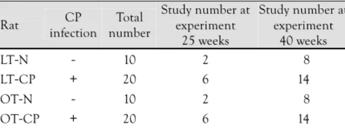
Table 1. Number of LETO rats and OLETF rats
Rat CP
infection Total number
Study number at experiment
25 weeks
Study number at experiment
40 weeks
LT-N - 10 2 08
LT-CP + 20 6 14
OT-N - 10 2 08
OT-CP + 20 6 14
LETO rat: long-evans tokushima otsuka rat, OLETF rat: otsuka long- evans tokushima fatty rat, CP: chlamydia pneumoniae, LT-N: LETO rats without chlamydia pneumoniae infection, LT-CP: LETO rats infected with chlamydia pneumoniae, OL-N: OLETF rats without chlamydia pneumoniae infection, OL-CP: OLETF rats infected with chlamydia pneumoniae
비해 통계학적으로 유의하게 높았다(p<0.05)(Table 2). 중 성지방과 공복시 혈당의 경우 LETO군 간에 통계학적 차이 는 보이지 않았지만, OLETF군에서는 클라미다아 폐렴균 감 염 쥐에서 유의한 상승을 보였다(p<0.05)(Table 2).
클라미디아 폐렴균 감염후 대동맥벽의 변화
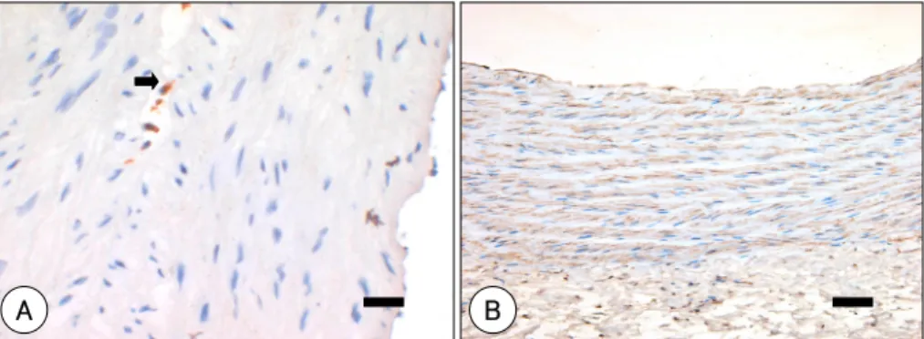
대동맥벽의 클라미디아 폐렴균 감염을 확인하기 위해 24 주령에 희생시킨 쥐의 대동맥에서 anti-Chalmydia pneumo- niae 단일클론 일차항체를 이용하여 면역 조직화학 염색을 시행한 결과 클라미디아 폐렴균에 감염되지 않은 쥐의 대동 맥에서는 항원이 발견되지 않았고(Fig. 1A), 감염시킨 쥐의 대동맥에서는 클라미디아 폐렴균의 항원을 분명하게 관찰 할 수 있었다(Fig. 1B). 24주령과 40주령에 희생시킨 쥐의
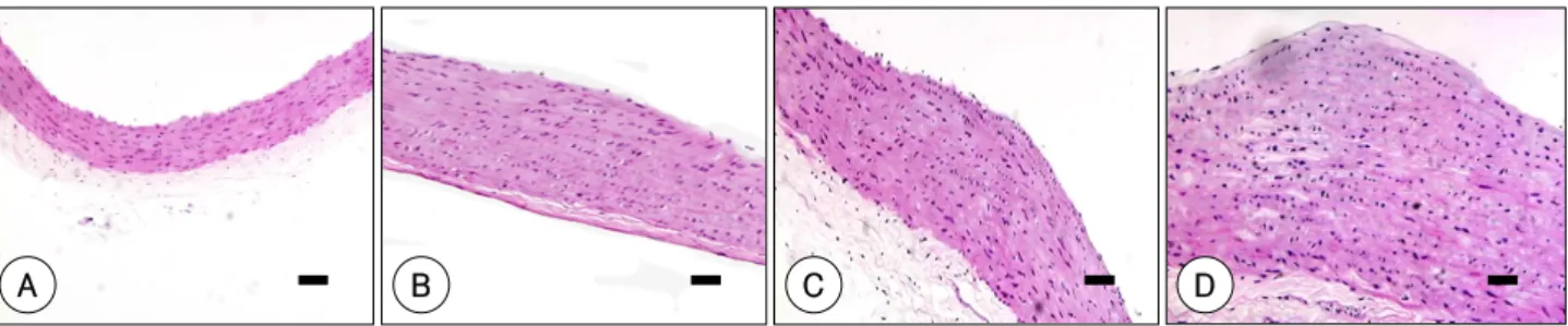
상행대동맥에서 hematoxylin-eosin 염색을 시행하여 각 군 의 병변의 특징을 살펴보고 병변의 면적을 측정 비교하였 다. 클라미디아 폐렴균에 감염되지 않은 LETO쥐에서는 내 막비후와 중막비후가 전혀 발생하지 않았으며, 그 외 다른 군에서는 24주령부터 혈관벽의 변화가 관찰되었다. 클라미 디아 폐렴균에 감염된 LETO군과 클라미디아 폐렴균 감염 에 관계없이 OLETF군에서는 24주령부터 내막과 중막의 비 후와 더불어 내탄력막의 분열이 관찰되었다. 중막쪽 혈관 평 활근 세포는 환상(circumferential)배열을 하고 있는 반면 내막쪽의 혈관 평활근 세포는 종축(longitudinal)배열을 하 고 있으며 불규칙 모양을 하고 있었다. 40주령에서도 클라 미디아 폐렴균에 감염되지 않은 LETO쥐에서는 내막비후와 중막비후가 전혀 발생하지 않았으며, 그 외 다른 군에서는 국소적인 내막의 비후가 보다 분명하게 발생하였으며, 크기 는 비교적 다양하여 클라미디아 폐렴균에 감염된 LETO쥐와 감염되지 않은 OLETF쥐에서는 대부분 융기(elevation)형태 로 관찰되었고 감염된 OLETF쥐에서는 분명한 결절(nodule) 의 형태로 관찰되었으며 기질의 침착도 다른 군에 비하여 풍부하였다(Fig. 2). 죽상동맥경화성 혈관 조직의 특징은 다 른 동물모델과 달리 대식구의 침윤이 거의 없으며, 고지질 식사로 인한 고지혈증에도 불구하고 지질의 침착도 이루어지 지 않았으며 병변을 이루는 대부분의 세포는 혈관 평활근 세포이었다. 이는 대식구와(Fig. 3A) 혈관 평활근 세포에 대 한 면역조직화학염색에서 관찰할 수 있었다(Fig. 3B). 영상 분석 프로그램을 이용한 죽상동맥경화병변의 면적을 측정한
Table 2. Serum lipid profiles and FBS at 40 weeks
LT-N LT-CP OT-N OT-CP
TC (mg/dL) 90±30 97±70 207±34 215±92*
TG (mg/dL) 35±11 28±10 114±67 134±68*
LDL (mg/dL) 52±20 57±60 143±36 145±85*
HDL (mg/dL) 31±20 35±50 043±80 45±4*
FBS (mg/dL) 94±20 96±40 126±14 138±7**
Lipid profile and FBS in OLETF groups were higher than those of LETO groups with statistical significances (p<0.05). But there were no statistical significance not only between LT-N and LT-CP, but also between OT-N and OT-CP, except for triglyceride and FBS. Values are mean±SD. TC: total cholesterol, TG: triglyceride, FBS: fasting blood sugar, LDL: low density lipoprotein cholesterol, HDL: high density lipoprotein cholesterol. *: significant difference between OT-N and OT-CP (p<0.05)
Fig. 1. Immunohistochemical stain with chlamydia-specific monoclonal antibody CF-2 in ascending aorta of rat at experiment 24 weeks. Scale bar: 20 μm. A: no reaction in ascending aorta of non Chlamydia pneumoniae infected rat. B: several areas of brown precipitate (arrow) in cytoplasm of smooth muscle cell. Fig. B showed presence of chlamydial antigen in ascending aorta of chlamydia pneumoniae infected rat.
B A
Fig. 2. H-E staining of rat ascending aorta at experiment 40 weeks. A: LT-N; no intima proliferation. B: LT-CP; mild intima proliferation. C:
OT-N; moderate intima proliferation. D: OT-CP; marked intima proliferation. Scale bar: 20 μm. LT-N: LETO rats not infected with chlamydia pneumonia, LT-CP: LETO rats infected with chlamydia pneumonia, OT-N: OLETF rats not infected with chlamydia pneumonia, OT-CP:
OLETF rats infected with chlamydia pneumonia.
A B C D
결과 클라미디아 폐렴균에 감염되지 않은 LETO쥐에서는 내 막비후와 중막비후가 전혀 발생하지 않아 내막면적이 0 mm2 이었고, 클라미디아 폐렴균에 감염된 LETO 쥐에서는 내막 면적이 3.29±1.23 mm2으로 클라미디아 폐렴균에 감염되 지 않은 OLETF쥐의 내막면적인 4.91±2.11 mm2과 유사한 정도로 죽상동맥경화병변이 발생함을 확인할 수 있었다. 클 라미디아 폐렴균에 감염된 OLETF 쥐는 내막면적이 9.20±
4.62 mm2로 다른 군과 비교하여 통계적으로 유의하게 죽 상동맥경화병변의 발생이 많이 이루어졌음을 보여 주었다 (Fig. 4).
고콜레스테롤 식이와 클라미디아 폐렴균 감염이 혈청 PAI-1, MCP-1, 그리고 CRP에 미치는 영향
PAI-1는 24주와 40주에 클라미디아 폐렴균 감염에 관 계없이 OLETF군이 LETO군과 비교해서 통계학적 유의성 을 가지고 상승되어 있었다(p<0.05)(Fig. 5). 그러나 MCP- 1는 실험 24주에 쥐 종류와 클라미디아 폐렴균 감염에 관 계없이 통계학적으로 차이가 없었고, 40주에서도 유의성을
찾지 못했다(Fig. 6). CRP도 24주와 40주에 네 군 사이에 유의성이 없었다(Fig. 7).
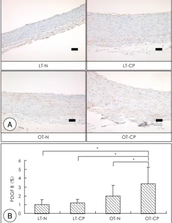
클라미디아 폐렴균이 혈관벽에서 PDGF-B의 발현에 미치는 영향 혈관평활근세포의 증식과 이주에 주로 관여하는 PDGF 의 발현을 알아보기 위해 OLETF쥐와 LETO쥐의 대동맥조 직에서 시행한 PDGF-B의 면역조직화학적 염색에서 클라 미디아 폐렴균이 감염되지 않은 LETO쥐에서는 거의 발현 이 되지 않았고, 감염된 LETO쥐는 감염되지 않은 OLETF 쥐와 유사한 정도로 혈관벽에서 PDGF-B의 발현이 증가되 어 있었다. 클라미디아 폐렴균에 감염된 OLETF쥐에서는 다 른 군과 비교하여 현저하게 PDGF-B의 발현이 증가되어 있었다(Fig. 8).
고 찰
본 연구에서 클라미디아 폐렴균의 감염은 비당뇨쥐인 LETO 쥐의 대동맥에서 죽상동맥경화병변을 발생시켰으며, 당뇨쥐 인 OLETF쥐에서는 감염되지 않은 쥐와 비교하여 죽상동맥
Fig. 3. Immunohistochemical stain in ascending aorta of rat at experiment 40 weeks. Scale bar: 20 μm. A: immunohistochemical stain with mouse anti-macrophage monoclonal antibody (arrow indicates the cytoplasm of macrophage). B: immunohistochemical stain with human anti-α smooth muscle actin monoclonal antibody (brown color indicates the smooth muscle actin).
A B
12
Area (mm2) 10
8
6
4
2
0 LT-N LT-CP OT-N OT-CP
Rat
*
*
*
*
*
Fig. 4. Atherosclerotic lesion in rat aorta at experiment 40 weeks.
Intima mean area of LT-N was 0 mm2, but that of LT-CP is 3.29±
1.23 mm2. There was statistical significance between 2 groups. Intima mean area of OT-N was 4.91±2.11 mm2. That of OT-CP was 9.20±
4.62 mm2. There also was statistical significance between 2 groups.
LT-N: LETO rats not infected with chlamydia pneumoniae, LT-CP:
LETO rats infected with chalmydia pneumoniae, OT-N: OLETF rats not infected with chlamydia pneumoniae, OT-CP: OLETF rats infected with chlamydia pneumoniae. *: p<0.05.
Fig. 5. ELISA of serum plasminogen activator inhibitor-1 (PAI-1) at experiment 24 and 40 weeks. There were statistical significances between LETO rats and OLETF rats at experiment 24 weeks and 40 weeks, irregardless of CP infection. LT-N: LETO rats not infected with chlamydia pneumoniae, LT-CP: LETO rats infected with chal- mydia pneumoniae, OT-N: OLETF rats not infected with chlamydia pneumoniae. OT-CP: OLETF rats infected with chlamydia pneu- moniae. *: p<0.05.
12
PAI1 (ng/dL) 10 8
6
4
2
0 24 weeks 40 weeks
* *
*
* 14
16
L-CP L+CP O-CP O+CP
경화증의 진행을 악화시켰음을 확인하였다. 현재까지 클라 미디아 폐렴균에 의하여 죽상동맥경화증이 발생한 동물모 델에는 New Zealand산 흰토끼와 마우스모델이 보고 된 바 있고,24) 본 연구에서는 비당뇨쥐인 LETO쥐에서 클라미디아 폐렴균에 의해 대동맥에 죽상동맥경화병변이 발생함을 확인 할 수 있었다. 감염되지 않은 LETO쥐에서는 고콜레스테롤 식이를 하여도 대동맥에서 전혀 죽상동맥경화증이 발생하지 않아 분명히 대조된 결과를 얻을 수 있었다. OLETF쥐에서 는 감염되지 않은 쥐에서도 대동맥에 죽상동맥경화병변이 발생하였는데, 클라미디아 폐렴균에 감염된 OLETF쥐에서 유의하게 죽상동맥경화병변이 더 넓게 발생되어 있는 것을 확인할 수 있어 클라미디아 폐렴균이 apolipoprotein E 결 핍 마우스에서 죽상동맥경화증의 진행을 악화시킨다는 기존
의 보고13)14)와 비슷한 결과를 보였다.
클라미디아 폐렴균은 세포내에서만 생존하는 세균으로 이
에 대한 항체는 20세까지 전체인구의 50%에서 나타나고 노 인인구에서는 80%정도 양성으로 나타난다. 한국인의 항체 양 성율은 30%로 외국인에 비해 낮은 것으로 알려져 있으며,25) 한국인에 있어 클라미디아 폐렴균과 죽상동맥경화증의 연관 성에 대한 혈청학적 연구도 많이 이루어지지 않았고 보고들 도 상반되어 죽상동맥경화군과 정상 대조군 사이에 클라미디 아 폐렴균 항체 양성률이 차이가 없다는 보고와26) 1 : 128 이상의 고역가군에서 연관성이 있다는 보고가4) 있다.
클라미디아 폐렴균이 죽상동맥경화증의 발생과 진행에 관 여하는 기전에 대한 지금까지의 연구들을 보면 호흡기 등을 통해 흡입된 클라미디아 폐렴균은 혈액내 단핵구를 감염시 키고 감염된 단핵구를 통해 혈관내에서 혈관조직내 세포들 을 감염시킨다. 본 연구에서도 anti-Chalmydia pneumoniae 단일클론 일차항체인 CF-2를 이용하여 시행한 면역조직화 학 염색에서 클라미디아 폐렴균이 감염되지 않은 쥐의 대 동맥과 달리 감염된 쥐의 대동맥에서는 분명한 발현을 보여 클라미디아 폐렴균에 의한 혈관조직의 감염을 확인할 수 있 었다. 감염된 혈관벽내 대식구는 과다한 저밀도 지단백을 포 식하여 거품세포가 되고 여러 염증성 사이토카인의 분비가 증
Fig. 6. ELISA of serum monocyte chemoattractant protein-1 (MCP- 1) at experiment 24 and 40 weeks. There were no statistical signifi- cances between 4 groups at experiment 24 weeks and 40 weeks. LT-N:
LETO rats not infected with chlamydia pneumoniae, LT-CP: LETO rats infected with chlamydia pneumoniae, OT-N: OLETF rats not infected with chlamydia pneumoniae, OT-CP: OLETF rats infected with chlamydia pneumoniae.
MCP1 (ng/dL) 20 15
10
5
0 24 weeks 40 weeks
25 30
LT-N LT-CP OT-N OT-CP
Fig. 7. ELISA of serum C-reactive protein (CRP) at experiment 24 and 40 weeks. There were no statistical significances between 4 groups at experiment 24 weeks and 40 weeks. LT-N: LETO rats not infected with chlamydia pneumoniae, LT-CP: LETO rats infected with chl- amydia pneumoniae, OT-N: OLETF rats not infected with chlamydia pneumoniae, OT-CP: OLETF rats infected with chlamydia pneu- moniae.
LT-N LT-CP OT-N OT-CP
CRP (ug/dL)
300
250
200
50
0 24 weeks 40 weeks
350 400
150
100
OT-N OT-CP
LT-CP LT-N
PDGF B (%)
0 1 2 3 4 5 6
LT-N LT-CP OT-N OT-CP
*
*
*
A
Fig. 8. Anti-human PDGF B of LETO rats and OLETF rats. A: im- munohistochemical stain with PDGF B in ascending aorta of rat at experiment 24 weeks (brown color indicates PDGF B). Scale bar:
20 μm. B: anti-human PDGF B (%). LETO rat: long-evans tokus- hima otsuka rat, OLETF rat: otsuka long-evans tokushima fatty rat, CP: chlamydia pneumoniae, LT-N: LETO rats not infected with chla- mydia pneumoniae, LT-CP: LETO rats infected with chlamydia pneu- moniae, OT-N: OLETF rats not infected with chlamydia pneumoniae, OT-CP: OLETF rats infected with chlamydia pneumoniae. *: p<0.05.
B
가된다.27) 감염된 혈관내피세포는 E-selectin, intercellular adhesion molecule 1 등의 세포유착인자(adhesion molec- ule) 발현이 유도되며 interleukin-6 등의 염증성 사이토카 인 분비가 촉진된다.8) 또한 클라미디아 폐렴균은 내피세포 에서 PDGF-B의 분비를 촉진하여 혈관 평활근세포의 증식 과 내막으로의 이주를 일으켜 신내막증식을 유도한다.28) 혈 관 평활근세포의 증식과 내막으로의 이주는 죽상동맥경화증 의 형성과정에서 중요한 역할을 수행하며, 특히 쥐에서 발생 하는 죽상동맥경화증의 조직학적 특징은 대부분이 혈관 평활 근세포로 이루어진 내막 과증식이므로18)19) PDGF-B가 쥐의 죽상동맥경화증 형성에서 중요한 역할을 수행할 것으로 추 정할 수 있었다. 본 연구에서도 클라미디아 폐렴균의 감염 은 당뇨쥐와 비당뇨쥐 모두에서 감염되지 않은 쥐와 비교하 여 혈관벽내 PDGF-B의 발현을 증가시키고 있었다. 하지만 감염되지 않은 당뇨쥐에서도 일정부분 PDGF-B의 발현이 관 찰되는 것으로 보아 PDGF-B의 발현 증가는 클라미디아 폐 렴균에 특이적인 현상은 아닐 것으로 생각된다.
본 연구는 또한 쥐의 죽상동맥경화증에 있어 인간과 다 른 동물모델의 죽상동맥경화증과는 다른 여러 특징들을 보 여 주었는데, 먼저 조직학적으로 당뇨쥐와 비당뇨쥐 모두의 죽상동맥경화병변에서 거품세포나 지질의 침착은 거의 관찰 할 수 없었으며 대부분이 혈관평활근세포의 증식과 이주에 의한 내막의 비후(intimal thickening)로 특징 지워지는 죽 상동맥경화병변이 관찰되었다. 24주령과 40주령의 쥐에서 시행한 혈청 ELISA 검사를 보면 쥐에서는 당뇨쥐와 비당뇨 쥐에 관계없이 단핵구의 혈관벽내 이주에 관여하는 강력한 사이토카인인 MCP-1의 혈중농도가 차이를 보이지 않았으 며, 전신염증의 지표이며 죽상동맥경화증 발생에 관여하는 중요한 급성기반응단백질인 CRP의 혈중농도도 차이를 보이 지 않았다. 하지만 PDGF-B의 발현은 증가되어 있고 내막 비후에 있는 대부분의 세포가 혈관 평활근세포인 점으로 보 아 인간이나 다른 동물과는 다른 기전으로 죽상동맥경화증 이 형성되는 것으로 생각되며 향후 이에 대한 연구가 더 필 요할 것으로 생각된다. PAI-1은 24주령부터 클라미디아 폐 렴균 감염과 관계없이 OLETF쥐에서 LETO쥐에 비해 유의 하게 상승되어 있었으며 40주령에서 유의하게 더 증가된 결과를 보이고 있다. PAI-1은 당뇨가 없는 환자의 죽상동 맥경화병변보다 제 2 형 당뇨환자의 죽상동맥경화병변에서 많 이 검출되는 것으로 보아,23) 클라미디아 폐렴균의 영향보다 는 당뇨에 의한 영향으로 증가되었을 것으로 생각된다.
결론적으로 본 연구는 클라미디아 폐렴균의 장기적인 감 염이 비당뇨쥐인 LETO쥐에서 죽상동맥경화증을 발생시키고 당뇨쥐인 OLETF쥐에서는 죽상동맥경화증의 진행을 악화시 키는 것을 분명히 보여 주었다. 또한 쥐에서 클라미디아 폐 렴균에 의한 죽상동맥경화증의 발생에는 PDGF-B에 의한 혈관 평활근세포의 증식과 내막으로의 이주에 의한 내막의 비후가 중요한 역할을 하고 있음을 보여 주었다.
요 약
배경 및 목적:
클라미디아 폐렴균(Chlamydia pneumonia, CP)이 죽상 동맥경화증의 진행을 촉진한다는 연구 보고는 많지만, 클라 미디아 폐렴균이 죽상동맥경화증을 유발시킨다는 연구는 일 부 동물모델에서 제한되어 보고되었다. 본 실험은 일반적으 로 죽상동맥경화증이 잘 발생하지 않는 쥐(rat)에서 클라미 디아 폐렴균이 죽상동맥경화증을 유발할 수 있는지, 또한 죽 상동맥경화증을 유발시킨다면 어떤 기전으로 유발시키는지 알아보고자 하였다.
방 법:
실험군인 11주령의 제 2 형 당뇨병 쥐인 Otsuka Long-Ev- ans Tokushima Fatty(OLETF) 쥐 30마리와 대조군인 Long- Evans Tokushima Otsuka(LETO) 쥐 30마리에게 고지방식 사를 시키면서, 11주령부터 2주 간격으로 3번에 걸쳐 클라 미디아 폐렴균을 복강 또는 비강을 통하여 접종하였다. 24 주령과 40주령에 plasminogen activator inhibitor-1(PAI- 1), monocyte chemoattractant protein-1(MCP-1), 그리고 C-reactive protein(CRP)에 대해 혈청 ELISA을 시행하였 다. 40주령에 상행대동맥에서 죽상동맥경화병변 면적을 분 석하였고 상행대동맥 혈관벽에서 클라미디아 폐렴균의 감염 여부를 면역 조직 화학 염색을 시행하여 확인하고 PDGF- B 활성도를 측정하였다.
결 과:
Specific monoclonal antibody를 사용한 면역 조직 화학 염색 시행결과 죽상동맥경화병변 내에서 클라미디아 폐렴균 감염을 확인하였다. 실험 24주와 40주에 시행한 혈청 ELISA 에서 PAI-1은 클라미디아 폐렴균 감염에 관계없이 OLETF 군이 LETO군에 비해 유의하게 높았으며(p<0.05), MCP-1 과 CRP는 두 군에서 유의한 차이는 없었다. CP를 접종한 LETO쥐와 OLETF쥐에서는 죽상동맥경화병변이 발견되었 지만, CP를 접종하지 않은 LETO쥐에서는 죽상동맥경화병 변이 발견되지 않았다. 특히 CP에 감염된 OLETF쥐는 다 른 군에 비해서 상행대동맥의 죽상동맥경화병변 면적이 더 넓었다(LT-N, 0 mm2; LETO-CP, 3.29±1.23 mm2; OT- N, 4.91±2.11 mm2; OT-CP, 9.20±4.62 mm2)(p<0.05).
특징적으로 쥐의 죽상동맥경화병변은 평활근세포가 주를 이 루었다. 쥐의 동맥혈관벽에서 죽상동맥경화병변의 면적은 PDGF-B의 염색정도와 양의 상관관계를 보였다(p<0.01).
결 론:
동맥 혈관벽에서 만성적인 클라미디아 폐렴균 감염은 고 지혈증이 동반된 LETO 쥐에서는 죽상동맥경화증을 유발시 키고, OLETF 쥐에서는 죽상동맥경화증을 촉진시켰으며, 이 러한 죽상동맥경화병변은 PDGF-B를 매개로 한 평활근세포 의 증식 및 이로 인한 내막증식에 기인한 것으로 유추된다.
중심 단어:죽상동맥경화증;대동맥;Chlamydia pneumo- niae;Inbred OLETF.
REFERENCES
1) Leinonen M, Saikku P. Evidence for infectious agents in cardi- ovascular disease and atherosclerosis. Lancet Infect Dis 2002;
2:11-7.
2) Liu C, Waters DD. Chlamydia pneumoniae and atherosclerosis:
from Koch’s postulates to clinical trials. Prog Cardiovasc Dis 2005;47:230-9.
3) Danesh J, Whincup P, Walker M, et al. Chlamydia pneumoniae IgG titers and coronary heart disease: prospective study and meta-analysis. BMJ 2000;321:208-13.
4) Lee J, Kim JH, Kim TY, et al. Association of Chlamydia pneu- moniae strain TWAR antibody and angiographically demonstrated coronary artery disaese. Korean Circ J 1999;29:1076-81.
5) Gibbs RG, Carey N, Davies AH. Chlamydia pneumoniae and vascular disease. Br J Surg 1998;85:1191-7.
6) Kalayoglu MV, Indrawati RP, Morrison SG, Yuan Y, Byrne GI.
Chlamydia virulence determinants in atherogenesis: the role of chlamydial lipopolysaccharide and heat shock protein 60 in macrophage-lipoprotein interactions. J Infect Dis 2000;181(Su- ppl 3):S483-9.
7) Moazed TC, Kuo CC, Grayston JT, et al. Evidence of systemic dissemination of Chlamydia pneumoniae via macrophages in the mouse. J Infect Dis 1998;177:1322-5.
8) Summersgill JT, Molestina RE, Miller RD, Ramirez JA. Intera- ctions of Chlamydia pneumoniae with human endothelial cells.
J Infect Dis 2000;181(Suppl 3):S479-82.
9) Kol A, Sukhova GK, Lichtman AH, Libby P. Chlamydia heat shock protein 60 localizes in human atheroma and regulates macrophage tumor necrosis factor-α and matrix metallopro- teinase expression. Circulation 1998;98:300-7.
10) Kol A, Bourcier T, Lichtman AH, Libby P. Chlamydial and hu- man heat shock protein 60s activate human vascular endothe- lium, smooth muscle cells, and macrophages. J Clin Invest 1999;
103:571-7.
11) Fong IW, Chiu B, Viira E, Jang D, Mahony JB. De Novo indu- ction of atherosclerosis by Chlamydia pneumoniae in a rabbit model. Infect Immun 1999;67:6048-55.
12) Muhlestein JB, Anderson JL, Hammond EH, et al. Infection with Chlamydia pneumoniae accelerates the development of athero- sclerosis and treatment with azithromycin prevents it in a rabbit model. Circulation 1998;97:633-6.
13) Moazed TC, Campbell LA, Rosenfeld ME, Grayston JT, Kuo CC. Chlamydia pneumoniae infection accelerates the progres-
sion of atherosclerosis in apolipoprotein E-deficient mice. J Infect Dis 1999;180:238-41.
14) Rothstein NM, Quinn TC, Madico G, Gaydos CA, Lowenstein CJ. Effect of azithromycin on murine arteriosclerosis exacer- bated by Chlamydia pneumoniae. J Infect Dis 2001;183:232-8.
15) Wright SD, Burton C, Hernandez M, et al. Infectious agents are notnecessary for murine atherogenesis. J Exp Med 2000;191:
1437-42.
16) Caligiuri G, Rottenberg M, Nicoletti A, Wigzell H, Hansson GK.
Chlamydia pneumoniae infection does not induce or modify atherosclerosis in mice. Circulation 2001;103:2834-8.
17) Adams CW. General pathology of atherosclerosis. In: Vascular Histochemistry. London: Lloyd-Luke Ltd;1967. p.35-82.
18) Reinila A. Ultrastructure of arteries in rats fed a high-fat chole- sterol diet. Arch Pathol Lab Med 1984;108:295-9.
19) Saito F, Kawaguchi M, Izumida J, Asakura T, Maehara K, Mar- uyama Y. Alteration in haemodynamics and pathological changes in the cardiovascular system during the development of type 2 diabetes mellitus in OLETF rats. Diabetologia 2003;46:1161-9.
20) Ross R, Glomset JA. Atherosclerosis and the arterial smooth muscle cell. Science 1973;180:1332-9.
21) Raines EW. PDGF and cardiovascular disease. Cytokine Growth Factor Rev 2004;15:237-54.
22) Jawien A, Bowen-Pope DF, Lindner V, Schwartz SM, Clowes AW. Pletelet-derived growth factor promotes smooth muscle mig- ration and intimal thickening in a rat model of balloon angio- plasty. J Clin Invest 1992;89:507-11.
23) Moazed TC, Kuo C, Grayston JT, Campbell LA. Murine models of Chlamydia pneumoniae infection and atherosclerosis. J Infect Dis 1997;175:883-90.
24) de Kruif MD, van Gorp EC, Keller TT, Ossewaarde JM, ten Cate H. Chlamydia pneumoniae infection in mouse models: relevance for atherosclerosis research. Cardiovasc Res 2005;65:317-27.
25) Lee J, Kim JK, Kim TY, et al. Association of Chlamydia pneu- moniae strain TWAR antibody and angiographically demonstrated coronary artery disease. Korean Circ J 1999;29:1076-81.
26) Kim KH, Jeong MH, Shin JH, et al. The role of chronic infection and inflammation in Korean patients with coronary artery dis- ease. Korean Circ J 2000;30:1107-16.
27) Kalayoglu MV, Byrne GI. Induction of macrophage foam cell formation by Chlamydia pneumoniae. J Infect Dis 1998;177:
725-9.
28) Coombes BK, Chiu B, Fong IW, Mahony JB. Chlamydia pne- umoniae infection of endothelial cells induces transcriptional activation of platelet-derived growth factor-B: a potential link to intimal thickening in a rabbit model of atherosclerosis. J Infect Dis 2002;185:1621-30.