Effect of dentin surface roughness on the shear bond strength of resin bonded restorations
Roodabeh Koodaryan1, Ali Hafezeqoran1*, Sajjad Poursoltan2
1Dental and Periodontal Research Center, Faculty of Dentistry, Tabriz University of Medical Sciences, Tabriz, Iran
2Dentist, Private Practice, Tabriz, Iran
PURPOSE. This study aimed to investigate whether dentin surface preparation with diamond rotary instruments of different grit sizes affects the shear bond strength of resin-bonded restorations. MATERIALS AND METHODS.
The buccal enamel of 60 maxillary central incisors was removed with a low speed diamond saw and wet ground with silicon carbide papers. The polished surfaces of the teeth were prepared with four groups of rotary diamond burs with super-coarse (SC), coarse (C), medium (M), and fine (F) grit sizes. Following surface preparation, 60 restorations were casted with nickel-chromium alloy and bonded with Panavia cement. To assess the shear bond strength, the samples were mounted on a universal testing machine and an axial load was applied along the cement-restoration interface at the crosshead speed of 0.5 mm/min. The acquired data was analyzed with one way ANOVA and Tukey post hoc test (α=.05). RESULTS. The mean ± SD shear bond strengths (in MPa) of the study groups were 17.75 ± 1.41 for SC, 13.82 ± 1.13 for C, 10.40 ± 1.45 for M, and 7.13 ± 1.18 for F. Statistical analysis revealed the significant difference among the study groups such that the value for group SC was significantly higher than that for group F (P<.001). CONCLUSION. Dentin surface roughness created by diamond burs of different grit sizes considerably influences the shear bond strength of resin bonded restorations.
[J Adv Prosthodont 2016;8:224-8]
KEY WORDS: Dentin; Resin-bonded bridge; Bonding; Shear strength; Dental instruments
INTRODUCTION
Since the introduction of resin bonded restorations by Rochette, these prostheses have been notably enhanced and changed. Despite successful clinical treatments, debonding of these restorations is regarded as the main problem of this treatment.1-4 In early resin-bonded prostheses, the weak bond of cement to metal was the main cause of debonding.
This deficiency was improved through several techniques, such as electrolytic or chemical etching of the casting sur- face, incorporation of macromechanical retention, applying silicoater, and air abrasion with aluminum oxide particles.
The current resin cements are chemically active and capable of creating strong bond with base metal alloys.5 Although adhesion to metal has been enhanced, the retention rate of resin bonded restorations is not satisfactory yet. Debonding still occurs commonly at the cement-dentin interface.1-3 Several studies have evaluated the effects of biomechanical properties and preparation design on the success of resin- bonded prostheses. They have consequently emphasized the resistance and retention forms as the determining factors of better clinical retention of resin-bonded restorations.4-9 Therefore, the design of these prostheses has changed from minimum or no preparation to extensive preparations with additional features such as boxes and grooves or occlusal rest seats. Unfortunately, extensive preparations increase the potential loss of enamel, which is highly crucial for bonding and adversely affects the retention.6,7,9,10
Corresponding author:
Ali Hafezeqoran
Dental and Periodontal Research Center, Faculty of Dentistry, Tabriz University of Medical Sciences, Golgasht St., Azadi Ave, Tabriz 51664, Iran
Tel. 98 41 33355965: e-mail, [email protected]
Received January 2, 2016 / Last Revision March 30, 2016 / Accepted March 31, 2016
© 2016 The Korean Academy of Prosthodontics
This is an Open Access article distributed under the terms of the Creative Commons Attribution Non-Commercial License (http://creativecommons.
org/licenses/by-nc/3.0) which permits unrestricted non-commercial use, distribution, and reproduction in any medium, provided the original work is properly cited.
Increasing the adhesive-tooth contact surface through managing the surface roughness is a technique to improve the retention. However, there are limited studies evaluating the effect of roughness created by different rotary instru- ments on the success of full coverage restorations. Ayad et al. studied the surface roughness produced by diamond, tungsten carbide, and tungsten carbide finishing burs and found that diamond burs increased the surface area by a larger degree when compared with carbide burs.11 In anoth- er study, Ayad et al. detected that crosscut carbide burs raised the retention of full coverage restoration cemented with zinc polycarboxylate by 46-55%. The study suggested considering retentive measures in preparation design when smooth surface is preferred.Roughening the preparation surface with rotary instruments increases the cement-tooth interlocking mechanism and improves the retention; conse- quently, the need for extra retentive measures declines.12
Due to the inconclusive literature about the effect of dentin roughness on the retention of crowns and also the lack of similar studies on resin-bonded restorations, it seems essential to determine the influence of dentin rough- ening by diamond rotary instruments on the retention of these restorations. Therefore, the present study aims to compare the effects of diamond rotary burs with different grit sizes on retention of resin-bonded restorations. The null hypothesis of this study is that different grit sizes of diamond rotary instruments do not affect the shear bond strength of resin-bonded restorations.
MATERIALS AND METHODS
In this experimental study, 60 maxillary central incisors of the same size were collected and stored in 1% chloramine- T solution (Solarbio Bioscience & Technology Co., Ltd., Shanghai, China) at 4°C for one month after extraction.
The teeth were intact and extracted because of periodontal diseases.
By using a dental surveyor, each tooth was embedded in a polymeric tube filled with auto-polymerizing acrylic resin (Unifast II, GC Corp., Tokyo, Japan) and was aligned paral- lel with the tube wall. The teeth were prepared for resin bonded restoration by cutting the incisal enamel. Then the proximal and buccal enamel surfaces of the teeth were cut with a low speed diamond saw (Diamant, Gmbh, D&Z, Berlin, Germany) and the buccal dentin was prepared as a rectangle (4 × 4 mm). A caliper with an accuracy of 0.1 mm (Mitutoya, Tokyo, Japan) was used to measure and con- trol the dimensions of all specimens. Once the samples were ready, their buccal dentin was ground with wet silicon carbide paper (Shanghai Hangli Co., Ltd., Shanghai, China).
Different grit sizes of silicon carbide papers were used in a sequence of 420, 600, 1000, and 1200 for 20 seconds.
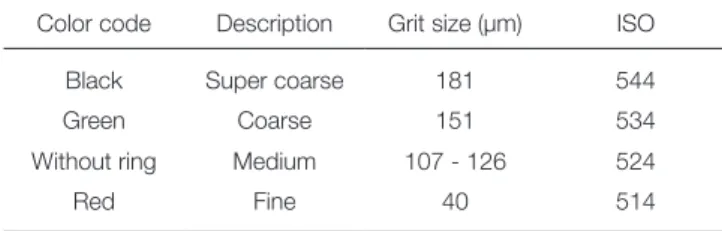
After standardization of the dentin roughness, samples were randomly classified into four groups and polished sur- face of each group was prepared by using diamond rotary burs (NTI-Kahla GmbH; Kahla, Germany) of different grit sizes. Diamond rotary instruments used to prepare den-
tin surfaces are listed in Table 1.
Based on the grit size of the diamond bur used, each group was named as SC (super-coarse with surface rough- ness of 181 µm), C (coarse: 151 µm), M (medium: 107-126 µm), and F (fine: 40 µm). The samples in group SC were prepared merely by very coarse diamond bur with surface roughness of 181 microns and a high-speed air turbine.
The samples in group C were prepared with super-coarse grit size diamond bur followed by a coarse grit bur (151 µm). The surfaces of samples in group M were prepared with super-coarse, coarse, and a medium grit size (107-126 µm) diamond burs sequentially. Group F was prepared with the same method of the previous group (M) and then treat- ed with a fine bur (40 µm).13
Having prepared the dentin surface, a custom acrylic resin tray (Unifast II: GC Crop., Tokyo, Japan) was con- structed for each sample, and the final impression was made with polyether impression material (Impregum; 3M ESPE AG, Seefeld, Germany). Employing lost-wax casting technique, 60 metal castings (4 × 4 mm, 1 mm thick) were made of nickel-chromium alloy (Verabond II: AalbaDent, Cordelia, CA, USA). After finishing and polishing of the castings, their internal surfaces were air-abraded with 50 µm aluminum oxide particles for 10 seconds. According to the manufacturer’s instruction, ED primer liquids A and B were mixed equally, applied on dentin surface, and air dried gen- tly. Then, equal amounts of Panavia cement pastes (Panavia F2; Kurary, Osaka, Japan) were mixed for 20 seconds and applied on the surface of the casting and bonded to the prepared dentin surfaces. Each sample was loaded in the universal testing machine (Hounsfield; Model H5-KS, Surray, UK) for 5 minutes with a load of 10 N. Cement excess was removed and, after 5 minutes, samples were cured for 40 seconds by light curing unit (Astralis 7; Ivoclar Vivadent AG, Lichtenstein, Germany). All samples were thermocycled in 5ºC to 55ºC water baths for a total of 1000 cycles with dwell and transfer times of 15 seconds.
In order to determine the shear bond strength, samples were mounted on the universal testing machine and an axial load was applied with a chisel-shaped rod at a crosshead speed of 0.5 mm/min along the casting-cement interface until failure occurred. The maximum load that caused debonding was recorded in MPa for each sample.
Table 1. Diamond rotary instruments used to prepare the samples
Color code Description Grit size (µm) ISO
Black Super coarse 181 544
Green Coarse 151 534
Without ring Medium 107 - 126 524
Red Fine 40 514
Under a stereomicroscope (Nikon; Tokyo, Japan) at 40
× magnification, the types of failure were evaluated and classified into three categories as the following: (i) adhesive failure with the cement remnant on the prepared dentin surfaces, (ii) adhesive failure with the cement remnant on the casting surface, and (iii) mixed failure with cement rem- nants on both prepared dentin and casting surfaces.
SPSS software (IBM SPSS version 17, Chicago, IL, USA) was used for data analysis. One-way ANOVA was performed to determine the effect of dentin roughness on shear bond strength, and Tukey’s post-hoc test was used to compare the differences among the study groups. The level of statistical significance was set at 5%.
RESULTS
The mean ± SD shear bond strength (in MPa) of the groups are summarized in Table 2. The shear bond strength of the samples prepared with super coarse grit diamond burs (group SC) was significantly higher than that with fine grit rotary instruments (group F) (P < .001).
Table 3 illustrates the failure mode distribution.
Regardless of the surface treatment, the most frequent fail- ure type was adhesive failure with the resin cement rem- nants remained on the castings. Mixed failure mode was detected in all groups; however, it was most seen in group SC (33%) followed by group C (20%).
DISCUSSION
The results of the present study showed that the bond strength of Panavia cement to dentin surfaces treated with super coarse grit diamond bur was higher than that with the fine grit diamond bur, and the difference was statistically significant. Thus, the null hypothesis of the study that den- tin roughness would not affect the retention of the resin- bonded restorations was rejected. Although there is no sim- ilar previous study about the bond strength of resin-bond- ed restorations, the result of this study supports several reported studies that have emphasized the important effect of dentin roughness on crown retention.
Ayad et al.11 demonstrated that dentin surface treatment affects the adhesive bond strength through changing the surface structure. In a similar study, Ayad et al. showed that higher dentin roughness increased the retention of the crowns cemented with zinc phosphate cements while the smooth surfaces of teeth created by finishing burs provided less retention. The study also reported that, regardless of the type of instrumentation, Panavia exhibited more reten- tive strength than zinc phosphate cement. Ayad concluded that the retention of casting restorations depended on both the type of cement and the bur used in dentin surface fin- ishing.12 Likewise, the study by Mowery et al. revealed that the rough surface of prepared dentin provided a higher bond strength, which might be attributed to the increased total surface area involved in the bond.14
Table 2. Mean shear bond strength values (in MPa) and standard deviations calculated for different surface instrumentation of the groups
Study group N Mean ± SD Minimum Maximum
Super Coarse (SC) 15 17.75 ± (1.41)a 15.41 20.73
Coarse (C) 15 13.82 ± (1.13)b 11.08 15.61
Moderate (M) 15 10.40 ± (1.45)c 6.31 12.83
Fine (F) 15 7.13 ± (1.18)d 4.68 8.53
Total 60
Values followed by distinct lowercase letters represent statistical differences between groups.
Table 3. Failure mode distributions of study groups after debonding
Study group N Cement remnants on dentin Mixed (Cement remnants on
dentin and casting) Cement remnants on casting
Super Coarse (SC) 15 2 (13.3%) 5 (33.3%) 8 (53.3%)
Coarse (C) 15 1 (6.6%) 3 (20%) 11 (73.3%)
Moderate (M) 15 0 (0%) 2 (13.3%) 13 (86.6%)
Fine (F) 15 0 (0%) 1 (6.6%) 14 (93.3%)
Intimate contact of two opposing surfaces is necessary to establish a stable bond between dentin and the resin cement. The degree to which the adhesive moistens the dentin surface depends on the cement viscosity, contact angle between the two surfaces, topography of dentin sur- face, and the grit size of the cutting instrument.15-20 When the dentin is cut, a smear layer of different thickness and density is formed on it, whose morphology and characteris- tics depend on the grit size of burs or abrasive instruments.
Accordingly, the greater the grit size of the abrasive rotary instrument, the thicker the smear layer.18,19,21,22 Debris and smear layer reduce the surface energy, which may lead to poor adhesion; for that reason, some investigations have recommended removing the smear layer to improve the bond strength.23-25
To interpret the results of this study, it should be consid- ered that the condition of the dentin surface after prepara- tion (including the roughness and smear layer characteristics) has antagonistic effects on bonding of resin cements.26-28 Increasing the roughness of dentin surface provides a larg- er surface area for physical interaction with resin cement, resulting in stronger chemical bond with the dentin. On the other hand, thicker smear layer formed by diamond burs may prevent proper interaction between high-viscosity resin cement and the underlying dentin.18,19,22,29-31 The negative effect of smear layer thickness on the bond strength was not observed in the present study. The ED primer in Panavia cement kit had the ability to etch through the smear layer and leave the underlying dentin partially demineralized, increasing dentin permeability to the functional monomer of resin cement.32,33 Moreover, bonding to the prepared surface of dentin might also have been influenced by other factors.
It seems that the pressure imposed on indirect restora- tions during cementation plays an important role in resin bonding and determines the efficacy of reaction between adhesive cement and dentin surface covered with smear lay- er. Some studies agree that the seating pressure applied on luting agents during cementation of indirect restoration improves their adaptation and bond strength to dentin.16,26,34 According to De Munck et al., viscosity of the material decreas- es under continuous shear rate.35 Besides, higher seating pressure reduces the number of pores left on the bond interface or inside the adhesive cement. Furthermore, inti- mate contact between the adhesive cement and the underly- ing dentin improves the physical interaction between the surfaces through affecting the factors involved in bond strength, including Van der Waals force, hydrogen bridges, and charge-transfers.16,29,35
In addition to the positive effect of the seating pressure on the bond strength, Chieffi et al. and other researchers have evaluated the effect of applying a constant pressure during the cement setting and concluded that it prevented water absorption and globule formation; therefore, a better adhesive surface would be achieved.34,36 As a result, decreas- ing infiltration of water from the underlying dentin into the primer-cement system might lead to better coupling of the
resin to dentin and improve bond strength. Thus, selecting the appropriate cement in combination with pressure appli- cation duration affected the bond strength significantly.36
With respect to the findings of this study, dentin sur- face roughness has the potential to improve the bond strength of resin bonded restoration. However, long-term in vitro and in vivo studies should be conducted to support clinical cases. Additionally, new miscellaneous adhesive cements with high variety must be evaluated of their specific performance.
CONCLUSION
According to findings of the current study, preparing tooth surface by using coarse diamond burs enhances the bond strength of resin-bonded restorations.
ORCID
Ali Hafezeqoran http://orcid.org/0000-0002-7756-3616 REfERENCES
1. Goodacre CJ, Bernal G, Rungcharassaeng K, Kan JY. Clinical complications in fixed prosthodontics. J Prosthet Dent 2003;
90:31-41.
2. Pjetursson BE, Tan WC, Tan K, Brägger U, Zwahlen M, Lang NP. A systematic review of the survival and complica- tion rates of resin-bonded bridges after an observation peri- od of at least 5 years. Clin Oral Implants Res 2008;19:131-41.
3. Priest G. An 11-year reevaluation of resin-bonded fixed par- tial dentures. Int J Periodontics Restorative Dent 1995;15:
238-47.
4. Wyatt CC. Resin-bonded fixed partial dentures: what's new? J Can Dent Assoc 2007;73:933-8.
5. Doh RM, Lee KW. Dislodgement resistance of modified res- in-bonded fixed partial dentures utilizing tooth undercuts: an in vitro study. J Adv Prosthodont 2009;1:85-90.
6. Chow TW, Chung RW, Chu FC, Newsome PR. Tooth prepa- rations designed for posterior resin-bonded fixed partial den- tures: a clinical report. J Prosthet Dent 2002;88:561-4.
7. el-Mowafy O, Rubo MH. Retention of a posterior resin- bonded fixed partial denture with a modified design: an in vi- tro study. Int J Prosthodont 2000;13:425-31.
8. Keulemans F, Shinya A, Lassila LV, Vallittu PK, Kleverlaan CJ, Feilzer AJ, De Moor RJ. Three-dimensional finite element analysis of anterior two-unit cantilever resin-bonded fixed dental prostheses. ScientificWorldJournal 2015;2015:864389.
9. Shimizu H, Takahashi Y. Retainer design for posterior resin- bonded fixed partial dentures: a technical report. Quintessence Int 2004;35:653-4.
10. el-Mowafy O, Rubo MH. Resin-bonded fixed partial den- tures-a literature review with presentation of a novel ap- proach. Int J Prosthodont 2000;13:460-7.
11. Ayad MF, Rosenstiel SF, Hassan MM. Surface roughness of dentin after tooth preparation with different rotary instru- mentation. J Prosthet Dent 1996;75:122-8.
12. Ayad MF, Rosenstiel SF, Salama M. Influence of tooth sur- face roughness and type of cement on retention of complete cast crowns. J Prosthet Dent 1997;77:116-21.
13. Li YQ, Wang H, Wang YJ, Chen JH. Effect of different grit sizes of diamond rotary instruments for tooth preparation on the retention and adaptation of complete coverage resto- rations. J Prosthet Dent 2012;107:86-93.
14. Mowery AS Jr, Parker M, Davis EL. Dentin bonding: the ef- fect of surface roughness on shear bond strength. Oper Dent 1987;12:91-4.
15. Al-Omari WM, Mitchell CA, Cunningham JL. Surface rough- ness and wettability of enamel and dentine surfaces prepared with different dental burs. J Oral Rehabil 2001;28:645-50.
16. Goracci C, Cury AH, Cantoro A, Papacchini F, Tay FR, Ferrari M. Microtensile bond strength and interfacial proper- ties of self-etching and self-adhesive resin cements used to lute composite onlays under different seating forces. J Adhes Dent 2006;8:327-35.
17. Hipilito V, Azevedo LC, Piveta FC, Filho WSV, Netto CA, Alonso RCB, Alpino PHP. Effect of dentinal surface prepa- ration on the bonding of self-adhesive luting cements. J Adhes Sci Technol 2014;28:1907-24.
18. Hosoya Y, Shinkawa H, Suefiji C, Nozaka K, García-Godoy F. Effects of diamond bur particle size on dentin bond strength. Am J Dent 2004;17:359-64.
19. Peerzada F, Yiu CK, Hiraishi N, Tay FR, King NM. Effect of surface preparation on bond strength of resin luting cements to dentin. Oper Dent 2010;35:624-33.
20. Reifeis PE, Cochran MA, Moore BK. An in vitro shear bond strength study of enamel/dentin bonding systems on enamel.
Oper Dent 1995;20:174-9.
21. Ayad MF, Johnston WM, Rosenstiel SF. Influence of dental rotary instruments on the roughness and wettability of hu- man dentin surfaces. J Prosthet Dent 2009;102:81-8.
22. Barros JA, Myaki SI, Nör JE, Peters MC. Effect of bur type and conditioning on the surface and interface of dentine. J Oral Rehabil 2005;32:849-56.
23. Brännström M, Glantz PO, Nordenvall KJ. The effect of some cleaning solutions on the morphology of dentin pre- pared in different ways: an in-vivo study. ASDC J Dent Child 1979;46:291-5.
24. Pashley DH, Tao L, Boyd L, King GE, Horner JA. Scanning electron microscopy of the substructure of smear layers in human dentine. Arch Oral Biol 1988;33:265-70.
25. Tagami J, Tao L, Pashley DH, Hosoda H, Sano H. Effects of high-speed cutting on dentin permeability and bonding. Dent Mater 1991;7:234-9.
26. Escribano N, de la Macorra JC. Microtensile bond strength of self-adhesive luting cements to ceramic. J Adhes Dent 2006;8:337-41.
27. Marshall GW Jr, Marshall SJ, Kinney JH, Balooch M. The dentin substrate: structure and properties related to bonding.
J Dent 1997;25:441-58.
28. Perdigão J. Dentin bonding-variables related to the clinical situation and the substrate treatment. Dent Mater 2010;26:
e24-37.
29. Burgess JO, Ghuman T, Cakir D. Self-adhesive resin cements.
J Esthet Restor Dent 2010;22:412-9.
30. McInnes PM, Wendt SL Jr, Retief DH, Weinberg R. Effect of dentin surface roughness on shear bond strength. Dent Mater 1990;6:204-7.
31. Wahle JJ, Wendt SL Jr. Dentinal surface roughness: a compar- ison of tooth preparation techniques. J Prosthet Dent 1993;69:160-4.
32. Van Meerbeek B, Yoshihara K, Yoshida Y, Mine A, De Munck J, Van Landuyt KL. State of the art of self-etch adhe- sives. Dent Mater 2011;27:17-28.
33. Yang B, Ludwig K, Adelung R, Kern M. Micro-tensile bond strength of three luting resins to human regional dentin.
Dent Mater 2006;22:45-56.
34. Chieffi N, Chersoni S, Papacchini F, Vano M, Goracci C, Davidson CL, Tay FR, Ferrari M. Effect of the seating pres- sure on the adhesive bonding of indirect restorations. Am J Dent 2006;19:333-6.
35. De Munck J, Vargas M, Van Landuyt K, Hikita K, Lambrechts P, Van Meerbeek B. Bonding of an auto-adhesive luting material to enamel and dentin. Dent Mater 2004;20:
963-71.
36. Chieffi N, Chersoni S, Papacchini F, Vano M, Goracci C, Davidson CL, Tay FR, Ferrari M. The effect of application sustained seating pressure on adhesive luting procedure.
Dent Mater 2007;23:159-64.