Introduction
Peripheral artery disease (PAD) is comorbid in 10.6% of patients with ischemic heart disease.
1) The difficulty in accessing the arterial system may complicate ventricular tachycardia (VT) ablation proce- dures in patients with PAD. The radial artery is the preferred access route for patients undergoing percutaneous coronary intervention, in terms of the patients’ comfort and in terms of a decreased risk for vascular complications.
2)3) Herein, we present a case of a success- ful catheter ablation of VT by radial artery access in a post-myocar- dial infarction patient with PAD, who had previously failed ablation via a transseptal approach.
Case
A 73 year-old man with prior anterior myocardial infarction was admitted for management of an electrical storm (ES) occurring 20
Jun Kim, MD, Dongcheul Han, MD, Changbae Sohn, MD, Jeong Su Kim, MD, and Yong Hyun Park, MD
Division of Cardiology, Pusan National University Yangsan Hospital, Yangsan, Korea
Herein, we present a case of a successful catheter ablation of ventricular tachycardia (VT) using a radial artery approach in a post-myocar- dial infarction patient, who had an implantable cardioverter-defibrillator and peripheral artery disease. Although the patient did not use anti- arrhythmic drugs, the patient experienced no recurrence of VT during the following 3-year period. (Korean Circ J 2012;42:632-637) KEY WORDS: Catheter ablation; Ventricular tachycardia; Ventricular premature complexes; Radial artery.
Received: December 16, 2011 Revision Received: February 8, 2012 Accepted: February 13, 2012
Correspondence: Jun Kim, MD, Division of Cardiology, Pusan National University Yangsan Hospital, 20 Geumo-ro, Mulgeum-eup, Yangsan 626- 770, Korea
Tel: 82-55-360-2357, Fax: 82-55-360-2204 E-mail: [email protected]
• The authors have no financial conflicts of interest.
This is an Open Access article distributed under the terms of the Creative Commons Attribution Non-Commercial License (http://creativecommons.
org/licenses/by-nc/3.0) which permits unrestricted non-commercial use, distribution, and reproduction in any medium, provided the original work is properly cited.
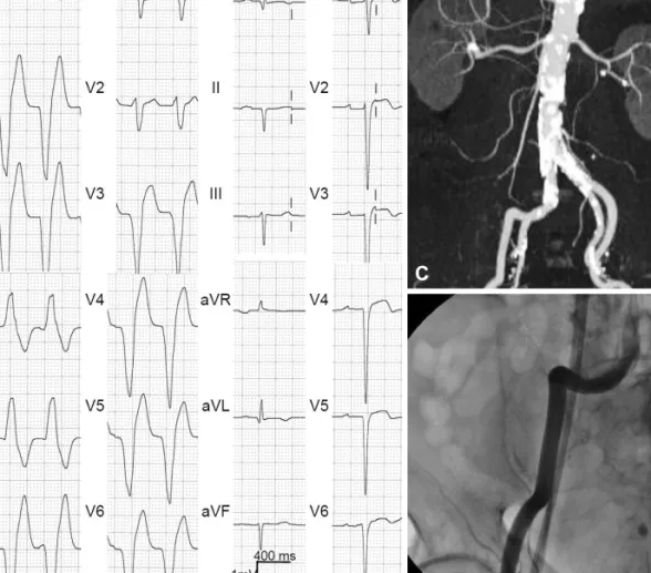
months after the implantation of an implantable cardioverter-de- fibrillator (ICD). His medical conditions included an anterior myocar- dial infarction that had occurred 15 years prior, heart failure, periph- eral arterial disease, non-disabling stroke, hypertension, gout, and dia- betes mellitus. He underwent implantation of dual chamber ICDs due to sustained VT (Fig. 1A and B). At the time, the left ventricular (LV) ejection fraction was 36%, and a LV aneurysm was noted. Co- ronary angiography showed a 50% stenosis in the mid-portion of the left anterior descending artery, 80% stenosis at the distal left circumflex artery, and 40% stenosis in the proximal right coronary artery. His medications included a beta-blocker, amiodarone, an an- giotensin-converting enzyme inhibitor, aspirin, and diuretics. Com- puted tomography angiography revealed extensive calcification from the distal abdominal aorta to the bilateral common iliac artery and the tortuous right common iliac artery (Fig. 1C and D).
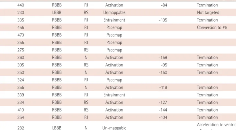
The patient underwent catheter ablation for medically refractory, frequent premature ventricular complexes (60% of total beats) that were originating from the left apico-inferior septum. The ablation was performed by a transseptal approach due to the deterioration of heart failure, which had begun 13 months before presentation (Fig. 2).
Before the occurrence of the ES, there was only one episode of VT treated with anti-tachycardia pacing. Sixteen episodes of VT with a cycle length of 430 to 455 ms occurred per day, and 3 episodes were terminated by cardioversion (20 J).
After obtaining informed consent from the patient, he underwent catheter ablation of VT via a transseptal approach, using an external irrigation catheter (power-controlled mode, 30 Watts, 45°C, 17 mL/
min). During the procedure, VT was still inducible by programmed