• 검색 결과가 없습니다.

A Case of Osteosarcoma after Treatment of Endodermal Sinus Tumor

N/A
N/A
Protected

Academic year: 2021

Share "A Case of Osteosarcoma after Treatment of Endodermal Sinus Tumor"

Copied!
5
0
0

로드 중.... (전체 텍스트 보기)

전체 글

(1)

내배엽동종양 치료 후 발생한 골육종 1예

진다희

1

ㆍ이 윤

1

ㆍ이정화

1

ㆍ이광철

1

ㆍ김철환

2

고려대학교 의과대학

1

소아과학교실,

2

병리학교실

A Case of Osteosarcoma after Treatment of Endodermal Sinus Tumor

Dahee Jin, M.D.

1

, Yoon Lee, M.D.

1

, Jung Hwa Lee, M.D.

1

, Kwang Chul Lee, M.D.

1

and Chul Hwan Kim, M.D.

2

Departments of

1

Pediatrics,

2

Pathology, Korea University College of Medicine, Seoul, Korea

Endodermal sinus tumor is a type of germ cell tumor that is relatively common in children. An important problem concerns secondary neoplasms after treatment. We re- port a case of osteosarcoma that developed five years after treatment of intrapelvic endo- dermal sinus tumor. The patient was a seven-year-old girl who presented with right thigh and knee joint pain. The patient had been diagnosed with endodermal sinus tumor five years previously and treated with a regimen consisting of high dose cisplatin, etoposide, bleomycin and surgery but without radiotherapy. We detected a mass shadow on the right distal femur that proved to be osteoblastic osteosarcoma by incisional biopsy. The patient received surgical treatment after chemotherapy that included high dose methotrexate. The follow-up bone scan revealed no abnormal uptakes. There has been no evidence of recurrence eighteen months of follow-up after chemotherapy.

pISSN 2233-5250 / eISSN 2233-4580 http://dx.doi.org/10.15264/cpho.2014.21.1.41 Clin Pediatr Hematol Oncol 2014;21:41∼45

Received on March 17, 2014 Revised on April 7, 2014 Accepted on April 15, 2014

Corresponding Author: Kwang Chul Lee Department of Pediatrics, Korea University Anam Hospital, 73, Inchon-ro, Seongbuk-gu, Seoul 136-705, Korea

Tel: +82-2-920-5090 Fax: +82-2-922-7476 E-mail: kwcl5609@korea.ac.kr

Key Words: Endodermal sinus tumor, Osteosarcoma, Second primary neoplasm

Introduction

Endodermal sinus tumor (EST) is a type of germ cell tu- mor arising from a common progenitor germ cell [1]. After the introduction of regimen including high dose cisplatin, bleomycin, etoposide (PEB), the prognosis of EST in chil- dren has markedly improved [2,3].

Although the prognosis has improved and the life span after primary tumor has been extended, secondary neoplasm remains a major problem in the field of the oncology.

Various types of secondary neoplasms have been re- ported [4-7], the type depending on several risk factors in-

cluding genetic predisposition, type of primary neoplasms, treatment, etc. There are only a few cases reported after treatment of EST. Here we report a case of osteosarcoma after EST treated with bleomycin, etoposide and cisplatin.

Case Report

A seven-year-old girl presented with right thigh and knee joint pain. Physical examination revealed diffuse swelling and mild tenderness around the right knee joint. The distal sensory and motor functions were intact.

On review of the patient’s past medical history, we found

that she had visited our hospital due to constipation and

(2)

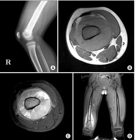
Fig. 1. (A) A simple X-ray film image,

(B) axial image on MRI scan, T1- weighted image, (C) T2-weighted image and (D) coronal image, contrast-enhanced T1-weighted image.

abdominal distension at eighteen months of age. She had been diagnosed with EST stage 3 that had appeared in the form of a 9 cm-sized intrapelvic lump. The alpha-fetoprotein (AFP) level was 1,210 ng/mL (reference range, 0.8-87 ng/mL). She was successfully treated with six cycles of high dose PEB regimen but without radiotherapy. She had regu- larly visited the outpatient clinic at three month intervals without evidence of recurrence. The AFP had decreased by 1.15 ng/mL 40 months after completion of chemotherapy.

There was no family history of malignancies.

Simple X-ray film showed a mass shadow on the right distal femur with osteosclerotic and permeative osteolytic lesion. An amorphous osteoid mineralization, a codman’s triangle and periosteal reaction with hair-on-end were also noticed (Fig. 1A).

A complete blood cell count and biochemical analysis of

the blood revealed no abnormal findings, except elevated lactate dehydrogenase. (726 IU/L). With suspicion of bone malignancy, a magnetic resonance imaging (MRI) study was done. A solitary mass was found in the patient’s right distal femur with low to intermediate signal intensity on T1 weighted images and heterogeneous signal intensity on T2 weighted images. It was also accompanied by some soft tissue components and cortical lifting (Fig. 1B-D).

Incisional biopsy was done and histologic studies re- vealed osteoblastic osteosarcoma (Fig. 2).

The patient received neoadjuvant chemotherapy based on

Pediatric Oncology Group Study (POG-8651), including high

dose methotrexates (12 g/m

2

of body surface area) and leu-

covorin rescue, doxorubicin, cisplatin, cyclophosphamide,

bleomycin and dactinomycin [10]. The patient also received

wide excision and bone reconstruction with allo-bone and

(3)

Fig. 2. (A) An osteoid is found between individual tumor cells as lacelike pattern. ×200. Hematoxylin and eosin stain. (B) The tumor

cells show marked nuclear pleomorphism and hyperchromasia with eosinophilic cytoplasm. ×400. Hematoxylin and eosin stain.

cement prosthesis and continued with adjuvant chemotherapy.

On a follow-up bone scan 20 months from the operation, there were no abnormal uptakes. There has been no evidence of recurrence 16 months after completion of chemotherapy.

Discussion

Germ cell tumor is one of the common childhood neoplasms. About 20% of germ cell tumors are malignant and they can arise in various sites such as ovary, testis and the sacrococcygeal area. The term germ cell tumor includes several histologic types, including EST, also called yolk sac tumor, germinoma (dysgerminoma or seminoma), embryonal carcinoma and choriocarcinoma. Serologic markers such as alpha-fetoprotein and human choriogonadotropin are useful for the diagnosis and the follow-up of germ cell tumors [1].

Using PEB regimen, the prognosis of malignant germ cell tumors in children has improved dramatically over the past few decades. Malignant germ cell tumors excepting those found in extracranial sites have shown survival rates rang- ing from 85% to 95% [11]. However, increased survival rates may result in a greater incidence of secondary neoplasms.

There have been some reports of secondary tumors diag- nosed after treatment for germ cell tumor [12,13].

Osteosarcoma is one of the most common secondary ne- oplasms among childhood cancers. Hawkins et al. reported that 0.9% of patients surviving at least 3 years after diag-

nosis of all types of childhood cancer developed bone can- cers within 20 years [9]. Le Vu B et al. reported that the incidence of secondary osteosarcoma 30 years after diag- nosis of the first cancer was 1.23% [14].

There are many risk factors which play a role in the eti- ology of second malignancies. The genetic predisposition makes a person more susceptible to development of secon- dary malignancies than those without such abnormalities.

In some cases, a second neoplasm is a manifestation of a family cancer syndrome. A family history of cancer was as- sociated with secondary sarcomas and the relative risk (RR) was 1.6 (95% confidence interval (CI) 1.1-2.3) [5]. The sib- lings had 1.5 times greater risk compared with that of the general population [6]. But our patient does not have any suspicious genetic predisposition of cancer.

A certain type of first primary cancer has a higher risk

of secondary cancer than others. Henderson et al. reported

that the RR of secondary sarcoma after soft tissue sarcoma

and Hodgkin lymphoma was 8.8 (95% CI 4.6-16.8) and 5.5

(95% CI 3.0-10.9), respectively [5]. Another group of inves-

tigators reported much higher RRs of secondary cancer after

primary sarcoma. The risk after soft tissue sarcoma was twen-

ty-five-fold greater compared with the general population

and ten-fold higher after bone cancer, and twelve-fold higher

after Hodgkin lymphoma [6]. Furthermore Le Vu B claimed

that the risk of osteosarcoma is 1,515-fold greater with the

initial treatment of bilateral retinoblastoma than that found

(4)

in the general populations [14]. There were only limited re- ports dealing with secondary cancer after primary germ cell tumor. There were no established RRs either.

Radiotherapy was consistently reported as a strong risk factor and the risk increases with greater cumulative doses of radiation exposure. And concurrent chemotherapy wor- sens the risk significantly [4-6,9,14]. Radiotherapy increases the risks of various secondary solid tumors at numerous sites. Henderson et al. reported that radiation was asso- ciated with increased risk of secondary sarcoma. The RR of development of secondary sarcoma after radiotherapy was 3.1 (95% CI 1.5-6.2) [5]. But our patient did not receive radiotherapy. So radiotherapy had nothing to do with the development of her osteosacoma.

Anthracyclines [4-6] and alkylating agents [5,6,9] are also known as risk factors for secondary neoplasms in several studies. One report showed that anthracyclines were asso- ciated with secondary sarcoma and the odds ratio was 3.5 (95% CI 1.6-7.7). But this report showed no significant rela- tionship between secondary sarcoma and other chemo- therapeutic agents including alkylating agents and epi- podophyllotoxins [4]. Another study suggested that the risk was related to the dose of anthracycline or alkylating agent [5]. Hawkins et al. revealed elevated RR of secondary bone cancer after exposure to alkylating agents at a dose greater than 20,000 mg/m

2

, four-fold greater than that without ex- posure [9].

A combination of these factors including individual ge- netic predisposition, previous radiotherapy and chemo- therapy would play a role in development of the secondary osteosarcoma. But our patient did not receive radiotherapy, the strongest risk factor, nor had a family history of malignancy. The patient’s primary tumor, EST is not com- monly recognized as strong risk factor for secondary tumor.

Although exposure to alkylating agents is the sole well known risk factor in the present case, it does not necessa- rily mean that the primary tumor has nothing to do with osteosarcoma.

Despite extensive review of literature, we failed to find similar reports of secondary osteosarcoma after treatment of a germ cell tumor. But we found a case report about extra- skeletal osteosarcoma of the mediastinum after treatment of

a mediastinal germ cell tumor. Ulusakarya et al. reported that the secondary tumor in their case was a histologic transformation of the teratomatous component of primary germ cell tumor to extraskeletal osteosarcoma [12]. In Korea, a case of renal cell carcinoma after treated for germ cell tumor has been reported [13]. The vincristine, bleomy- cin, cisplatin based regimen that the patient received was similar to our PEB regimen.

Secondary osteosarcomas are reported to have a poor prognosis compared with primary osteosarcomas. However, there are no specialized guidelines or statements for secon- dary osteosarcoma [15]. Our patient received surgery and treatment of osteosarcoma with multiple agents including methotrexate, doxorubicin and cisplatin, a treatment strategy which has so far proved successful.

In summary, we report a rare case of osteosarcoma which occurred five years after treatment of EST with PEB regimen and surgery but no radiotherapy. The prevalence of secondary neoplasms has increased with improved sur- vival of children with malignant neoplasms. Plans for the treatment of tumors should be designed to decrease the risk of secondary cancer. Close monitoring is also needed for cancer survivors who received radiotherapy and chemo- therapy, including anthracyclines or alkylating agents.

References

1. Davidoff AM, Hebra A, Bunin N, Shochat SJ, Schnaufer L.

Endodermal sinus tumor in children. J Pediatr Surg 1996;31:

1075-8.

2. Billmire DF. Malignant germ cell tumors in childhood. Semin in Pediatr Surg 2006;15:30-6.

3. Rescorla FJ, Sawin RS, Coran AG, Dillon PW, Azizkhan RG.

Long-term outcome for infants and children with sacrococcy- geal teratoma: a report from the Childrens Cancer Group. J Pediatr Surg 1998;33:171-6.

4. Henderson TO, Rajaraman P, Stovall M, et al. Risk factors as- sociated with secondary sarcomas in childhood cancer survi- vors: a report from the childhood cancer survivor study. Int J Radiat Oncol Biol Phys 2012;84:224-30.

5. Henderson TO, Whitton J, Stovall M, et al. Secondary sarco- mas in childhood cancer survivors: a report from the Childhood Cancer Survivor Study. J Natl Cancer Inst 2007;99:

300-8.

6. Meadows AT, Friedman DL, Neglia JP, et al. Second neo-

(5)

plasms in survivors of childhood cancer: findings from the Childhood Cancer Survivor Study cohort. J Clin Oncol 2009;

27:2356-62.

7. Travis LB. The epidemiology of second primary cancers.

Cancer Epidemiol Biomarkers Prev 2006;15:2020-6.

8. Bechler JR, Robertson WW Jr, Meadows AT, Womer RB.

Osteosarcoma as a second malignant neoplasm in children.

J Bone Joint Surg Am 1992;74:1079-83.

9. Hawkins MM, Wilson LM, Burton HS, et al. Radiotherapy, al- kylating agents, and risk of bone cancer after childhood cancer. J Natl Cancer Inst 1996;88:270-8.

10. Goorin AM, Schwartzentruber DJ, Devidas M, et al. Presurgical chemotherapy compared with immediate surgery and adjuvant chemotherapy for nonmetastatic osteosarcoma: Pediatric Oncology Group Study POG-8651. J Clin Oncol 2003;21:1574-80.

11. Buyukpamukcu M, Varan A, Küpeli S, et al. Malignant sacro-

coccygeal germ cell tumors in children: a 30-year experience from a single institution. Tumori 2013;99:51-6.

12. Ulusakarya A, Terrier P, Regnard JF, de Montpreville V, Munck JN. Extraskeletal osteosarcoma of the mediastinum af- ter treatment of a mediastinal germ-cell tumor. Am J Clin Oncol 1999;22:609-14.

13. Nam CH, Lee HS, Choi SR, et al. Secondary renal cell carcino- ma in a child treated for germ cell tumor. Korean J Pediatr Hematol Oncol 2004;11:86-91.

14. Le Vu B, de Vathaire F, Shamsaldin A, et al. Radiation dose, chemotherapy and risk of osteosarcoma after solid tumours during childhood. Int J Cancer 1998;77:370-7.

15. Bielack SS, Kempf-Bielack B, Heise U, Schwenzer D, Winkler

K. Combined modality treatment for osteosarcoma occurring

as a second malignant disease. Cooperative German-Austrian-

Swiss Osteosarcoma Study Group. J Clin Oncol 1999;17:1164.

수치

Fig. 1. (A) A simple X-ray film image, (B) axial image on MRI scan, T1-  weighted image, (C) T2-weighted  image and (D) coronal image,  contrast-enhanced T1-weighted image.
Fig. 2. (A) An osteoid is found between individual tumor cells as lacelike pattern. ×200

참조

관련 문서

•Targeted therapies are a cornerstone of precision cancer medicine , a form of medicine that uses. information about a person’s tumor genes and proteins to prevent,

Methods: I conducted a retrospective analysis of clinical records of children aged between 4 and 19 years who were treated with flunarizine for headache at

With the increase of the dental implant procedure, there are many cases of Maxillary Sinus Floor Elevation Surgery(MSFES). Additionally, its side effect has

Methods to overcome insufficient bone due to poor bone quality, the pneumatization of a maxillary sinus and other anatomical limitations of implant placement

Subjects with a smoking period of 1 to 3 years had a high smoking cessation success rate (p=0.024), and the lower the average daily smoking amount, the higher the

Long-term outcomes of short dental implants supporting single crowns in posterior region: a clinical retrospective study of 5-10 years.. Osteotome sinus

The OSFE (osteotome sinus floor elevation) technique has been used for maxillary sinus augmentation.. The implants were clinically and radiographically followed

Patient had laparoscopic surgery on the adnexal tumor and excised tissue was removed through Douglas pouch incision by single surgeon.. Results: The mean age