273 臨床耳鼻:第 26 卷 第 2 號 2015
• • • • • • • • • • • • • • • • • • • • • • • • • • • • • • • • • • • • • • • • • • • • • • • • • • • • • • • • • • • • • • • • • • • • • • • • • • • • • • • • • • • • • • • • • • • • • • • • • • • • • • • • • • • • • • • • • • • • • • • • • • • • • • • • • • • • • • • • • • • • • • • • • • • • • • • • • • • • • • • • • • • • • • • • • • • • • • • • • • • • • • • • • • • • • • • • • • • • • • • • • • • • • • • • • • • • • • •
J Clinical Otolaryngol 2015;26:273-276
서 론
이소치는 치아영역 외에 다양한 곳에서 발견될 수 있 으며 그 빈도는 유치의 0.3~0.8%, 영구치의 1.0~3.5% 정 도로 알려져 있다.1) 이소치는 하악골의 구상돌기, 경구 개, 비강, 상악동 등에서 발견된 경우가 보고된바 있으 며, 상악동 내의 이소치는 무증상을 보이는 경우가 많 으나 상악동염의 증상을 유발할 수 있다.2-4) 이소치가 발 생하는 원인에 대해서는 여러 가지 가설이 있으나 아직
명확히 밝혀진 것은 없으며, 치아의 형태와 그와 인접한 해부학적 환경 또는 외상, 감염, 병리학적 상태, 그리고 발 생학적 장애 등이 관계가 있을 것으로 생각하고 있다.2,4,5) 저자들은 비폐색과 화농성 비루를 주소로 내원한 46세 여자환자에서 진균구 감염을 동반한 상악동 이소치를 경험하였기에 문헌 고찰과 함께 보고하는 바이다.
증 례
46세 여자환자가 20일 전부터 지속된 우측 비폐색과 화농성 비루, 안면부 압통을 주소로 내원하였다. 환자는 내원 4개월 전 자긍근종절제술을 받은 과거력 외에 전 신질환 및 부비동 및 치아의 수술, 외상의 병력은 없었다.
비내시경 검사상 우측 중비도에서 점액농성 비루가 보이고 그 외 비강 내 소견 및 치아의 상태는 정상이었다.
내원 당시 시행한 부비동 전산화단층촬영상 우측 상악 동 내부에 균일한 연조직 음영이 채워져 있었고 우측 구
상악동 진균구 속의 이소치 1예
대진의료재단 분당제생병원 이비인후-두경부외과학교실,1 병리학교실2
김강현1
·
이상민1·
한은미2·
김홍중1A Case of Ectopic Tooth in Fungal Ball of the Maxillary Sinus
Kang Hyeon Kim, MD1, Sang Min Lee, MD1, Eun Mee Han, MD2 and Hong Joong Kim, MD1
1Department of Otorhinolaryngology-Head and Neck Surgery; 2Pathology, Bundang Jesaeng General Hospital, Daejin Medical Center, Seongnam, Korea
- ABSTRACT -
Ectopic eruption of a tooth into dental structures is a common entity whereas ectopic eruption of a tooth in oth- er sites is uncommon. Only few cases of ectopic tooth eruption in the maxillary sinus have been reported due to its rarity. A fungal ball usually occurs in the maxillary sinus and it is relatively common disease. However it is rare ectopic tooth and fungal sinusitis occur simultaneously. We report a case of patient presenting with a history of nasal obstruction and purulent rhinorrhea, who was found to have a tooth structure in fungal ball of maxillary sinus. (J Clinical Otolaryngol 2015;26:273-276)
KEY WORDS:Ectopic tooth eruptionㆍSupernumerary toothㆍSinusitisㆍMaxillary sinusㆍAspergillus.
증 례
논문접수일 :2015년 18월 20일 논문수정일 :2015년 11월 25일 심사완료일 :2015년 11월 25일
교신저자 :김홍중, 13590 경기도 성남시 분당구 황새울로341 번길 23
대진의료재단 분당제생병원 이비인후-두경부외과학교실 전화 :(031) 779-0260・전송 :(031) 779-0265
E-mail:[email protected]
J Clinical Otolaryngol 2015;26:273-276
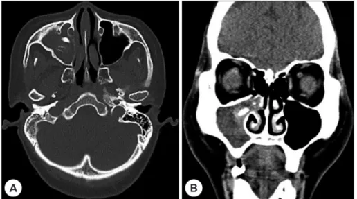
274 상돌기가 내측으로 편위된 소견이 보였다. 상악동 연조 직 음영 중심부에는 약 1.5 cm 크기의 석회화가 동반된 고밀도 음영이 관찰되었다(Fig. 1). 이를 토대로 우측 상악동에 발생한 진균구로 진단하고 우측 상악동 병변 에 대하여 전신마취 하에 내시경적 부비동 접근법을 통 한 진균구의 제거를 시행하였다. 진균구의 제거 중 치 아조직으로 생각되는 강한 경도를 갖는 1 cm 크기의 석 회화 조직이 발견되어 제거하였으며 잔여 진균구를 제 거한 후 수술을 종료하였다. 제거된 석회화 조직은 약 1.0×0.5 cm의 크기를 가지며 표면은 백색의 광택을 보 였다(Fig. 2). 술 후 시행한 병리 조직학적 검사에서 석
회화 조직은 치아조직으로 확인되었고 진균구는 국균 종에 의한 것으로 확인되었다(Fig. 3). 환자는 수술 후 2 일째 합병증 없이 퇴원하였고, 이후 4개월간 외래에서 추적관찰 하는 동안 수술 전에 보였던 비폐색 및 농성 비루는 호전된 양상을 보였다.
고 찰
이소치의 병인에 대하는 아직까지 명확하게 밝혀진 것 이 없으나 앞선 연구들은 환자의 외상 또는 감염의 과거 력, 병리학적 상태, 그리고 발생학적 장애 여부 등이 관 계가 있을 것으로 보고 있다.2,5)
태아는 발생 6주째부터 유치의 발달이 시작되고 출 생 후 5개월에서 10개월 사이에 영구치의 형성을 위해 외배엽층의 증식이 시작된다. 구강 상피층과 간엽조직 (mesenchymal tissue) 사이 여러 단계의 상호작용이 영 구치 형성에 중요한 역할을 하는데 이 과정에서 비정상 적인 상호작용이 발생할 경우 이소치가 발생한다고 알 려져 있다.6)
부비동 내의 이소치는 일생 동안 무증상일 수도 있으 나 때로는 다양한 질환과 증상을 유발할 수 있으며 재 발성 또는 만성 부비동염, 패혈증, 비루관 폐쇄, 부비동 개구연합 폐쇄, 두통과 안면마비감 등이 보고되고 있
A B
Fig. 1. Preoperative axial (A) and coronal (B) computed tomography scans. CT scans show soft tissue density lesion in the right maxillary sinus with slight bulging contour at the right uncinate process and high density oval shaped lesion about 1.5 cm with calcifications in the center of the right maxillary sinus.
Fig. 2. Gross pathologic specimen of the extracted mass shows a tooth like structure with a glossy surface (about 1.0×0.5 cm sized).
김강현 외 : 상악동 진균구 속의 이소치
275
다.2,7-11) 환자가 증상을 보이는 경우는 반드시 수술적으
로 제거해야 하며 일부 저자들은 Caldwell-Luc 수술을 추천하고 있으나 본 증례의 환자는 내시경적 부비동 수 술만으로 이소치와 함께 발생한 진균구의 제거까지 성 공적으로 시행하였다.2,9)
진균성 부비동염이 발생하면 진균에 의한 비부비동 점막의 단순한 자극에서부터 생명에 위협을 주는 질환 까지 임상적 양상이 다양하게 나타난다.17) 부비동 내의 점액섬모운동 능력이 손상되면 호흡기를 통해 들어온 진균의 배출이 원활하지 못해 진균과 상피세포의 접촉 시간이 증가하게 되면서 병원성을 갖게 된다. 이로 인 해 진균에서 분비되는 단백질분해효소에 의한 국소적 염증반응이 유발되어 결국 진균의 조직 내 침투를 용이 하게 하여 여러 질병을 일으키게 된다.12-14) 보고된 바에 의하면 진균에 의한 부비동 감염은 국균종에 의한 경우 가 가장 흔하다고 하며, 본 증례에서 관찰된 진균도 형 태학상 국균종과 일치하였다. 이는 이소치로 인해 발생 한 비강 점막의 손상이 장기간 동안 방치되고 결국 상악 동 내 점액섬모의 손상을 초래하여 진균구가 동반된 것으 로 생각된다.
상악동 진균구 속에서 치아 조직이 보고된 증례는 국 내에서 현재까지 2예가 보고되고 있다.15,16) 그 중 1예는 구순구개열이 동반한 환자에서 발생한 이소치가 진균 구와 함께 상악동에서 발견된 경우이고,15) 나머지 1예는 과거 좌측 상악 제1대구치에 대한 신경치료를 받은 과 거력이 있는 환자에서 좌측 상악동 내에서 진균구와 함 께 이소치가 발견된 경우였다.16) 본 증례에서는 기존의
증례와 달리 상기와 같은 병력 없이 상악동 진균구 내 에서 치아 조직이 발견되었다. 특이 과거력이 없는 진 균구 부비동염 환자가 전산화 단층 촬영에서 내부에 고 음영의 영역이 확인될 경우 석회화에 의한 가능성 외에 도 이소치의 존재 유무에 대해서도 고려해봐야 할 것으 로 생각된다.
중심 단어:이소치・과잉치・부비동염・상악동・국 균종.
REFERENCES
1) Scheiner MA, Sampson WJ. Supernumerary teeth: a re- view of the literature and four case reports. Aust Dent J 1997;42(3):160-5.
2) Goh YH. Ectopic eruption of maxillary molar tooth--an un- usual cause of recurrent sinusitis. Singapore Med J 2001;
42(2):80-1.
3) Pracy JP, Williams HO, Montgomery PQ. Nasal teeth. J Laryngol Otol 1992;106(4):366-7.
4) Kim DH, Kim JM, Chae SW, Hwang SJ, Lee SH, Lee HM.
Endoscopic removal of an intranasal ectopic tooth. Int J Pediatr Otorhinolaryngol 2003;67(1):79-81.
5) Raghoebar GM, Boering G, Vissink A, Stegenga B. Erup- tion disturbances of permanent molars: a review. J Oral Pathol Med 1991;20(4):159-66.
6) Ramanojam S, Halli R, Hebbale M, Bhardwaj S. Ectopic tooth in maxillary sinus: Case series. Ann Maxillofac Surg 2013;3(1):89-92.
7) Prabhu SP, Padwa BL, Robson CD, Rahbar R. Dentigerous cyst associated with a displaced tooth in the maxillary si- nus: an unusual cause of recurrent sinusitis in an adoles- cent. Pediatr Radiol 2009;39(10):1102-4.
8) Buchanan MA, Prince SE, Prinsley PR. Frontal mucocele caused by an ectopic maxillary tooth. J Laryngol Otol 2008;
122(12):1384-5.
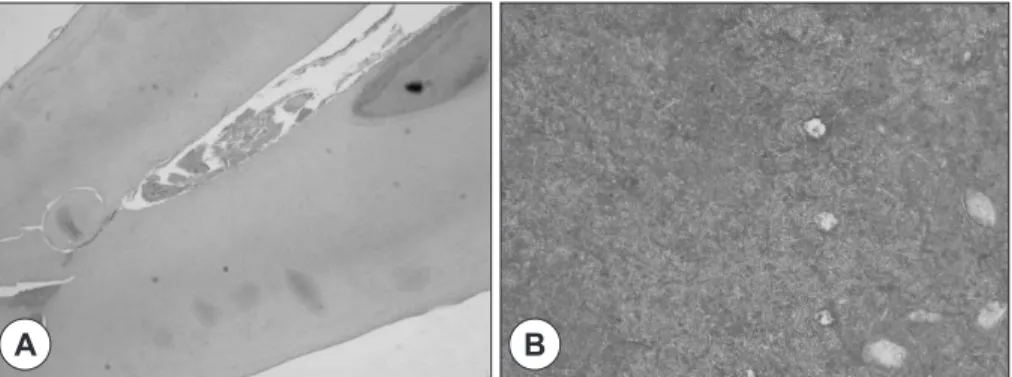
Fig. 3. A : Histopathology specimen of the calcified mass shows enamel-like structure, clinically it seems like ectopic tooth (H-E stain ×40). B : At higher magnification, the organisms are thin, uniform in size and shows branching, sep- tated hyphae and parallel arrangement in one direction (H-E stain ×200).
A B
J Clinical Otolaryngol 2015;26:273-276
276 9) Buyukkurt MC, Tozoglu S, Aras MH, Yolcu U. Ectopic
eruption of a maxillary third molar tooth in the maxillary sinus: a case report. J Contemp Dent Pract 2005;6(3):104- 10) Lamb JF, Husein OF, Spiess AC. Ectopic molar in the max-10.
illary sinus precipitating a mucocele: a case report and literature review. Ear Nose Throat 2009;88(8):E6-E11.
11) Weber BP, Kempf HG, Mayer R, Braunschweig R. Ectopic teeth in the area of the paranasal sinuses. Hno 1993;41(6):
317-20.
12) Tomee JF, Wierenga AT, Hiemstra PS, Kauffman HK.
Proteases from Aspergillus fumigatus induce release of pro- inflammatory cytokines and cell detachment in airway ep- ithelial cell lines. J Infect Dis 1997;176(1):300-3.
13) Shen HD, Lin WL, Tam MF, Wang SR, Tsai JJ, Chou H, et al. Alkaline serine proteinase: a major allergen of Asper-
gillus oryzae and its cross-reactivity with Penicillium ci- trinum. Int Arch Allergy Immunol 1998;116(1):29-35.
14) Tomee JF, Hiemstra PS, Heinzel-Wieland R, Kauffman HF. Antileukoprotease: an endogenous protein in the in- nate mucosal defense against fungi. J Infect Dis 1997;176(3):
740-7.
15) Lee JH, Jo MH, Choi KH. A case of ectopic tooth with su- perimposed fungal infection in nasal cavity of cleft lip and palate patient. Korean J Otorhinolaryngol-Head Neck Surg 2012;55(12):802.
16) Jung SY, Park EH, Bae JH, Lee SS. A case of supernu- merary tooth within fungus ball in the maxillary sinus. Ko- rean J Otorhinolaryngol-Head Neck Surg 2010;53(3):184.
17) Shin SH. The role of fungi in chronic rhinosinusitis. J Clin- ical Otolaryngol 2004;15(2):328-31.