“Extended” Distal Pancreatectomy with Segmental Resection of Both Splenic Vessels; Extended Warshaw’s
Procedure
Purpose: We would like to assess the safety and feasibility of extended spleen-preserving distal pancreatectomy with segmental resection of both splenic vessels (SPDP-SRSV) in patients with large, benign and borderline malignant pancreas body tumors.
Methods: We encountered seven extended SPDP-SRSV cases from January 2006 to March 2010. Among them, three were excluded due to combined pylorus-reserving pancreaticodu- odenectomy (PPPD). For the extended surgical technique, the pancreas was divided above the confluence of the superior mesenteric vein-splenic vein-portal vein (SMV-SV-PV), and vascular control was achieved at the origin of the splenic artery and the junction of the splenic vein with the SMV. The segments of both splenic vessels were then extracted along with the specimen.
Results: All the patients were female with a median age of 57 years (range: 24∼70 years).
The median tumor size was 5.5 cm (range: 5∼11 cm), the median operation time was 362 minutes (range: 337∼441 min), the median estimated blood loss was 150 ml (range: 50∼300 ml) and the median hospital stay was 9 days (range: 7∼20 days). One patient underwent robot-assisted extended Warshaw procedures. No mortality was noted, but one partial intestinal obstruction occurred and this was resolved with conservative management. On the recent follow-up, the CT scans showed no evidence of tumor recurrence or spleen infarction, but newly developed perigastric varix was noted, but it was without variceal bleeding.
Conclusion: SPDP-SRSV with division of the pancreatic neck portion above the confluence of the SMV-SV-PV in patients with large, benign and borderline malignant pancreatic body tumors appears to be an ideal approach because of the expected long-term survival and preserving the role of the spleen.
Dong Hyun Kim, M.D.
4, Chang Moo Kang, M.D.
1,2,3, Ho Kyoung Hwang, M.D
1,2,3, Woo Jung Lee, M.D.
1,2, Hoon Sang Chi, M.D.
1,21
Department of Surgery, Yonsei University College of Medicine,
2
Clinic of Pancreatic and Biliary Cancer, Institute of Gastroenterology, Yonsei University Health System,
3
Young Yonsei Pancreatic Tumor Study Group,
4Department of Surgery, Yonsei University Wonju College of Medicine
Corresponding Author Chang Moo Kang
Department of Surgery, Yonsei University College of Medicine, 250, Seongsanno, Seodaemun-gu, Seoul 120-750, Korea
Tel: +82-2-2228-2135 Fax: +82-2-313-8289 E-mail: [email protected]
Key Words : Spleen-preserving, Distal Pancreatectomy, Pancreas body tumors
Received: 2010. 7. 29.
Accepted: 2010. 10. 1.
Introduction
During recent years, laparoscopic SPDP has been widely applied for benign and borderline malignant tumors requiring distal pancreatectomy. The rationale for spleen- preserving procedures comprises prevention of posto- perative infection-related complications including overwhel-
ming postsplenectomy infection syndrome (OPSI)1-3 as well as a possible advantage regarding reduced carcinoge- nesis.2,4 In general, there are two surgical techniques for preservation of the spleen in distal pancreatectomy. First, the spleen can be safely preserved by simply conserving both splenic artery and vein during distal pancreatectomy;
however, this technique is time- and labor-consuming, and may be difficult in cases of large left-sided pancreatic
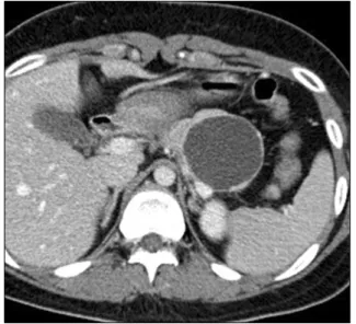
Fig. 1. Preoperative image in case number 2. About large 6 cm-sized pancreatic cyst was noted in the body of the pancreas of a 24-year-old patient. Tumor was closely abutted with both splenic artery and vein.
tumors and pancreatic pathologies associated with chronic pancreatitis. The other technique, the so-called “Warshaw procedure”, combines distal pancreatectomy with segmen- tal resection of both splenic vessels, leaving the spleen perfused with the intact left gastroepiploric vessels and short gastric vessels,5,6 This technique is quick and relatively easy, omitting individual vascular control of small tributary vessels between the pancreas and both splenic vessels.
However, spleen-related postoperative complications, such as splenic infarction and abscess, need to be considered and the potential risk of postoperative gastric varix and bleeding is another controversial issue.7,8
Crippa et al.9 called distal pancreatectomy an “extended”
distal pancreatectomy when the pancreas was divided at the level of SMV-SV-PV confluence. Thus, when the pancreas is divided at the level of the SMV-SV-PV confluence and the splenic artery and vein are securely controlled at the origin of the vessels, we specifically call this, an “extended” Warshaw procedure instead. We recently encountered four cases of extended Warshaw procedure in large benign and borderline malignant tumor of the pancreas, In this study, we retrospectively evaluated the surgical outcome of this technique and discussed the technical feasibility of this procedure.
Methods
This was a retrospective study design. Between January 2006 and March 2010, a total of 7 patients underwent
“extended” Warshaw procedure due to large benign and borderline malignant tumor of the pancreas (Fig. 1).
Among them, three patients underwent combined PPPD for complete removal of an intraductal papillary mucin- producing tumor involving the entire pancreas and were excluded from the current study. General characteristics of patients were reviewed and perioperative surgical out- comes, including spleen perfusion and perigastric fundal varix, were summarized (Table 1).
Operative techniques: The techniques for minimally
invasive surgery and conventional open surgery are similar.
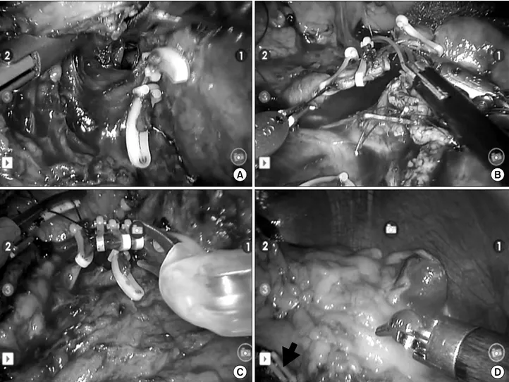
In brief, the gastrocolic ligament was divided to expose the entire pancreas. The lower border of the pancreatic neck portion was carefully dissected to separate it from the portal vein and the pancreatic neck area. After making a complete window between the pancreas and the portal vein (Fig.
2A), the pancreas was divided by endo-GIA. The splenocolic and gastrosplenic ligaments were not divided and the spleen was not mobilized. The upper part of the remnant pancreas was carefully dissected to isolate the origin of the splenic artery. It seems to be easier to approach the origin of the splenic vessels after division of the pancreatic neck portion. Vascular control can be obtained at the origin of the splenic artery and the junction of the splenic vein joined with the superior mesenteric vein (Fig. 2B, C). The coronary vein may or may not be divided during the operation. The distal part of the splenic vessel was simultaneously divided with vascular endo-GIA near the splenic hilar area, and the left gastroepiploric vessels were conserved (Fig. 2D). The pancreas was easily deta- ched from the retroperitoneal space. Sometimes, the spleen perfusion was rechecked by intraoperative ultrasound in
Fig. 2. Operative view of case number 2 (Robot-assisted extended Warshaw procedure). Complete window between SMV-SV-PV confluence and pancreatic neck portion was made (A). The origin of splenic artery and splenic vein at the junction with SMV were securely ligated and divided (B, C). Spleen was well preserved with good splenic perfusion and division site (black arrow) of distal part of splenic vessels was noted (D).
Doppler mode. Two closed suction drains were placed around the surgical field.
Results
1. Preoperative characteristics and perioperative surgical outcome
One patient underwent robot-assisted extended Warshaw procedures (Fig. 2, 3A, B). All patients were female with a median age of 57 years (range, 24∼76 years), a median tumor size of 5.2 cm (range, 2.5∼11 cm), a median operation time of 349.2 minutes (range, 200∼441 min), a
median estimated blood loss of 125 ml (range, 50∼300 ml), and a median hospital stay of 8.5 days (range, 7∼20 days) postoperatively. Chronic inflammatory change of the pancreas was noted in all patients. One patient showed about 2-cm sized low attenuated spleen lesion at CT scan taken on postoperative 7th day, but had no symptom related to this finding. One patient experienced postoperative partial intestinal obstruction, but it was successfully treated by conservative management. No mortality was noted (Table 1).
Fig. 3. Surgical specimen and follow up CT scan in case number 2. Final pathologic report was mucinous cyst adenoma (A and B) and follow-up CT scan showed naturally developed collateral venous circulation around gastric fundus (C and D).
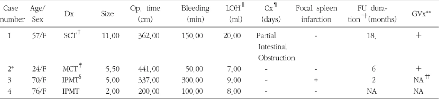
Table 1. Perioperative clinicopathologic characteristics
Case Age/ Op. time Bleeding LOH
∥Cx
¶Focal spleen FU dura-
Dx Size GVx**
number Sex (cm) (min) (ml) (days) infarction tion
‡‡(months)
1 57/F SCT
†11.00 362.00 150.00 20.00 Partial - 18. +
Intestinal Obstruction
2* 24/F MCT
‡5.50 441.00 50.00 7.00 - - 6 +
3 70/F IPMT
§5.00 337.00 300.00 9.00 - + 2 NA
††4 76/F IPMT 2.00 200.00 100.00 8.00 - - NA NA
*Robot-assisted, the others were open surgery;
†SCT=serous cystic tumor;
‡MCT=mucinous cystic tumor;
§IPMT=intraductal
papillary mucin-producing tumor;
∥LOH=length of hospital stay following operation;
¶Cx=complication; **GVx=perigastric varix
noted on FU CT scan;
††NA=not available, but will be followed up 6 month later;
‡‡FU=follow-up
2. Short-term follow-up results
During the recent follow-up period (6∼34 months), all patients appeared in good health condition. Follow-up CT scan (except last patient) showed no evidence of tumor recurrence and spleen infarction, but newly developed perigastric varix was noted (Fig. 3C, D). However, no one experienced clinically relevant variceal bleeding (Table 1).
Discussion
Advanced surgical techniques can provide patients with safe and adequate management of their disease entities.
Surgeons should consider patients’ quality of life when treating patients with benign or borderline malignant tumor of the pancreas because long-term survival is highly expected, unlike for those with pancreatic cancers.
Recently, benign and borderline pancreatic tumors without specific clinical symptoms seem to be discovered more frequently. Function-preserving minimally invasive surgery seems to be an ideal approach for these tumors.
In the past, the spleen was removed when performing distal pancreatectomy due to its anatomic intimacy with the tail of the pancreas and the technical difficulty associated with conserving both splenic vessels. However, the clinical significance of the spleen has been emphasized1 and many surgeons have recently been trying to preserve the spleen whenever possible in cases of benign or borderline malignant pancreatic pathologies requiring distal pancrea- tectomy.
For the purpose of increasing the technical feasibility and reducing the operation time, another spleen-preserving technique has been introduced that involves segmental resection of both splenic artery and vein. In this technique, surgeons do not need to individually control small tributary vessels between pancreas and splenic vessels to conserve the spleen. En-block resection of the distal pancreas with the segment of both splenic vessels reduces surgical time and technical efforts and spares the spleen. The spleen can
survive by perfusion of short gastric vessels and left gastroepiploric vessels splenorenal and splenocolic vessels.
Spleen-related postoperative morbidity and perigastric varix are complications to consider after Warshaw procedure.
One patient was noted to have a small area of spleen infraction on postoperative CT scan, but it was not clinically significant. In addition, follow-up study showed newly developed perigastric varix in two patients, but there were no evidence of gastrointestinal bleeding from the fundal varix. In fact, Warshaw also noted that perigastric venous collateral development following the Warshaw procedure was a natural compensating process and did not have significant clinical adverse impact, indicating the safety and efficacy of the original technique.6
Most cases of distal pancreatectomy with segmental resection of splenic vessels may require division some- where in the body or tail of the pancreas. In this report, we report four consecutive patients who underwent
“extended” Warshaw procedure dividing pancreatic neck portion above SMV-SV-PV confluence with vascular control at the origin of the splenic artery and splenic vein. We observed no severe complication or mortality. One patient experienced postoperative partial obstruction that was managed conservatively. In addition, a young female patient successfully underwent minimally invasive approach (robot-assisted). Therefore, we suggest that the “extended”
Warshaw procedure is also a feasible and safe surgical approach. Large benign or borderline malignant pancreatic body pathologies associated with chronic pancreatitis are thought to be good indications for this technique.
In summary, extended SPDP with segmental resection of both splenic vessels is feasible and can be safely applied to large benign and borderline malignant pancreatic body pathology considering the expected long-term survival and the role of the spleen.