Protective Effect of
Sasa Quelpaertensisand
p-Coumaric Acid on Ethanol-induced Hepatotoxicity in Mice
Sang Il Lee1,2, Sang Mi An1,2, Gyeong In Mun1,2, Seung Jin Lee1,2, Kwon Moo Park3, Sun Hong Park4, and Yong Chool Boo1,2*
1Department of Molecular Medicine, 2Cell and Matrix Research Institute, and
3Department of Anatomy, Kyungpook National University School of Medicine, Daegu 700-422, Korea
4Onc Co. Ltd., Jeju 690-756, Korea
Received May 29, 2008; Accepted August 18, 2008
Excessive alcohol use causes oxidative stress in the liver, and antioxidant therapy has been an attractive approach for the treatment of ethanol-induced liver damage. The present study examined the hepatoprotective effect of Sasa quelpaertensis Nakai (Korean name, Jeju-Joritdae) in C57BL/6 mice intoxicated with ethanol. Mice were intraperitoneally administered with ethanol alone, or together with test materials three times at 12-h intervals. At 3 h after the last dosing, hepatotoxicity was assessed based on serum activities of aspartate aminotransferase and alanine aminotransferase, and hepatic contents of thiobarbituric acid-reactive substances and glutathione.
Sasa quelpaertensis extract mitigated the acute ethanol hepatotoxicity as effectively as silymarin.
Its n-butanol fraction was more active than methylene chloride or aqueous fraction. p-Coumaric acid, a major constituent of S. quelpaertensis, was found to effectively prevent the ethanol-induced hepatotoxicity. These data suggest that S. quelpaertensis and p-coumaric acid could be useful for the prevention of liver disease caused by alcohol abuse.
Key words: alcohol hepatotoxicity, Sasa quelpaertensis Nakai, p-coumaric acid
The liver is the organ where alcohol (ethanol) metabolism mainly occurs, and excessive alcohol use can lead to acute and chronic hepatic lesions, termed ALD [Mendez-Sanchez et al., 2005]. Alcohol increases the hepatic lipid synthesis causing an accumulation of lipids within the hepatocytes, the predominant cell type of the liver. This condition, known as fatty liver (steatosis), is reversible; however, continuous drinking may lead to steatohepatitis, in which the fatty liver is accompanied by inflammation (hepatitis). Steatohepatitis can precede the development of liver cirrhosis and end-stage liver disease.
Ethanol-induced oxidative stress appears to play an
important etiological role in the pathogenesis of ALD [Albano, 2006; Dey and Cederbaum, 2006]. In liver cells, ethanol is metabolized by three major pathways: alcohol dehydrogenase in the cytosol, aldehyde dehydrogenase in the mitochondria, and cytochrome P450 2E1 in the endoplasmic reticulum. These pathways result in the production of ROS, ethanol radical, and other toxic metabolites causing oxidative stress. Other sources of ROS, i.e., NADPH oxidase, xanthine oxidase, and nitric oxide synthase also play a role in the ethanol-induced oxidative stress. Furthermore, ethanol administration decreases the concentration level of the hepatic antioxidants including vitamin E and GSH [Albano, 2006; Koch et al., 2004]. Ethanol-induced depletion of GSH may be related to the increased oxidation into GSSG or conjugation to other metabolites including acetaldehyde [Videla et al., 1981; Volkel et al., 2005].
Supporting the importance of antioxidant defense, augmentation of GSH by its precursors including N- acetyl-L-cysteine, S-adenosyl-L-methionine, and L-2- oxothiazolidine-4-carboxylic acid has been shown to inhibit the ethanol-induced liver injury in animal models [Iimuro et al., 2000; Fernandez-Checa et al., 2002; Izu et
*Corresponding author
Phone: +82-53-420-4946; Fax: +82-53-426-4944 E-mail: [email protected]
Abbreviations: ALD, alcoholic liver disease; ALT, alanine ami- notransferase; AST, aspartate aminotransferase; BW, body weight; GSH, glutathione; GSSG, glutathione disulfide; MC, methylene chloride; RNS, reactive nitrogen species; ROS, reac- tive oxygen species; TBARS,2-thiobarbituric acid-reactive sub- stances
doi:10.3839/jabc.2008.026
al., 2006; Wang et al., 2006]. Ethanol-induced hepatotoxicity has also been prevented by a synthetic hydrophilic vitamin E analog, raxofelast [Altavilla et al., 2005] and by phytochemicals with potent antioxidant activities including (-)-epigallocatechin gallate [Lee et al., 2008], silymarin [Song et al., 2006] and resveratrol [Kasdallah-Grissa et al., 2007]. Therefore, supplementation of natural antioxidants is one of the attractive approaches for the prevention of ethanol-induced hepatotoxicity [Dhiman and Chawla, 2005; Xu et al., 2005].
Sasa quelpaertensis Nakai (Korean name, Jeju- Joritdae) is a type of bamboo grass widely distributed in Jeju Island, Korea. Its culms and leaves long been used as traditional medicines in the treatment of liver diseases, and a recent study has demonstrated that the plant is a rich source of phenolic antioxidants including p-coumaric acid [An et al., 2008]. Other species of the bamboo grasses have been reported to have anti-ulcer, anti-cancer, and anti-oxidant effects [Otani et al., 1990; Tsunoda et al., 1998; Kurokawa et al., 2006;], suggesting that S.
quelpaertensis could provide a beneficial effect in the prevention of ALD by mitigating the oxidative stress. To test this hypothesis, the present study examined the potential hepatoprotective and antioxidant effects of the crude S. quelpaertensis extract, its solvent fractions, and p-coumaric acid, one of the major constituents, in ethanol-intoxicated mice using silymarin as a positive control [Song, et al., 2006]. Serum activities of AST and ALT, as well as the hepatic contents of TBARS and GSH, were determined to monitor hepatotoxicity and oxidative stress.
Materials and Methods
Preparation of S. quelpaertensis extract and fingerprint HPLC analysis. The culms and leaves of S.
quelpaertensis Nakai were harvested in July 2007, from Jeju Island, Korea. The plant materials were continuously extracted with 30% aqueous ethanol at room temperature and evaporated under reduced pressure to obtain an extract with approximately 4% yield. The extract (36 g) was suspended in water and partitioned successively with MC and BuOH, followed by evaporation under reduced pressure to yield an MC fraction (0.95 g), a BuOH fraction (2.42 g), and a H2O fraction (31 g). The Amersham Biosciences AKTA HPLC system (GE Healthcare Bio- Sciences AB, Uppsala, Sweden) equipped with a UV-Vis detector was used for fingerprint HPLC analysis of S.
quelpaertensis extract (Fig. 2). The column was a Waters XTerra RP-18 (5-µm, 4.6 mm×150 m, Milford, MA).
The mobile phase consisted of 0.5% formic acid (A) and acetonitrile (B). The gradient was programmed as follows:
0~6 min, 100% A; 6~10 min, a linear gradient from 0 to 12% B; 10~35 min, a linear gradient from 12 to 21% B;
35~40 min, a linear gradient from 21 to 25% B; 40~
50 min, a linear gradient from 25 to 100% B; 50~60 min, 100% B. The flow rate was 1.0 mL/min, and the detector was set at 280 nm.
Animal experiments. Experiments were performed on 9-week-old female C57BL/6 mice (Daehan Biolin Co., Chungbuk, Korea) in accordance with the guideline of Kyungpook National University. The animals were maintained under controlled environmental conditions (23±1oC, 55±5% humidity, 12-h light/dark cycle) with free access to water and an ad libitum standard laboratory diet (Superfeed, Kangwon, Korea). After acclimation the mice were randomly divided into five or six groups of 5~9 animals for each experiment and received ethanol at 2,500 mg/kg BW by intraperitoneal injection at 12-h intervals for a total of three doses. This ethanol dose was chosen due to the consistent production of moderate toxicity in the preliminary experiments. Ethanol was injected as a 30% (v/v) solution to prevent the peritoneal irritation. Mice of the test groups received three doses of 30% aqueous ethanol solution containing the test materials including extract, fraction, silymarin, and p- coumaric acid at different concentrations to allow simultaneous administration of the test materials, without additional injections. The solutions were prepared immediately prior to the administration to avoid any potential contamination or ethanol evaporation. Control mice received an isocaloric glucose solution according to the same protocol mentioned above. At 3 h after the last injection, the mice were anesthetized with isoflurane and euthanized to collect the blood and the liver samples.
Serum enzyme assay. Blood taken by a heart puncture was placed into the serum separator tubes and let stand for 30 min at room temperature. The serum was separated from the blood cells by centrifugation at 600 rpm for 5 min. Serum activities of AST (EC 2.6.1.1.) and ALT (EC 2.6.1.2.) were determined by using the Vitros 250 chemistry system (Johnson & Johnson, Rochester, NY).
Histochemical examination. Liver tissues were cut and fixed with 4% paraformaldehyde in phosphate- buffered saline. The fixed tissues were embedded in paraffin. Tissues sections of 6-µm were stained with hematoxylin and eosin, and observed with an Eclipse 80i microscope (Nikon Instruments Inc., Melville, NY).
Lipid peroxidation assay. The analysis of TBARS as a marker of the lipid peroxidation was carried out as described previously [Mihara and Uchiyama, 1978].
Briefly, the liver tissue was homogenized in 9.0 mL of 50 mM Tris-Cl buffer (pH 7.4) containing 180 mM KCl and 10 mM EDTA. One milliliter of the tissue homogenate
was mixed with 3.0 mL of 1.0% phosphoric acid and 1.0 mL of 0.9% 2-thiobarbituric acid, followed by heating in a boiling water bath for 45 min. After cooling, 4.0 mL of BuOH was added, and the mixture was centrifuged at 10,000 rpm for 15 min. The absorbance of the BuOH layer was determined at 532 nm. TBARS concentration was calculated using 1,1,3,3-tetramethoxypropane as the standard.
Determination of GSH plus GSSG concentration.
For the extraction of GSH plus GSSG, the tissue homogenate was mixed with an equal volume of 10%
meta-phosphoric acid, and the mixture was centrifuged at 10,000 rpm for 15 min. The resulting supernatants were used for the determination of GSH plus GSSG using the enzymatic recycling method [Anderson, 1985]. The reaction mixture (100 mL) containing 143 mM sodium phosphate (pH 7.5), 6.3 mM EDTA, 0.6 mM 5,5'-dithiobis[2- nitrobenzoic acid], 0.3 mM NADPH, 0.25 units/mL GSH reductase, and 2.0µL sample was incubated at 37oC for 20 min. The formation of 2-nitro-5-thiobenzoic acid was monitored by absorption at 415 nm and converted into the GSH concentration using a calibration curve with known amounts of GSH.
Statistical analysis. All data are presented as the means±S.D. The statistical analyses were performed using the Sigma Stat 3.1 software program. Significant differences among the groups were determined by a one-
way ANOVA. Duncan’s multiple-range test was performed if differences were identified between the groups at
p< 0.05.
Results
In the present study, mice were given 30% aqueous ethanol solution at 2,500 mg/kg BW by intraperitoneal injection, total three times at 12-h intervals. This protocol was chosen, because moderate liver toxicity was consistently observed in most animals. The animals were euthanized at exactly 3 h after the last dosing, because toxicity parameters appeared to change time-dependently.
To examine the potential effect of S. quelpaertensis
extract on the ethanol hepatotoxicity, the extract was dissolved in 30% aqueous ethanol to be administered along with ethanol. Control mice received an isocaloric glucose solution according to the same protocol. S.
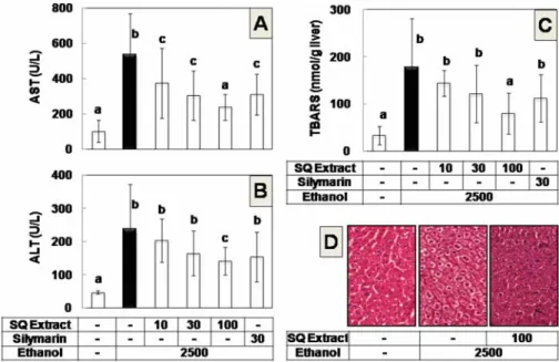
quelpaertensis extract appeared to attenuate the hepatotoxicity of the ethanol as monitored by various markers of the liver injury (Fig. 1). As expected, the serum activities of AST and ALT were markedly increased by ethanol treatment (Fig. 1A and B). In contrast, the biochemical changes were significantly reduced in the animals co-treated with S. quelpaertensis
extract at 100 mg/kg BW, whereas lower doses of S.
quelpaertensis extract (10 and 30 mg/kg BW) and
Fig. 1. S. quelpaertensis extract attenuates ethanol hepatotoxicity in mice. Mice were intraperitoneally administered with 2,500 mg/kg BW of ethanol alone (n=9), or together with S. quelpaertensis (SQ) extract at 10 mg/kg BW (n=8), 30 mg/kg BW (n=9), 100 mg/kg BW (n=9) or together with silymarin at 30 mg/kg BW (n=7) three times at 12-h intervals. Control mice received isocaloric glucose (n=8). At 3 h after third administration, mice used for the analyses of serum activities of AST (A) and ALT (B), hepatic lipid peroxidation (C), and histochemical examination of liver sections with hematoxylin- eosin staining (D). Dose unit is mg/kg BW. Data are means±S.D. Columns not sharing the same letter are significantly different from each other (p<0.05).
silymarin (30 mg/kg BW), used as a positive control, affected only one of these two markers (Fig. 1A and B).
Ethanol treatment also increased the lipid peroxidation of the liver tissues as determined by TBARS; however, S.
quelpaertensis extract significantly inhibited the peroxidation at 100 mg/kg BW (Fig. 1C). Histochemical examination of the tissue sections from mice that received ethanol showed increased content of lipid vacuoles and a loss of the membrane integrity that represent steatosis and necrosis, respectively (Fig. 1D). The histological changes were suppressed by S. quelpaertensis extract at 100 mg/
kg BW (Fig. 1D). The results confirmed the protective effect of S. quelpaertensis extract against ethanol hepatotoxicity.
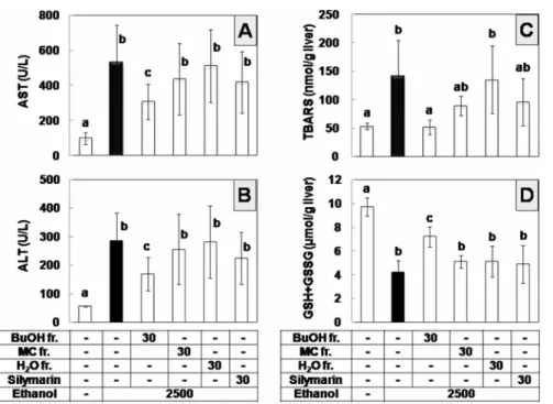
In the following experiment, H2O, BuOH, and MC fractions of S. quelpaertensis extract were compared in an attempt to characterize the active principle(s) attenuating ethanol hepatotoxicity. Fingerprint HPLC profiles of S.
quelpaertensis extract and its fractions are shown in Fig.
2. A previous study has identified p-coumaric acid a major phenolic compound of S. quelpaertensis extract [An et al., 2008], and thus this compound was used here as a reference (Fig. 2). Mice were given an aqueous fraction, BuOH fraction or MC fraction of S.
quelpaertensis extract at 30 mg/kg BW along with 2,500 mg/kg BW ethanol, three times at 12-h intervals. Upon comparison with the mice that received ethanol only or isocaloric glucose (control), mice that received the BuOH
fraction appeared to be the most active (Fig. 3). The BuOH fraction prevented the increase of serum activities of AST and ALT caused by ethanol, whereas the aqueous or MC fraction failed to show a significant effect (Fig. 3A and B). The BuOH fraction also significantly inhibited the hepatic lipid peroxidation (Fig. 3C) and GSH depletion (Fig. 3D); however, the other fractions and silymarin were not inhibitory at the tested doses.
Because the BuOH fraction appeared to contain p- coumaric as the major phenolic compound (Fig. 2), the influence of this compound on the ethanol hepatotoxicity was further examined. In this experiment, p-coumaric acid was administered alone or together with ethanol, and the results showed that p-coumaric acid treatment at 15 and 30 mg/kg BW did not alter the serum AST and ALT activities nor induce the hepatic lipid peroxidation or GSH depletion in the absence of ethanol, but inhibited the ethanol hepatotoxicity in a dose-dependent manner (Fig.
4), suggesting that p-coumaric acid may be an active component of S. quelpaertensis byproviding hepatoprotective effect against acute ethanol toxicity.
Discussion
Many herbal medicines have long been used in the treatment of ALD; however, controversy remains as to their efficacy and safety [Dhiman and Chawla, 2005; Xu,
et al., 2005]. Although S. quelpaertensis has also been
Fig. 2. Fingerprint HPLC of S. quelpaertensis extract. S. quelpaertensis (SQ) extract and its H2O, BuOH and MC fractions, prepared as in A, were analyzed by HPLC at 280 nm (B). p-Coumaric acid, one of the major compounds, was used as a reference.
used as a traditional medicine for various purposes including ALD, its hepatoprotective effect has not yet been supported by direct evidence. In the present study, S.
quelpaertensis was demonstrated to attenuate hepatotoxicity and oxidative stress caused by repeated ethanol administration in mice. Our group also identified p-coumaric acid as a Fig. 3. Effect of solvent fractions derived from S. quelpaertensis extract on the ethanol hepatotoxicity in mice. Mice were intraperitoneally administered with 2,500 mg/kg BW of ethanol alone (n=7), together with BuOH fraction (n=7), MC fraction (n=6), H2O fraction (n=5) at 30 mg/kg BW or together with silymarin at 30 mg/kg BW (n=6), three times at 12-h intervals. Control mice received isocaloric glucose (n=5). At 3 h after third administration, mice were used for the analyses of serum activities of AST (A) and ALT (B), hepatic lipid peroxidation (C), and GSH plus GSSG contents (D). Dose unit is mg/kg BW. Data are means±S.D. Columns not sharing the same letter are significantly different from each other (p<0.05).
Fig. 4. p-Coumaric acid attenuates the ethanol hepatotoxicity in mice. Mice were intraperitoneally administered with 2,500 mg/kg BW of ethanol alone (n=7) or together with p-coumaric acid at 15 mg/kg BW (n=5) or 30 mg/kg BW (n=6) three times at 12-h intervals. Other mice were given water (n=5) or p-coumaric acid at 15 mg/kg BW (n=5) or 30 mg/kg BW (n=5). At 3 h after the third administration, mice were used for the analyses of serum activities of AST (A) and ALT (B), hepatic lipid peroxidation (C), and GSH plus GSSG content (D). Dose unit is mg/kg BW. Data are means±S.D.
Columns not sharing the same letter are significantly different from each other (p<0.05).
bioactive constituent of S. quelpaertensis, inhibiting oxidative stress and hepatotoxicity in mice intoxicated with ethanol.
Crude S. quelpaertensis extract at 100 mg/kg BW and purified BuOH fraction at 30 mg/kg BW significantly inhibited the ethanol-induced changes of all four markers of hepatotoxicity and oxidative stress, whereas S.
quelpaertensis extract at 30 mg/kg BW and silymarin at 30 mg/kg BW showed a less dramatic effect (Figs. 1 and 3). In addition, p-coumaric acid consistently prevented the ethanol-induced changes of all four markers at 30 mg/
kg BW and showed a minor preventive effect at 15 mg/kg BW (Fig. 4). Therefore, it was suggested that the hepatoprotective effect of the S. quelpaertensis extract is comparable with that of silymarin, and the purified BuOH fraction and p-coumaric acid are more active than silymarin.
No apparent sign of toxicity was observed when mice were treated with S. quelpaertensis extract or its fractions.
In addition, p-coumaric acid did not cause any hepatotoxicity and oxidative stress at 15 and 20mg/kg BW, demonstrating its potential as a hepatoprotective agent with low toxicity.
p-Coumaric acid is a common dietary phenolic acid widely distributed in plants [Scalbert and Williamson, 2000]. Its beneficial effect attenuating oxidative stress has been previously demonstrated in other animal models. p- Coumaric acid lowered the low-density lipoprotein cholesterol oxidation in the rat blood [Zang et al., 2000]
and mitigated oxidative stress induced by doxorubicin in the rat heart [Abdel-Wahab et al., 2003]. The potent hepatoprotective effect of p-coumaric acid has previously been demonstrated in rats intoxicated with carbon tetrachloride [Perez-Alvarez et al., 2001]. Therefore, the hepatoprotective effect of p-coumaric acid against ethanol toxicity observed in the present study (Fig. 4) provides additional evidence for its antioxidant property attenuating oxidative stress.
The contents of p-coumaric acid in crude S.
quelpaertensis extract and BuOH fraction were estimated to be 1.2 and 12%, respectively (Fig. 2), suggesting p- coumaric acid is very likely one of the major compounds providing hepatoprotective activity. However, it may not be the only active compound, because the activity of BuOH fraction was almost comparable to that of the pure
p-coumaric acid. Therefore, the hepatoprotective effect of
S. quelpaertensis extract could be attributed to multiple active principles including p-coumaric acid enriched in BuOH fraction.
The mechanisms by which S. quelpaertensis extract could mitigate oxidative stress in the liver are of interest.
Numerous enzymes including ethanol-inducible cytochrome P450 2E1 are involved in the catalytic reaction from
ethanol to acetaldehyde and cause overproduction of ROS and free radicals [Morimoto et al., 1995]. Potential mechanisms may include inhibition and activation of ethanol metabolism, and/or direct scavenging of the overproduced ROS/free radicals by S. quelpaertensis
extract. Alternatively, S. quelpaertensis extract could enhance the existing antioxidant defense against oxidative stress. Interestingly, p-coumaric acid has been known to induce hepatic antioxidant enzymes and phase II enzymes that play critical roles in defense against the oxidant insults [Yeh and Yen, 2006a, b]. Further studies, including analysis of the ethanol metabolites and the metabolic enzymes, are needed to define the exact mechanisms involved in the hepatoprotective effect of S.
quelpaertensis extract and p-coumaric acid.
In conclusion, the present study demonstrated that the crude extract and the purified fraction of S. quelpaertensis, as well as its major component, p-coumaric acid, are potentially useful hepatoprotective agents against ethanol intoxication.
Acknowledgments. This work was supported by a grant from the Industrial Technology Development Program of the Ministry of Commerce, Industry and Energy, Republic of Korea. This work was also supported by the Brain Korea 21 Project 2006.
References
Abdel-Wahab MH, El-Mahdy MA, Abd-Ellah MF, Helal GK, Khalifa F, and Hamada FM (2003) Influence of p- coumaric acid on doxorubicin-induced oxidative stress in rat's heart. Pharmacol Res 48, 461-465.
Albano E (2006) Alcohol, oxidative stress and free radical damage. Proc Nutr Soc 65, 278-290.
Altavilla D, Marini H, Seminara P, Squadrito G, Minutoli L, Passaniti M, Bitto A, Calapai G, Calo M, Caputi AP, and Squadrito F (2005) Protective effects of antioxidant rax- ofelast in alcohol-induced liver disease in mice. Pharma- cology 74, 6-14.
An SM, Lee SI, Choi SW, Moon SW, and Boo YC (2008) p-Coumaric acid, a constituent of Sasa quelpaertensis Nakai, inhibits cellular melanogenesis stimulated by alpha-melanocyte stimulating hormone. Br J Dermatol
159, 292-299.
Anderson ME (1985) Determination of glutathione and glu- tathione disulfide in biological samples. Methods Enzy- mol 113, 548-555.
Dey A and Cederbaum AI (2006) Alcohol and oxidative liver injury. Hepatology 43, S63-74.
Dhiman RK and Chawla YK (2005) Herbal medicines for liver diseases. Dig Dis Sci 50, 1807-1812.
Fernandez-Checa JC, Colell A, and Garcia-Ruiz C (2002) S- Adenosyl-L-methionine and mitochondrial reduced glu-
tathione depletion in alcoholic liver disease. Alcohol 27, 179-183.
Iimuro Y, Bradford BU, Yamashina S, Rusyn I, Nakagami M, Enomoto N, Kono H, Frey W, Forman D, Brenner D, and Thurman RG (2000) The glutathione precursor L-2-oxothiazolidine-4-carboxylic acid protects against liver injury due to chronic enteral ethanol exposure in the rat. Hepatology 31, 391-398.
Izu H, Shobayashi M, Manabe Y, Goto K, and Iefuji H (2006) S-adenosylmethionine (SAM)-accumulating sake yeast suppresses acute alcohol-induced liver injury in mice. Biosci Biotechnol Biochem 70, 2982-2989.
Kasdallah-Grissa A, Mornagui B, Aouani E, Hammami M, El May M, Gharbi N, Kamoun A, and El-Fazaa S (2007) Resveratrol, a red wine polyphenol, attenuates ethanol-induced oxidative stress in rat liver. Life Sci 80, 1033-1039.
Koch OR, Pani G, Borrello S, Colavitti R, Cravero A, Farre S, and Galeotti T (2004) Oxidative stress and antioxi- dant defenses in ethanol-induced cell injury. Mol Aspects Med 25, 191-198.
Kurokawa T, Itagaki S, Yamaji T, Nakata C, Noda T, Hirano T, and Iseki K (2006) Antioxidant activity of a novel extract from bamboo grass (AHSS) against ischemia-reperfusion injury in rat small intestine. Biol Pharm Bull 29, 2301-2303.
Lee SI, Kim HJ, and Boo YC (2008) Effect of green tea and (-)-epigallocatechin gallate on ethanol-induced toxic- ity in HepG2 cells. Phytother Res 22, 669-674.
Mendez-Sanchez N, Almeda-Valdes P, and Uribe M (2005) Alcoholic liver disease. An update. Ann Hepatol 4, 32- Mihara M and Uchiyama M (1978) Determination of mal-42.
onaldehyde precursor in tissues by thiobarbituric acid test. Anal Biochem 86, 271-278.
Morimoto M, Hagbjork AL, Wan YJ, Fu PC, Clot P, Albano E, Ingelman-Sundberg M, and French SW (1995) Modulation of experimental alcohol-induced liver disease by cytochrome P450 2E1 inhibitors. Hepatology
21, 1610-1617.
Otani K, Yanaura S, Yuda Y, Kawaoto H, Kajita T, Hirano F, Osawa F, and Inouye S (1990) Histo-chemical studies on the anti-ulcer effect of bamboo grass in rats. Int J
Tissue React 12, 319-332.
Perez-Alvarez V, Bobadilla RA, and Muriel P (2001) Struc- ture-hepatoprotective activity relationship of 3,4-dihy- droxycinnamic acid (caffeic acid) derivatives. J Appl Toxicol 21, 527-531.
Scalbert A and Williamson G (2000) Dietary intake and bio- availability of polyphenols. J Nutr 130, 2073S-2085S.
Song Z, Deaciuc I, Song M, Lee DY, Liu Y, Ji X, and McClain C (2006) Silymarin protects against acute etha- nol-induced hepatotoxicity in mice. Alcohol Clin Exp Res 30, 407-413.
Tsunoda S, Yamamoto K, Sakamoto S, Inoue H, and Nagasawa H (1998) Effects of Sasa Health, extract of bamboo grass leaves, on spontaneous mammary tumouri- genesis in SHN mice. Anticancer Res 18, 153-158.
Videla LA, Fernandez V, Fernandez N, and Valenzuela A (1981) On the mechanism of the glutathione depletion induced in the liver by acute ethanol ingestion. Subst Alcohol Actions Misuse 2, 153-160.
Volkel W, Alvarez-Sanchez R, Weick I, Mally A, Dekant W, and Pahler A (2005) Glutathione conjugates of 4- hydroxy-2(E)-nonenal as biomarkers of hepatic oxida- tive stress-induced lipid peroxidation in rats. Free Radic Biol Med 38, 1526-1536.
Wang AL, Wang JP, Wang H, Chen YH, Zhao L, Wang LS, Wei W, and Xu DX (2006) A dual effect of N-acetylcys- teine on acute ethanol-induced liver damage in mice.
Hepatol Res 34, 199-206.
Xu BJ, Zheng YN, and Sung CK (2005) Natural medicines for alcoholism treatment: a review. Drug Alcohol Rev
24, 525-536.
Yeh CT and Yen GC (2006a) Induction of hepatic antioxi- dant enzymes by phenolic acids in rats is accompanied by increased levels of multidrug resistance-associated protein 3 mRNA expression. J Nutr 136, 11-15.
Yeh CT and Yen GC (2006b) Modulation of hepatic phase II phenol sulfotransferase and antioxidant status by phe- nolic acids in rats. J Nutr Biochem 17, 561-569.
Zang LY, Cosma G, Gardner H, Shi X, Castranova V, and Vallyathan V (2000) Effect of antioxidant protection by p-coumaric acid on low-density lipoprotein cholesterol oxidation. Am J Physiol Cell Physiol 279, C954-960.