Anti-obesity Effect of Ethanol Extracts from Silkworm (Bombyx mori) Pupae Powder Fermented with Cordyceps militaris in the Primary Adipocytes and High Fat Diet-induced Obesity Model Mice
Ji Eun Kim1†, Mi Rim Lee1†, Jun Young Choi1, Jin Ju Park1, Hye Ryeong Kim1, Bo Ram Song1, Young Whan Choi2, Kyung Mi Kim3 and Dae Youn Hwang1*
1Department of Biomaterials Science, College of Natural Resources & Life Science/Life and Industry Convergence Research Institute, Pusan National University, Miryang 627-706, Korea
2Department of Horticultural Bioscience, College of Natural Resources & Life Science, Pusan National University, Miryang 50463, Korea
3Novarex Co., Chungju 28126, Korea
Received March 20, 2018 /Revised May 19, 2018 /Accepted May 27, 2018
Silkworm (Bombyx mori) pupae have been widely worked in wound dressing, hepatoprotective activ- ity, antigenotoxicity, control of glucose level and anti-cancer activity. To investigate the anti-obesity activity of ethanol extract of silkworm pupae powder fermented with Cordyceps militaris (ESfC), the free glycerol release and cAMP concentration as well as fat accumulation were measured in the pri- mary adipocytes of SD (Sprague Dawley) rats and high fat diet (HFD)-treated C57BL/6 mice treated with 12 weeks. Firstly, the presence of the cordycepin with lipid lowering effect was confirmed in ESfC using HPLC analysis. The level of free glycerol and cAMP concentration was significantly in- creased in the primary adipocytes treated with high dose of ESfC (400 μg/ml) although these levels were consistently maintained in other dose ESfC treated groups. In HFD-induced obesity model, the increased fat weight and size of adipocytes in HFD+Vehicle treated group was recovered in HFD+ESfC treated group. Also, the liver weight and the number of lipid droplets were higher in HFD+Vehicle treated group than No treated group. But, this level was significantly decreased in HFD+ESfC treated group compared with HFD+Vehicle treated group. Furthermore, a similar recovery was detected on the phosphorylation of periliphin and HSL, and ATGL expression. Overall, the results of the present study provide some scientific evidences that ESfC can stimulate lipolysis in primary adipocytes and prevent fat accumulation in HFD-treated obesity model, and therefore have the potential for use as anti-obesity agents to treat obese patient.
Key words : Adipocytes, Cordyceps militaris, fat accumulation, obesity, silkworm pupae
†Authors contributed equally.
*Corresponding author
*Tel : +82-55-350-5388, Fax : +82-55-350-5389
*E-mail : [email protected]
This is an Open-Access article distributed under the terms of the Creative Commons Attribution Non-Commercial License (http://creativecommons.org/licenses/by-nc/3.0) which permits unrestricted non-commercial use, distribution, and reproduction in any medium, provided the original work is properly cited.
Journal of Life Science 2018 Vol. 28. No. 7. 786~794 DOI : https://doi.org/10.5352/JLS.2018.28.7.786
서 론
비만(Obesity)은 섭취한 에너지 중 체내에서의 대사활동으 로 소비하고 남은 에너지가 지방조직(Fat tissue)에서 중성지 방(Triglyceride, TG)으로 전환되어 축적된 상태로 정의되며 [17], 유전적, 영양적, 환경적 및 사회적 요인 등 다양한 원인들 이 관여하는 복잡한 만성질환(Chronic disease)이다[2]. 현재 까지 비만에 대한 정확한 기전은 알려져 있지 않으나, 비만은 고지혈증(Hyperlipidemia)이나 고혈압(Hypertension), 심혈 관계질환(Cardiovascular disease), 당뇨(Diabetes), 호흡기질
환(Respiratory disease) 등을 발생시킬 수 있다고 알려져 있으 며[3, 4, 17], 비만과 연관된 질병의 이해와 치료를 위해 지방세 포의 분화와 지방대사에 대한 연구가 활발히 진행되고 있다[1, 13, 22]. 특히, 비만의 유발과 발병 과정에서 중성지방의 지방 분해(lipolysis)와 합성(lipogenesis) 사이의 불균형이 중요한 역할을 한다[5]. 지방조직의 lipid droplet에서 중성지방의 분 해와 이로 인한 glycerol의 가수분해는 지방의 축적을 조절할 수 있다[21, 29]. 또한, 지방분해 메커니즘 중 하나인 cAMP-de- pendent protein kinase A (PKA) 경로는 hormone sensitive lipase (HSL)의 인산화와 신호전달에 관여한다[6, 8].
한편, 누에번데기(Silkworm (Bombyx mori) pupae)는 오랫 동안 중국, 일본 등 여러 나라에서 영양분을 제공하기 위하여 음식으로 사용되고 있다[40]. 누에번데기는 탄수화물, 단백질, 미네랄, 비타민 등 인체에 필수적인 성분들을 많이 포함하고 있으며, 특히 다양한 아미노산[45]과 palmitic acid, oleic acid, linoleic acid, stearic acid 등 다양한 지방산(fatty acid)을 다량 함유하고 있다[19]. 이러한 누에번데기는 창상드레싱[15, 46], 간보호와 세포사억제 활성[7], 항유전독성[31], 포도당과 지방
농도 조절[12, 14], 항암활성[16, 34], 에스트로겐활성[40], 저항 성운동을 통한 근육량증가 유도[20] 등의 기능이 있는 것으로 연구되었다. 이러한 연구들 중에서, 지방대사와 관련된 연구 는 일부 논문에서 보고되어 있다. 누에번데기는 돌연변이나 간독성 없이 지방의 합성을 효과적으로 억제하였으며[28, 49], 수영운동을 실시한 랫드에서 혈청지질의 농도와 지방축적을 감소시켰다[33]. 하지만 누에번데기 가루를 붉은동충하초를 이용하여 발효시킨 후 얻어진 추출물의 항비만 효능에 대한 연구는 현재까지 진행된바 없다.
한편, 동충하초(Vegetable worms)는 다양한 곤충이나 벌레 에 기생하였다가 이를 숙주로 하여 자실체를 형성하거나 충체 상에 포자를 형성하는 여러 가지 버섯의 총칭으로 우리나라에 서는 보통 누에를 숙주로 눈꽃동충하초(Paecilomyces tenuipes) 를 접종하여 생산한다[18, 23, 35, 37]. 동충하초의 유용성 성분 은 cordycepin, cordycepic acid, 아미노산, 다당류, 비타민 등 으로 보고된 바 있고[9, 25], 효능으로는 항암작용, 면역증강제, 혈당강하, 콜레스테롤 저해 효과 등이 알려져 있다[25]. 특히 동충하초의 지표성분 중 하나인 cordycepin은 3T3-L1 세포주 에 분화유도배지와 함께 처리하였을 때 C/EBPβ, PPAR-γ의 mRNA 및 단백질의 생성을 저하시켜 항비만 효능이 있다고 보고된 바 있다[38]. 이러한 우수성 때문에 다양한 균주를 이용 하여 유용한 추출물을 개발하기 위한 조건을 확립하고 효능을 평가하는 연구들에 많은 관심이 집중되고 있다.
따라서, 본 연구에서는 누에번데기 가루의 붉은동충하초 발 효추출물인 ESfC의 항비만효능을 평가하기 위하여, 일차지방 세포와 고지방사료로 유도된 비만모델동물에 ESfC를 처리하 여 지방분해와 지방합성에 연관된 중요인자들을 분석하였다.
이러한 결과는 ESfC는 일차지방세포에서 free glycerol re- lease, cAMP농도를 증가를 유도하였고, HFD유발 비만모델동 물의 지방과 간조직에서 지방의 축적을 효과적으로 억제함을 제시하고 있다.
재료 및 방법
ESfC 제조
누에번데기 가루는 정읍농업협동조합(정읍, 한국)으로부터 구매하였고, 붉은동충하초균은 부산대학교 생명환경생화학 과 이상몽 교수로부터 제공받아 실험에 사용하였다. ESfC 제 조를 위해, 먼저 누에번데기 가루에 10% 동충하초균(v/w)을 첨가하여 혼합배양액을 준비하고 25℃에서 3주 동안 진탕배 양기에서 발효시켰다. 배양혼합물은 원심분리하여 수확한 후 추출은 70% EtOH을 용매로 하여 80℃조건에서 실시하였다.
발효혼합물은 용매와 일정한 비율(혼합물/용매 비율, 1:10)로 혼합하고, JAC 초음파장비(Jinwoo Co., Seoul, Korea)를 사용 하여 1시간 동안 초음파 처리하였다. 초음파 처리된 추출물은 3,000 rpm에서 10분간 원심분리하여 상층액을 수확하였다. 다
음으로 동일한 용매 9 ml를 침전물에 첨가하여 동일한 조건에 서 초음파처리하고 상층액을 재수집하였다. 상기과정을 반복 한 후, 수집된 상층액은 0.4 μm 필터로 여과하고 진공증발장치 를 이용하여 농축시켰다. 마지막으로 ESfC는 순환추출장비 (IKA Labortechnik, Staufen, Germany)를 사용하여 동결건조 시켰고, DMSO에 40 mg/ml로 용해시켜 -20℃에서 보관하였 다.
HPLC (High Pressure Liquide Chromatograph) 분석 HPLC는 진공가스제거기(vacuum degasser), 단일용매전 달펌프, 온도조절 컬럼(thermostatted column compartment), 20 μl 시료주입기(injector), 검출기(diode-array detector)로 구 성된 Agilent 1200 liquid chromatography system (Agilent Technologies, USA)를 이용하여 실시하였다. HPLC 칼럼은 Luna C18 (250 mm × 4.6 mm, 5 μm particle size)을 사용하였 고, 이동상(mobile phase)용매는 (A) 0.1% formic acid와 (B) acetonitrile를 사용하였다. 이동상의 경사도 조건은 다음과 같 다: 0-5분, 10% B; 5-6분, 10-15% B; 6-40분, 15% B; 40-41분, 15-30% B; 41-50분, 30% B; 50-55분, 30-10% B; 55-60분, 10%
B. 유속은 0.5 ml/min이고, 흡광도 254 nm에서 검출하였다.
시료는 주입전에 0.45 μm필터막를 이용하여 필터하였고, 관찰 된 피크(peak)는 표준물질의 피크와 정체시간(retention times) 을 비교하여 확인하였다.
일차지방세포(primary adipocytes)의 분리 및 배양 지방조직은 이전에 보고된 바와 같이[48] 8주령 랫드를 CO2
가스로 안락사시킨 후 복부와 고환 주변으로부터 분리하여 1% BSA (MP Biomedicals, Illkirch, France)를 함유한 KRBH 용액(Krebs ringer bicarbonate/HEPES solution: 25 mM NaHCO3, 125 mM NaCl, 5 mM glucose, 2.5 mM KCl, 1.25 mM NaH2PO4, 2 mM CaCl2, 1 mM MgCl2, 25 mM HEPES)에 서 세척하였다. 세척된 지방조직은 0.1%의 type I collagenase (Worthington Biochemical Co., Freehold, NJ, USA)와 1%
BSA를 함유한 KRBH용액에 옮긴 후 가위로 잘게 절단하여 shaking incubator (JSR, Gongju-City, Korea) 에서 37°C 1시간 동안 배양하였다. 지방혼합물은 100 μm nylon mesh로 필터한 후 1% BSA (bovine serum albumin)를 함유한 KRBH으로 3번 세척하여 일차지방세포를 분리하였다. 분리된 지방세포는 3%
BSA를 함유한 KRBH으로 세포를 희석하여 24-well plate에 1x10⁵ 세포/well만큼 분주하였다. 세포는 1시간 동안 CO2 배 양기에서 안정화시킨 후 ESfC를 처리하여 지방대사인자 분석 을 실시하였다.
Free glycerol 방출량 측정
Glycerol level을 측정하기 위하여, 랫드의 일차지방세포는 2×10⁵ 세포/ml만큼 분주하고 positive control로 Isoproterenol
(10 μM)과 다양한 농도의 ESfC를 처리한후 CO2 배양기에서 24시간 배양한 세포와 배양액을 수확하였다. 배양액의 상층액 은 65℃ 항온수조에서 15분간 방치한 후, 1,000 rpm에서 3분 동안 원심분리하여 수확하였다. 준비된 상층액(10 μl)과 glycerol reagent (200 μl)를 혼합하여 200 rpm에서 15분 동안 진탕배양 하여 540 nm에서 Molecular Devices VERSA max plate read- er (Sunnyvale, CA, USA)를 이용하여 흡광도를 측정하였다.
cAMP 농도 측정
cAMP의 농도는 cAMP ELISA kit (STA-500, Cell Biolabs Inc., San Diego, USA)를 이용하여 제조사의 권장법에 따라 측정하였다. 먼저, 랫드의 일차지방세포를 24 well plate에 4×10⁵ 세포/ml만큼 분주하고, No 처리군, Vehicle 처리군, ESfC 처리군(100, 200, or 400 μg/ml), ISOP 처리군(10 μM)으 로 분류하여, 각 well에 다양한 물질로 처리하고 24시간 동안 배양하여 상층액을 준비하였다. 준비된 배양액과 Acetylation reagent (10 μl)를 혼합하여 acetylation반응을 유발하고, 혼합 시료 (50 μl), peroxidase cAMP tracer conjugate (25 μl), Rabbit anti-cAMP polyclonal antibody (50 μl)를 goat anti-rabbit an- tibody coated plate에 첨가하여 2시간 동안 상온에서 반응하 였다. 반응액을 제거하고, substrate solution (100 μl)를 첨가하 여 상온에서 10분 동안 추가반응한 후 stop solution (100 μl)로 반응을 종료시켰다. 최종적으로 반응액의 흡광도를 VersaMax Elisa Microplate Reader (molecular Devices, Sunnyvale, CA, USA)를 이용하여 450 nm에서 측정하였다.
실험동물의 준비 및 동물실험 설계
본 연구는 부산대학교 동물윤리위원회(PNU-IACUC)로부 터 과학성과 윤리성에 대한 심사를 거쳐 승인(승인번호: PNU- 2017-1519)받아 수행하였다. 수컷 C57BL/6N 마우스(5 주령) 은 Samtako BioKorea Inc. (Osan, Korea)에서 구입하여 물을 자유급식하도록 하였다. 모든 실험동물은 12시간의 조명주기 (08:00~20:00)로 specified pathogen-free (SPF) 상태에서 온도 22±1℃, 상대습도 50±5%의 조건으로 관리하였고, 식품의약품 안전처(FDA) (인증 번호-000231)와 국제실험동물인증협회 (AAALAC International, 인증번호: 001525)의 인증을 획득한 부산대학교 청정실험동물센터(PNU-Laboratory Animal Re- sources Center)에서 사육하였다.
먼저, 실험동물은 No 처리군과 HFD 처리군으로 분류하고, 일반사료와 비만을 유발시키기 위하여 HFD (Research Diets, Inc., New Brunswick, USA)를 자유급식하였다. HFD처리군 은 다시 Vehicle 처리군(HFD+Vehicle 처리군), ESfC 처리군 (HFD+ESfC 처리군)으로 분류하여 Vehicle 처리군은 dH2O를 0.2 ml 경구투여 하였고, ESfC 처리군은 10 mg/ml의 ESfC를 12주간 경구투여 하였다. 최종투여 후 실험동물은 이산화탄소 가스로 안락사하여 지방조직과 간조직을 적출하여 무게를 측
정하고 실험에 사용하기 전까지 -70℃에 보관하였다.
지방 및 간무게의 측정
실험동물의 비만도를 측정하기 위해 지방과 간의 무게를 측정하였다. 무게의 측정은 안락사 후에 1x PBS에 세척하여 티슈에 물기를 제거한 후 화학저울(Mettler toledo, Greifen- see, Switzerland)을 이용하여 측정하였다.
조직분석
실험동물로부터 적출한 지방조직과 간조직은 10% 포르말 린용액에 48시간 고정한 후, 적당한 크기로 절단하여 카세트 에 넣은 후 조직처리기(Shandon CitaDLEl 1000, Shandon Scientific Ltd., Cheshire, UK)를 이용하여 고정시켰다. 고정된 조직은 Paraffin Embedding Station (Leica Microsystems Nussloch GmbH, Nussloch, Germany)을 사용하여 함침시킨 후 Rotary Microtome (Leica Microsystems Nussloch GmbH) 을 사용하여 박절하였다. 조직 내 변화를 알아보기 위하여 4 μm의 크기로 박절된 조직을 Hematoxylin과 Eosin (H&E) 염 색을 실시하였고, Leica Application Suite (Leica Microsys- tems, Wetzlar, Germany)를 이용하여 분석하였다. 지방조직 내 지방세포의 면적은 Image J (National Institues of Health, Bethesda, MD, USA)를 이용하여 측정하였다.
Western blot
중성지방 대사에 관여하는 주요 효소의 발현을 분석하기 위하여, 실험동물로부터 간 조직을 추출하여 50 mg을 PRO- PREP Protein Extraction Solution (iNtRON Biotechnology, 17081)을 첨가하여 분쇄한 후 13,000 rpm에서 5분 동안 원심분 리하여 단백질을 분리하고, SMARTTM BCA Protein Assay Kit (iNtRON Biotechnology, 21071)을 이용하여 단백질을 정 량하여 Western blot 분석에 사용하였다. 4-20% SDS-PAGE gel에 30 μg의 단백질을 전기영동한 후 ECL membrane (Amersham Life Science, RPN2020D)에 전이하고, 3% skin milk용액에서 1시간 동안 블로킹하였다. 각 membrane은 an- ti-perilipin antibody (Cell Signaling Technology, Danvers, MA, USA, #9349), anti-p-perilipin antibody (Cell Signaling Technology, #9621), anti-HSL antibody (Cell Signaling Tech- nology, #4107), anti-p-HSL antibody (Cell Signaling Tech- nology, #4126), anti-ATGL antibody (Cell Signaling Technol- ogy, #2138) and anti-β-actin antibodies (Sigma-Aldrich Co.,
#4967S) 로 4℃에서 밤새 배양한 후 HRP-conjugated secon- dary antibody를 첨가하여 ECL Kit (Amersham Life Science, RPN2108)를 이용하여 발광량을 측정하였다.
통계분석
No와 다른 처리군, Vehicle 처리군과 ESfC 처리군간의 통
A
B
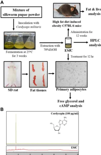
Fig. 1. Experimental scheme for anti-obesity effect of ESfC and identification of coerdycepin. (A) After the fermentation of silkworm pupae powder with Cordyceps militaris, ESfC were used to analysis primary adipocytes and C57BL/6N mice feed with high fat diet. (B) Chromatograms of the cordycepin were obtained by high performance liquid chromatography of ESfC. The peak height/area reflect the concentration of cordycepin in ESfC.
A
B
C
Fig. 2. Analysis of adipocyte size, free glycerol release and cAMP concetration. (A) Morphology of primary adipo- cytes were observed at 200x magnification after ESfC treatment for 24 hr. (B) Released glycerol was measured in the supernatant of primary adipocytes treated with ESfC (100, 200 and 400 μg/ml) that were fermented for 3 weeks. (C) After the collection of supernatants from the primary adipocytes treated with 100, 200 and 400 μg/ml of the ESfC, cAMP concentration was measured with cAMP ELISA kit, which has a detection range of 1 to 1,000 pmol/ml cAMP. The data shown represent the means ± SD of three replicates. *, p<0.05 relative to the Vehicle treated group. ISOP; isoproterenol.
계적 유의성은 일원분산분석(Windows 용 SPSS, Release 10.10, Standard Version)을 이용하여 다중비교를 위해 Turkey post hoc t-검정을 사용하여 분석하였다. 모든 값은 평균(mean) ± 표준편차(SD)로 표기하였으며, p<0.05의 값은 유의성을 갖는 것으로 간주하였다.
결 과
ESfC에 Cordycepin의 확인
지방분해 효능을 갖는 지표물질이 ESfC에 존재를 확인하기 위하여, cordycepin의 농도를 HPLC를 이용하여 분석하였다.
그 결과, HPLC의 그래프에서 표준 cordycepin에 해당하는 피 크가 8분경에 ESfC에서 관찰되었다(Fig. 1B). 이러한 결과는
ESfC는 cordycepin을 함유하고 있으며, 항비만 효과를 유도할 가능성이 높음을 제시하고 있다.
ESfC가 일차지방세포에서 지방분해작용에 미치는 영향 ESfC가 일차지방세포의 지방분해작용에 미치는 영향을 확 인하기 위하여, SD랫드로부터 분리된 일차지방세포에 ESfC를
A
B
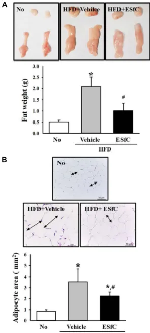
Fig. 3. Weight and histological structure of abdominal fat. (A) Weight of fat collected from abdominal region were measured using electrical balance. (B) The abdominal fat collected from the C57BL/6N mice was fixed in 4% for- malin and stained with H&E solution. The area of the adipocytes was measured in triplicate using a Leica Application Suite and is regarded as the cell size. The data shown represent the means ± SD of three replicates.
*, p<0.05 relative to the No treated group. #, p<0.05 rela- tive to the HFD+Vehicle treated group.
농도별로 처리한 후 지방세포 크기, free glycerol 농도, cAMP 농도의 변화를 측정하였다. 그 결과, 지방세포의 크기는 ESfC 를 처리한 그룹에서 농도 의존적으로 감소하였다(Fig. 2A).
Free glycerol 농도는 400 μg/ml ESfC처리그룹에서만 유의적 으로 증가하였으며, 100 μg/ml과 200 μg/ml ESfC 처리군에서 는 유의적인 변화가 관찰되지 않았다(Fig. 2B). 또한 cAMP농 도도 free glycerol 농도와 유사한 변화를 나타내었다. ESfC 400 μg/ml 처리군에서는 cAMP 농도가 유의적으로 증가하였 으나 100 μg/ml과 200 μg/ml 처리군에서는 유의적인 변화가 관찰되지 않았다(Fig. 2C). 따라서 이러한 결과는 ESfC는 고농 도에서 cAMP 신호전달과정을 동반하여 지방분해를 촉진하 는 활성이 있음을 제시하고 있다.
ESfC가 비만동물의 지방조직에서 지방축적에 미치는 영향 ESfC가 지방조직에서 지방축적에 미치는 영향을 평가하기 위하여, 12주간의 HFD와 ESfC를 C57BL/6마우스에 투여하고 지방조직의 변화를 관찰하였다. 그 결과, 복부지방의 무게는 정상사료를 섭취한 No 처리군에 비하여 HFD+Vehicle그룹에 서 320% 증가하였으나 HFD+ESfC 처리군에서는 80%만 증가 하였다(Fig. 3A). 또한, 지방세포의 크기도 유사한 경향을 나타 내었다. HFD+Vehicle 처리군에서는 지방세포의 크기가 375%
증가하였지만 HFD+ESfC 처리군에서는 163%만 증가하였다 (Fig. 3B). 따라서 이러한 결과는 ESfC는 C57BL/6마우스의 복 부지방조직에서 지방의 축적을 억제하는 효과를 나타냄을 제 시하고 있다.
ESfC가 비만동물의 간조직에서 지방축적 및 분해에 미치 는 영향
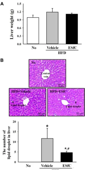
ESfC가 비만동물의 간조직에서 지방축적과 분해에 미치는 영향을 평가하기 위하여, 12주간의 HFD와 ESfC를 C57BL/6 마우스에 투여하고 간조직의 변화를 관찰하였다. 그 결과, 간 의 무게는 No 처리군에 비하여 HFD+Vehilce 처리군에서 증 가하였고 HFD+ESfC 처리군에서 감소하는 경향을 나타내었 으나 통계적 유의성은 없었다(Fig. 4A). 또한, 간조직에서 lipid drop의 축적도 유사한 경향을 나타내었다. HFD+Vehilce 처리 군은 급격하게 증가된 lipid drop의 수를 나타내었지만 HFD+ESfC 처리군은 유의적인 감소를 나타내었다(Fig. 4B).
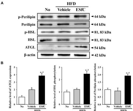
더불어, 지방분해 활성을 관찰하기 위하여 중성지방 대사와 연관된 주요단백질인 perilipin, HSL, ATGL의 변화를 관찰하 였다. 그 결과, perilipin과 HSL의 인산화는 No 처리군과 HFD +Vehicle 처리군에서는 유의적인 변화가 없었지만 HFD+
ESfC 처리군에서는 유의적으로 증가하는 경향을 나타내었다.
또한, ATGL의 발현양은 No 처리군에 비하여 HFD+Vehicle 처리군에서 약간 증가하였지만 HFD+ESfC 처리군에서는 급 격히 증가하였다(Fig. 5). 이러한 결과는 ESfC는 perilipin의 인산화를 유도하여 ATGL과 HSL의 발현과 활성을 증가시켜 간조직에서 지방분해를 촉진함을 제시하고 있다.
고 찰
지방분해활성과 지방축적억제활성을 갖는 새로운 비만 치 료제는 체내의 지방축적은 암, 심혈관질환, 고지혈증, 당뇨, 고혈압 등을 포함한 다양한 만성질환의 주요 원인 중의 하나 로 고려되기 때문에 오랫동안 많은 연구자들의 관심을 받아왔
A
B
Fig. 4. Weight and histological structure of liver. (A) Weight of liver collected from abdominal region were measured using electrical balance. (B) After the collection from the abdominal cavity, the liver tissue was fixed in 4% for- malin and stained with H&E solution. The number of lipid pore was counted in triplicate using a Leica Application Suite. The data shown represent the means
± SD of three replicates. *, p<0.05 relative to the No treat- ed group. #, p<0.05 relative to the HFD+Vehicle treated group.
다[24, 26]. 지금까지 많은 연구들은 norepinephrine, theophyl- line, forskolin, isoproterenol, dibutyryl-cAMP (DBcAMP) 등 여러 지방분해촉진제가 지방세포에서 지방분해를 촉진할 수 있다고 보고하고 있다[27]. 이와 더불어 다양한 천연물치료제 가 새로운 활성물질을 분리하고 부작용이 없고 안전한 항비만 치료제를 개발하기 위한 전략으로 알려져 있다[30]. 따라서 본 연구에서는 ESfC의 지방분해 촉진과 지방축적 억제 효능을 평가하기 위하여 SD랫드로부터 분리된 일차지방세포에서 free glycerol release, cAMP 농도를 분석하였고, 고지방사료 로 유발된 비만모델동물에서 지방조직과 간조직의 지방축적
을 분석하였다. 이러한 결과는 ESfC가 비만을 억제하는 우수 한 천연물로서 가능성이 있음을 보여주는 과학적인 증거를 제시하고 있다.
현재까지, cordycepin은 ribose의 3번 탄소위치에 OH그룹 이 없는 adenosine 유도체로서 Cordyceps militaris로부터 처음 에 추출되었다[36]. Cordycepin은 일부 백혈병세포(leukemic cell)에 독성을 나타내지만[39], 실험동물에서 우수한 항우울 제효과를 나타내었다[42]. 하지만 지금까지 대부분의 연구는 cordycepin의 지방대사기능에 집중되어 있다. Cordycepin은 3T3-L1세포의 지방합성과정을 억제하였고[43], hyperlipidermic 햄스터와 랫드에서 지방프로파일의 조절과 지방간 생성을 억 제하였다[44, 46, 50]. 본 연구에서는 cordycepin이 함유된 ESfC을 이용하여 지방분해활성과 지방축적억제 효능을 일차 지방세포와 HFD유도 비만모델(C57BL/6 마우스)을 이용하여 분석하였으며, 이러한 결과는 cordycepin의 비만조절 기전에 대한 부가적인 과학적 근거를 제시하고 있다.
누에번데기 오일의 중성지방감소 효과와 지방간 생성억제 등의 효능에 대한 연구는 일부 보고되었지만[14, 42], 누에번데 기 가루를 이용한 연구는 매우 적다. 누에번데기 가루를 5주 동안 SD랫드에 투여한 결과, 복부지방량은 29.5% 감소하였고, 지방대사에 관여하는 단백질인 PPAR-γ, L-FABP, CPF-1의 발 현은 유의적으로 증가하였다[33]. 이러한 실험결과는 비록 분 석된 지방대사 관련 단백질의 종류에 차이가 있지만 본 실험 의 결과와 매우 유사하다. 본 실험에서는 복부지방의 축적량 감소가 240%로 매우 컸으며, perilipin, HSL, ATGL의 유의적 인 발현증가를 제시하고 있다. 하지만 복부지방량의 차이는 실험과정에서 고지방사료 급여와 일반사료 급여에 의해 유도 된 것으로 사료된다.
한편, 지방분해는 중성지방이 가수분해를 통해 glycerol과 자유지방산으로 분해를 포함하며, 이러한 과정은 세포질 내 2차대사전달인자인 cAMP의 중계를 통한 PKA의 인산화가 중 요하게 작용한다[6] 인산화된 PKA는 perilipin과 HSL의 인산 화를 촉진시키며 최종적으로 ATGL과 p-HSL이 중성지방의 분해를 유도한다[10]. 다양한 천연물질은 지방분해과정에 주 요 인자들의 변화를 유도한다. 녹차추출물(Green tea extract) 은 고지방사료와 함께 Swiss마우스 투여할 때, 장간막지방조 직(mesenteric adipose tissue)에서 HSL과 perilipin발현 증가 를 유도하였고[10], Cyclopia maculata와 Cyclopia subternata의 열수추출물은 3T3L-1세포에서 HSL과 perilipin발현를 증가시 켰다[32]. 본 연구에서는 HFD와 ESfC를 12주간 C57BL/6마우 스에 투여하고, 지방조직과 간조직에서 지방의 축적을 관찰하 였으며, 대부분의 결과는 이전의 결과와 매우 유사하였다. 따 라서 본 연구의 결과는 중성지방의 가수분해를 촉진하는 새로 운 천연추출물에 대한 효능과 작용기전에 대한 과학적 근거를 제시하고 있다.
결론적으로 본 연구에서는 ESfC가 지방분해와 지방축적에
A
B
Fig. 5. Expression level of proteins related with lipolysis. Changes in the expression level of p-perilipin, perilipin, p-HSL, HSL, ATGL and β-actin were examined in the liver tissue using a specific antibody. The data shown represent the means ± SD of three replicates. *, p<0.05 relative to the No treated group. #, p<0.05 relative to the HFD+Vehicle treated group.
미치는 영향을 평가하기 위하여 일차지방세포와 비만모델동 물에서 지방대사에 ESfC를 처리하여 효능과 작용기전을 평가 하였다. 이러한 결과는 비록 추가적인 약리작용기전에 대한 연구가 필요하지만 ESfC가 지방분해를 촉진하고 지방축적을 억제하는 치료제로서 사용될 가능성이 있음을 제시하고 있다.
감사의 글
본 연구는 농림수산식품기술기획평가원(116027-032-HD030) 으로부터 지원을 받아 수행하였으며, 이에 감사드립니다.
References
1. Albu, J., Allixon, D., Boozer, C. N., Heymsfield, S., Kissileff, H., Kretser, A., Krumhar, K., Leibel, R., Nonas, C., Pi- Sunyer, X., VanItallie, T. and Wedral, E. 1997. Obesity sol- utions report of a meeting. Nutr. Rea. 55, 150-156.
2. An, S., Han, J. I., Kim, M. J., Park, J. S., Han, J. M., Baek, N. I., Chung, H. G., Choi, M. S., Lee, K. T. and Jeong, T.
S. 2010. Ethanolic extracts of Brassica campestrisspp. rapa roots prevent high-fat diet-induced obesity via beta (3)-adre- nergic regulation of white adipocyte lipolytic activity. J.
Med. Food 13, 406-414.
3. Ann, J. Y., Eo, H. Y. and Lim, Y. S. 2015. Mulberry leaves (Mours albla L.) ameliorate obesity-induced hepatic lipo- genesis, fibrosis, and oxidative stress in high-fat diet-fed mice. Genes Nutr. 10, 46-58.
4. Apostolopoulou, M., Savopoulos, C., Michalakis, K., Coppack, S., Dardavessis, T. and Hatzitolios, A. 2012. Age, weight and obesity. Maturitas 71, 115-119.
5. Azman, K. F., Amom, Z., Azlan, A., Esa, N. M., Ali, R. M., Shah, Z. M. and Kadir, K. K. 2012. Antiobesity effect of Tamarindus indicaL. pulp aqueous extract in high-fat diet-in- duced obese rats. J. Nat. Med. 66, 333-342.
6. Carmen, G. Y. and V´ıctor, S. M. 2006. Signaling mecha- nisms regulating lipolysis. Cell Signal. 18, 401-408.
7. Cha, J., Kim, Y., Moon, H. and Cho, Y. 2012 Hepatoprotective effects on alcoholic liver disease of fermented silk worms with Bacillus subtilis and Aspergillus Kawachii. Int. J. Food.
Sci. Nut. 63, 537-547.
8. Chen, J. and Li, X. 2007. Hypolipidemic effect of flavonoids from mulberry leaves in triton WR-1339 induced hyper- lipidemic mice. Asia Pac. J. Clin. Nutr. 16, 290-294.
9. Cho, H. J., Shim, M. J., Choi, E. C. and Kim, B. K. 1988.
Studies on constituents of higher fungi of korea (L57) com- parison of various antitumor constituents of coriolus versi-
color. Kor. J. Mycol. 16, 162-174.
10. Cunha, C. A., Lira, F. S., Rosa Neto, J. C., Pimentel, G. D., Souza, G. I., da Silva, C. M., de Souza, C. T., Ribeiro, E. B., Sawaya, A. C., Oller do Nascimento, C. M., Rodrigues, B., de Oliveira Carvalho, P. and Oyama, L. M. 2013. Green tea extract supplementation induces the lipolytic pathway, at- tenuates obesity, and reduces low-grade inflammation in mice fed a high-fat diet. Mediators Inflamm. 2013, 1-8.
11. Gabriela, P. C. M., Camila, O. S., Scherolin, de O. M., Thais, F. L., Bruno, L. S. P., José, C. R., Adelino, S. R. S., José, R.
P., Dennys, E. C., Eduardo, R. R., Bruno, R., Fabio, S. L.
and Claudio, T. S. 2015. Topiramate effects lipolysis in 3T3-L1 adipocytes. Biomed. Rep. 3, 827-830.
12. Gavia, M. H., Couto, R. C., Oyama, L. M., Couto, G. E., Silveira, V. L., Ribeiro, E. B. and Nascimento, C. M. 2003.
Diets rich in polyunsaturated fatty acids effect on hepatic metabolism in rats. Nutrition 19, 144-149.
13. Grundy, S. M. 1998. Multifactorial causation of obestiry: im- plications for prevention. Am. J. Clin. Nutr. 67, 563S-572S.
14. Harris, W. S., Ginsberg, H. N., Arunakul, N., Shachter, N.
S., Windsor, S. l., Adams, M., Berglund, L. and Osmundsen, K. 1997. Safety and efficacy of Omacor in severe hyper- triglyceridemia. J. Cardiovasc. Risk 4, 385-391.
15. Hudson, S. M. 1998. Applications of chitin and chitosan as fiber and textile chemicals. In: Domard A, Roberts GAF, Varum KM (eds) Advances in chitin science, vol 2. Lyon (France), Jacques Andre, pp 590-599.
16. Hursting, S. D., Thornfquist, M. and Henderson, M. M. 1990.
Types of dietary fat and the incidence of cancer at five sites.
Prev. Med. 19, 242-253.
17. Jeon, J. R., Kim, J. Y., Lee, K. M. and Cho, D. H. 2005. Anti- obese effects of mixture contained pine needle, black tea and green tea extracts. J. Kor. Soc. Appl. Biol. Chem. 48, 375-381.
18. Kamata, N., Sato, H. and Himazu, M. 1997. Seasonal changes in the infection of pupae of the beech caterpillar, Quadricalcarifera punctatella (motsch.) (Lep., Notodontidae), by Cordyceps militaris Link (Clavicipitales, Clavicipitaceae) in the soil of the Japanese beech forest. J. Appl. Entomol. 121, 17-21.
19. Kim, Y., Park, J., Zhoh, C., Hong, S. and Ryu, K. 2010. The component analysis of male silkworm’s extract. J. Kor. Soc.
Esthe. Cosm. 5, 37-45.
20. Kim, Y. S., Kim, H. B., Jeon, B. D., Lee, D. W. and Ryu, S. P. 2016. Increased muscle mass after resistance exercise training and ingestion of silkworm pupae powder (Bombyx mori L.) in ICR mice. Kor. J. Appl. Entomol. 55, 1-9.
21. Kim, H. J. and Kang, C. H. and Kim, S. K. 2012. Anti-adipo- genic effect of Undaria pinnatifida extracts by ethanol in 3T3-L1 adipocytes, J. Life Sci. 22, 1052-1056.
22. Kim, J. B. 2001. New horizon in atherosclerosis research;
Insights in to fat cell differentiation and insulin sensitivity with ADD1/SREBP1 and PPARγ. Korea J. Lipidol. 9, 79-83.
23. Kobayahi, Y. and shimizu, D. 1983. Ionography of vegetable wasp and plant warms. Hoikusa Pub. Comp Ltd. Osaka pp.280
24. Kobayashi, Y., Miyazawa, M., Kamei, A., Abe, K. and Kojima,
T. 2010. Ameliorative effects of mulberry (Morus alba L.) leaves on hyperlipidemia in rats fed a high-fat diet: in- duction of fatty acid oxidation, inhibition of lipogenesis, and suppression of oxidative stress. Biosci. Biotechnol. Biochem.
74, 2385-2395.
25. Kuo, Y. C., Lin, C. Y., Tsai, W. J., Wu, C. L., Chen, C. F.
and Shiao, M. S. 1994. Growth inhibitors against tumor cells in Cordyceps sinensis other than cordycepin and polysaccha- rides. Cancer Invest. 12, 611-615.
26. Lafontan, M. and Langin, D. 2009. Lipolysis and lipid mobi- lization in human adipose tissue. Prog. Lipid Res. 48, 275-297.
27. Lean, M. E. 2000. Pathophysiology of obesity. Proc. Nutr.
Soc. 59, 331-336.
28. Lee, H., Park, D., Yang, G., Bae, D. K., Yang, Y. H., Kim, T. K., Kim, D., Kyung, J., Yeon, S., Koo, K. C., Lee, J. Y., Hwang, S. Y., Joo, S. S. and Kim, Y. B. 2012. Silk and silk- worm pupa peptides suppress adipogenesis in preadipocytes and fat accumulation in rats fed a high-fat diet. Eur. J. Nur.
51, 1011-1019.
29. Lee, J. K., Kang, M. G., Kim, Y. H. and Lee, J. S. 2012.
Screening of medicinal plants containing lipase inhibitor and optimal extraction conditions. Kor. J. Med. Crop Sci. 20, 1-7.
30. Mayer, M. A., Hcht, C., Puyü, A. and Taira, C. A. 2009.
Recent Advances in obesity pharmacotheraphy. Curr. Clin.
Pharacol. 4, 53-61.
31. Meetali, D., Dulal Chandra, B., Dipali, D. and Rajlakshmi, D. 2014. Antioxidant and antigenotoxic effects of pupae of the muga silkworm Antheraea assamensis. Food Biosci. 5, 108- 114.
32. Pheiffer, C., Dudhia, Z., Louw, J., Muller, C. and Joubert, E. 2013. Cyclopia maculata (honeybush tea) stimulates lip- olysis in 3T3-L1 adipocytes. Phytomedicine 20, 1168-1171.
33. Ryu, S. P. 2014. Silkworm pupae powder ingestion increases fat metabolism in swim-trained rats. J. Exerc. Nutr. Biochem.
18, 141-149.
34. Sasaki, S., Horacsek, M. and Kesteloot, H. 1993. An eco- logical study of the relationship between dietary fat intake and breast cancer mortality. Prev. Med. 22, 187-202.
35. Shen, Q. and Chen, S. 2001. Effect of Cordyceps militaris on the damage of rats induced by n-hexane. Zhong Yao Cai 24, 112-116.
36. Sun, Y., Wang, Y. H., Qu, K. and Zhu, H. B. 2011. Beneficial effects of cordycepin on metabolic profiles of liver and plas- ma from hyperlipidemic hamsters. J. Asian Nat. Prod. Res.
13, 534-546.
37. Sung, J. M., Lee, K. H., Yoo, Y. J., Choi, Y. S., Kim, S. H., Kim, Y. O. and Sung, G. H. 1998. Classification of Cordycpes species based on protein banding pattern. Kor. J. Mycol. 26, 1-7.
38. Takahashi, S., Tamai, M., Nakajima, S., Kato, H., Johno, H., Nakamura, T. and Kitamura, M. 2012. Blockade of adipocyte differentiation by cordycepin. Br. J. Pharmacol. 167, 561-575.
39. Taniguchi, S., Asano, N., Tomino, F. and Miwa, I. 1998.
Potentiation of glucose-induced insulin secretion by fag- omine, a pseudo-sugar isolated from mulberry leaves. Horm.
초록:일차 지방세포와 고지방식이로 유발한 비만모델동물에서 Cordyceps militaris로 발효시킨 누에 (Bombyx mori) 가루 에탄올 추출물의 항비만 효과
김지은1†․이미림1†․최준영1․박진주1․김혜령1․송보람1․최영환2․김경미3․황대연1*
(1부산대학교 생명자원과학대학 바이오소재과학과 & 생명융합연구센터, 2부산대학교 생명자원과학대학 원예생명
과학과, 3(주)노바렉스)
누에(Bombyx mori)번데기는 창상치료용 드레싱, 간보호활성, 항유전독성, 포도당농도 조절, 항암활성 등에 다양 하게 적용되고 있다. 본 연구에서는 붉은동충하초(Cordyceps militaris)를 이용하여 발효시킨 누에번데기분말의 에 탄올 추출물(Ethanol extract of silkworm pupae fermented with Cordyceps militaris, ESfC)의 항비만효능을 분석하 기 위하여, SD랫드로부터 분리된 일차지방세포(Primary adipocytes)에서 농도별로 ESfC를 처리하여 free glycerol release, cAMP농도를 측정하였고, 고지방사료(High fat diet, HFD)가 처리된 C57BL/6N 비만마우스에 ESfC를 투 여하여 지방조직과 간조직에서 지방축적(Fat accumulation)을 관찰하였다. 먼저, 지방감소효과를 나타내는 지표물 질로서 cordycepin의 존재는 HPLC분석을 통하여 ESfC에서 확인하였다. 또한, free glycerol수준과 cAMP농도는 고농도 ESfC가 처리된 일차지방 세포에서 유의적으로 증가하였지만 이들 수준은 저농도와 중농도의 ESfC로처리 세포에서는 유의적인 변화가 없었다. HFD-유도 비만모델에서, 고지방사료의 투여에 의해 유의적으로 증가된 지 방무게와 지방세포의 크기는 ESfC처리그룹에서 감소되었다. 간 무게와 지방방울(Lipid droplet)의 수는 고지방사 료의 투여에 의해 증가하였지만 ESfC투여에 의해 유의적으로 감소하였다. 또한, 유사한 회복효과는 perilipin과 HSL의 인산화 수준뿐만 아니라 ATGL의 발현수준에서도 관찰되었다. 따라서 이상의 결과는 ESfC가 지방일차세 포에서 지방분해를 촉진하고, HFS유도 비만모델에서 지방 축적을 억제하는 효과를 나타냄을 보여주고 있으며, 향후 비만환자에 처리하기 위한 약물로서 사용가능성을 제시하고 있다.
Metab. Res. 30, 679-683.
40. Wang, W., Shen, S., Chen, Q., Tanga, B., He, G., Ruan, H.
and Das, U. N. 2008. Hydrolyzates of silkworm pupae (Bombyx mori) protein is a new source of angiotensin I-con- verting enzyme inhibitory peptides (ACEIP). Curr. Pharm.
Biotechnol. 9, 307-314.
41. Wu, C., Guo, Y., Su, Y., Zhang, X., Luan, H., Zhang, X., Zhu, H., He, H., Wang, X., Sun, G., Sun, X., Guo, P. and Zhu, P. 2014. Cordycepin activates AMP-activated protein kinase (AMPK) via interaction with the c1 subunit. J. Cell Mol. Med.
18, 293-304.
42. Yang, J. W., Choi, E. M., Kwon, M. G. and Koo, S. J. 2005.
In vitro estrogenic activity of silkworm (Bombyx mori) pupa and herbs. J. East Asian Soc. Dietary Life 15, 315-322.
43. Yang, X., Huang, L., Hu, J. and LI, T. 2002. Effects of silk- worm pupa oil on serum lipids level and platelet function in rats. Wei Sheng Yan Jiu. 31, 249-251.
44. Yang, S. J., Park, N. Y. and Lim, Y. S. 2014. Anti-adipogenic effect of mulberry leaf ethanol extract in 3T3-L1 adipocytes.
Nutr. Res. Pract. 8, 613-617.
45. Yeo, Y. G., An, E. Y. and Ryu, S. 2103. Effect of pupa powder
diet on muscle and blood amino acids composition in rat.
Kor. J. Phy. Edu. 52, 807-818.
46. Youh, E. J., Seo, B. K., Nam, S. S. and Kang, S. K. 2010.
Anti-obesity effect of Rhizoma Atractylodis herbal acu- puncture in high fat diet-induced obese ICR mouse. J. Kor.
Acupunct. Moxibustion Soc. 27, 31-42.
47. Yusof, N. L., Wee, A., Lim, L. Y. and Khor, E. 2003. Flexible chitin films as potential wound-dressing materials: wound model studies. J. Biomed. Mater. Res. A 66A, 224-232.
48. Zhang, T., He, J., Xu, C., Zu, L., Jiang, H., Pu, S., Guo, X.
and Xu, G. Mechanisms of metformin inhibiting lipolytic response to isoproterenol in primary rat adipocytes. J. Mol.
Endocrinol. 42, 57-66.
49. Zhou, J. and Han, D. 2006. Safety evaluation of protein of silkworm (Antheraea pernyi) pupae. Food Chem. Toxicol. 44, 1123-1130.
50. Zimmermann, R., Strauss, J. G., Haemmerle, G., Schoiswohl, G., Birner-Gruenberger, R., Riederer, M., Lass, A., Neuberger, G., Eisenhaber, F., Hermetter, A. and Zechner, R. 2004. Fat mobilization in adipose tissue is promoted by adipose triglyceride lipase. Science 306, 1383-1386.